. 2023 Mar 2;83(5):731-745.e4.
doi: 10.1016/j.molcel.2022.12.033. Epub 2023 Jan 23.

Higher-order SPOP assembly reveals a basis for cancer mutant dysregulation

Affiliations

Higher-order SPOP assembly reveals a basis for cancer mutant dysregulation

Matthew J Cuneo et al. Mol Cell. .

Abstract

The speckle-type POZ protein (SPOP) functions in the Cullin3-RING ubiquitin ligase (CRL3) as a receptor for the recognition of substrates involved in cell growth, survival, and signaling. SPOP mutations have been attributed to the development of many types of cancers, including prostate and endometrial cancers. Prostate cancer mutations localize in the substrate-binding site of the substrate recognition (MATH) domain and reduce or prevent binding. However, most endometrial cancer mutations are dispersed in seemingly inconspicuous solvent-exposed regions of SPOP, offering no clear basis for their cancer-causing and peculiar gain-of-function properties. Herein, we present the first structure of SPOP in its oligomeric form, uncovering several new interfaces important for SPOP self-assembly and normal function. Given that many previously unaccounted-for cancer mutations are localized in these newly identified interfaces, we uncover molecular mechanisms underlying dysregulation of SPOP function, with effects ranging from gross structural changes to enhanced self-association, and heightened stability and activity.

Keywords: cryo-EM; endometrial cancer; filament; gain of function; higher-order oligomer; hypermorphic; oncogene; prostate cancer; structure; tumor suppressor.


Conflict of interest statement

Declaration of interests T.M. was a consultant for Faze Medicines, Inc. and is a member of the journal’s advisory board.

Figures
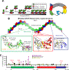
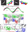
Figure 1.
Figure 1.. SPOP oligomer model cannot explain the molecular basis of cancer mutant dysfunction
(A) Left, cartoon schematic of concentration dependent SPOP self-association. SPOP dimerizes strongly through the BTB domain. The resulting BTB dimers further dimerize through the BACK domain resulting in linear higher-order oligomers with defined size distribution. A SPOP octamer is shown as example. (B) Model of a SPOP left-handed helical filament created through superposition of SPOP crystal structures (BTB dimer, PDB 3HQI; BACK dimer, PDB 4HS2). The molecular effect of cancer mutations located in the BTB/BTB (red box) and BACK/BACK (blue box) interfaces are readily rationalized in the structural model. In the MATH domain (green box), all but one prostate cancer mutations (purple) are localized to the substrate binding cleft and are known to reduce substrate binding. Most endometrial and other cancer mutations are outside of the substrate binding cleft in regions where no previous function has been identified. Only a few mutations can be rationalized in terms of MATH head-to-head dimerization or tail-to-tail interactions (grey circles). (C) The lollipop plot shows mutations identified in cancer patients in all three domains of SPOP (represented as a box plot). Prostate cancer mutations (magenta) are relatively frequent, occur in the substrate binding cleft and interfere with substrate binding. Intermediate-frequency endometrial cancer mutations (black) are clustered in regions of the MATH domain with no previously known function nor structural explanation by the filament model. Mutation data collated from cBioPortal.,
Figure 2.
Figure 2.. The cryo-EM structure of WT SPOP shows three previously unidentified protein-protein interfaces
(A) Representative 2D classes and sharpened cryo-EM maps of WT-SPOP oligomers in similar orientation (see also Figure S1). (B) The cryo-EM model of SPOP oligomers can be used to construct a left-handed helical filament by concatenation of tetramers. (C) The structure reveals three previously unidentified protein interfaces that mediate extensive MATH:MATH interactions (grey inset box) and “glue” the MATH domains and the C-terminus into the repeating unit of the filament (blue inset box). The linker connecting the MATH domains with the oligomerization domains appears in two distinct repeating conformations; one interacts specifically with the BTB and BACK domains of an adjacent monomer (Linker 1), whereas the other interacts only with an adjacent BTB domain (Linker 2). Alternating monomers are colored grey for clarity. (D) The MATH domains take on a continuous head-to-tail orientation along the filament with a repeating MATH:MATH interface (purple box). The β-extension interface (red box) mediates additional MATH:MATH self-association. Here, the N-terminus of SPOP, which was lacking from earlier studies, forms a well-ordered β-strand (β1) and a continuous β-sheet with the adjacent MATH domain as well as with what was in earlier crystal structures a disordered linker (now β9) following the structured MATH domain. (E) Close-up view of the glue pad, which anchors the MATH domain into the repeating oligomerization domains through a hydrophobic interface formed by three polypeptide chains. (Superscripts on residue names indicate the protein chains.)
Figure 3.
Figure 3.. SPOP cancer mutations line novel interfaces in the SPOP cryo-EM structure
Previously unexplainable mutations are found in the MATH:MATH and β-extension interfaces, and in the glue pad. A ridge of highly clustered mutations follows the face of the MATH domain that is positioned towards the interior of the filament (black callout box). This mutation ridge contains the cluster of charge-altering mutations that span the MATH:MATH interface (black and yellow). An additional cluster of cancer mutations is found in the glue pad (blue callout box), including the endometrial cancer mutation W22R and Y327C/Y327F in hepatocellular carcinoma and lung adenocarcinoma, respectively.
Figure 4.
Figure 4.. SPOP W22R forms dramatically altered filaments
(A) Representative 2D classes (left) and sharpened cryo-EM maps in similar orientation (right) of SPOP W22R double filaments with MATH hexamer assemblies, i.e., population 1 (see also Figure S3). (B) The cryo-EM model of population 1 of SPOP W22R can be used to construct a left-handed helical filament. Six ordered MATH domains (green surface, grey box) form a continuous assembly that link two individual SPOP filaments. The assembly is further held together by inter-monomer and inter-filament interactions of the substrate binding cleft in each MATH domain with a pseudo-SB motif sequence generated by mutation W22R (orange surface representation). Bottom, sequence alignment of N-terminal 30 amino acids of SPOP WT and W22R. Pseudo-SB motif in orange. (C) Close up view of MATH domains. The β2 strands intercalate across the double filament in a zipper-like fashion (β-zipper) and link together two SPOP filaments. The R22-containing pseudo-SB motif binds to adjacent monomers and further links the two filaments together. MATH monomers from one filament are colored grey, whereas MATH monomers from the other filament are colored green; the N-terminus bound in the SB binding cleft is colored orange. See also Figure S6 for representations of the connectivity. (D) Representative 2D classes (left) and sharpened cryo-EM maps in similar orientation (right) of SPOP W22R assemblies with MATH tetramer assemblies, i.e., population 2 (see also Figure S4). (E) Population 2 of SPOP W22R particles have an alternative arrangement with a tetramer repeating unit. MATH tetramers link together two filaments. Helical parameters are similar to the population 1 of the SPOP W22R double filament.
Figure 5.
Figure 5.. The E47K mutation leaves the structure of SPOP relatively unperturbed.
(A) Representative 2D classes (left) and sharpened cryo-EM maps in similar orientation (right) of SPOP E47K oligomers (see also Figure S7). (B) Left-handed helical filament constructed from the cryo-EM model of SPOP E47K; the helical parameters are similar to the WT filament, and the domains also have highly similar orientations relative to each other. (C) Ribbon representation of the MATH:MATH interface; monomer 1 is colored grey, monomer 2 green. A cluster of mutations that alter charge are found in the interface. The height of the labels indicates the number of identified mutations at the given position (e.g., K101I was found once, and D140G/N/V/Y was found 8 times). (D) Self-assembly of MATH:MATH interface mutants is enhanced. Relative molecular mass of WT SPOP and SPOP mutants determined from small-angle X-ray scattering intensity at zero scattering angle (Io). All data are normalized relative to WT SPOP at the lowest concentration. Lines are drawn to connect individual concentration measurements and guide the eye. Error bars indicate the error in P(r)-based I0 determination with Gnom. Dashed lines represent the 4.7-fold difference in molecular mass between WT and E47K at 10 μM. (For full SAXS curves see Figure S8.)
Figure 6.
Figure 6.. Endometrial cancer mutations enhance protein stability and therefore substrate turnover.
(A) E47K and E78K SPOP marginally increase ubiquitination activity in vitro, W22R does not. In vitro ubiquitination assays with CRL3SPOP were performed as described previously (Zhuang et al, 2009) using fluorescently labeled BRD3 as substrate, and WT SPOP or one of the characterized endometrial cancer mutants W22R, E47K and E78K SPOP. Top, ubiquitination efficiency was monitored by SDS–PAGE and fluorescent imaging. Bottom left and right, quantification of the decrease of unmodified BRD3 and of the increase of ubiquitinated BRD3 (Ubn-BRD3), respectively, as a function of time, in 3 independent assays. The mean value ± the S.D. are reported. (For additional conditions see Figure S8.) (B) SPOP endometrial cancer mutants enhance BRD3 ubiquitination in cells. Immunoblots showing BRD3-Flag ubiquitination in T-REX-293 cells transfected with the indicated constructs. 24 h post-transfection, cells were incubated with MG132 or DMSO at 20 μM for 4 hours. Cell lysates were subjected to His-Ub pulldown using nickel-NTA beads under denaturing conditions, followed by SDS–PAGE, and immunoblotting with anti-FLAG antibody. Input materials were subjected to immunoblotting using antibodies for Flag, MYC, and GAPDH (loading control). (C) SPOP endometrial cancer mutants increase the half-life of SPOP as determined by cycloheximide (CHX) chase assay in T-REX-293 cells. Left, a representative immunoblot analysis of CHX-treated T-REX-293 cells. Cells were transfected with the indicated SPOP constructs for 16 h and supplemented with 100 μg/ml of CHX for the indicated time periods. Right, the SPOP-Myc level at each time point relative to the level at time zero is the mean from three biological replicates and fit (solid line) to determine t1/2 values (inset box). Error bars are ± S.D.
Figure 7.
Figure 7.. SPOP filament structure provides insights into functional CRL assembly
(A) Updated box-plot of SPOP domains (top). Updated schematic of SPOP domain structure and SPOP filament highlighting the head-to-tail assembly of MATH domains and that every second MATH domain is locked into place on the BTB-BACK filament via the glue pad (bottom). As a consequence, alternate linkers have different conformations. (B) Model of the complete CRL3SPOP filament generated by superposition of X-ray crystal structures onto the cryoEM structure of the SPOP filament. The Cul3 N-terminal domain/SPOP complex (PDB ID 4EOZ) was used to orient the structure of the Cul1-Rbx1-Skp1-F-box-Skp2 SCF ubiquitin ligase complex. Each SPOP monomer can bind one Cullin without steric clashes. The substrate binding sites (orange surfaces) in the MATH domain are in a parallel orientation. (C) Model of the SPOP W22R filament in complex with Cullin, constructed as in panel (B). Binding of one Cullin per SPOP monomer creates steric clashes (grey inset); each SPOP dimer can bind one Cullin. The central hexameric MATH assembly creates two large anti-parallel continuous substrate binding sites (orange surface with black arrow showing direction of substrate chain), which may favor binding of multivalent substrates with closely spaced SPOP-binding motifs.

Comment in

Similar articles

Cited by

References

    1. Baek K, Scott DC, and Schulman BA (2021). NEDD8 and ubiquitin ligation by cullin-RING E3 ligases. Curr Opin Struct Biol 67, 101–109. 10.1016/j.sbi.2020.10.007. - DOI - PMC - PubMed
    1. Gao K, Jin X, Tang Y, Ma J, Peng J, Yu L, Zhang P, and Wang C (2015). Tumor suppressor SPOP mediates the proteasomal degradation of progesterone receptors (PRs) in breast cancer cells. Am J Cancer Res 5, 3210–3220. - PMC - PubMed
    1. Geng C, Kaochar S, Li M, Rajapakshe K, Fiskus W, Dong J, Foley C, Dong B, Zhang L, Kwon OJ, et al. (2017). SPOP regulates prostate epithelial cell proliferation and promotes ubiquitination and turnover of c-MYC oncoprotein. Oncogene 36, 4767–4777. 10.1038/onc.2017.80. - DOI - PMC - PubMed
    1. Geng C, He B, Xu L, Barbieri CE, Eedunuri VK, Chew SA, Zimmermann M, Bond R, Shou J, Li C, et al. (2013). Prostate cancer-associated mutations in speckle-type POZ protein (SPOP) regulate steroid receptor coactivator 3 protein turnover. Proc Natl Acad Sci U S A 110, 6997–7002. 10.1073/pnas.1304502110. - DOI - PMC - PubMed
    1. Janouskova H, El Tekle G, Bellini E, Udeshi ND, Rinaldi A, Ulbricht A, Bernasocchi T, Civenni G, Losa M, Svinkina T, et al. (2017). Opposing effects of cancer-type-specific SPOP mutants on BET protein degradation and sensitivity to BET inhibitors. Nat Med 23, 1046–1054. 10.1038/nm.4372. - DOI - PMC - PubMed

Publication types