. 2023 Jan 25;9(1):28.
doi: 10.1038/s41420-023-01309-0.

Gut microbiota and microbiota-derived metabolites promotes endometriosis

Affiliations

Gut microbiota and microbiota-derived metabolites promotes endometriosis

Sangappa B Chadchan et al. Cell Death Discov. .

Abstract

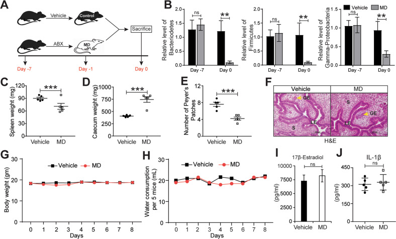
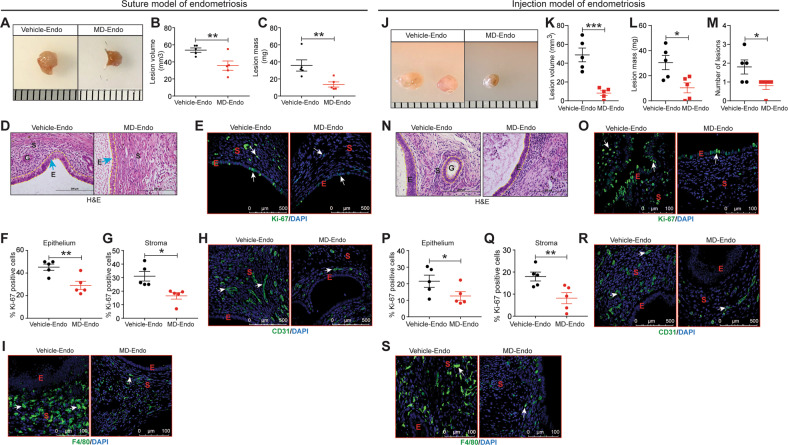
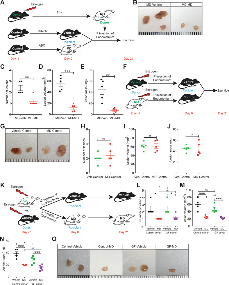
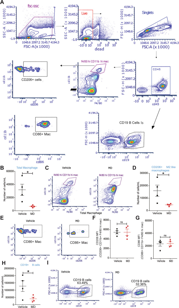
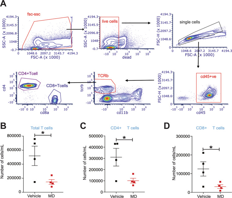
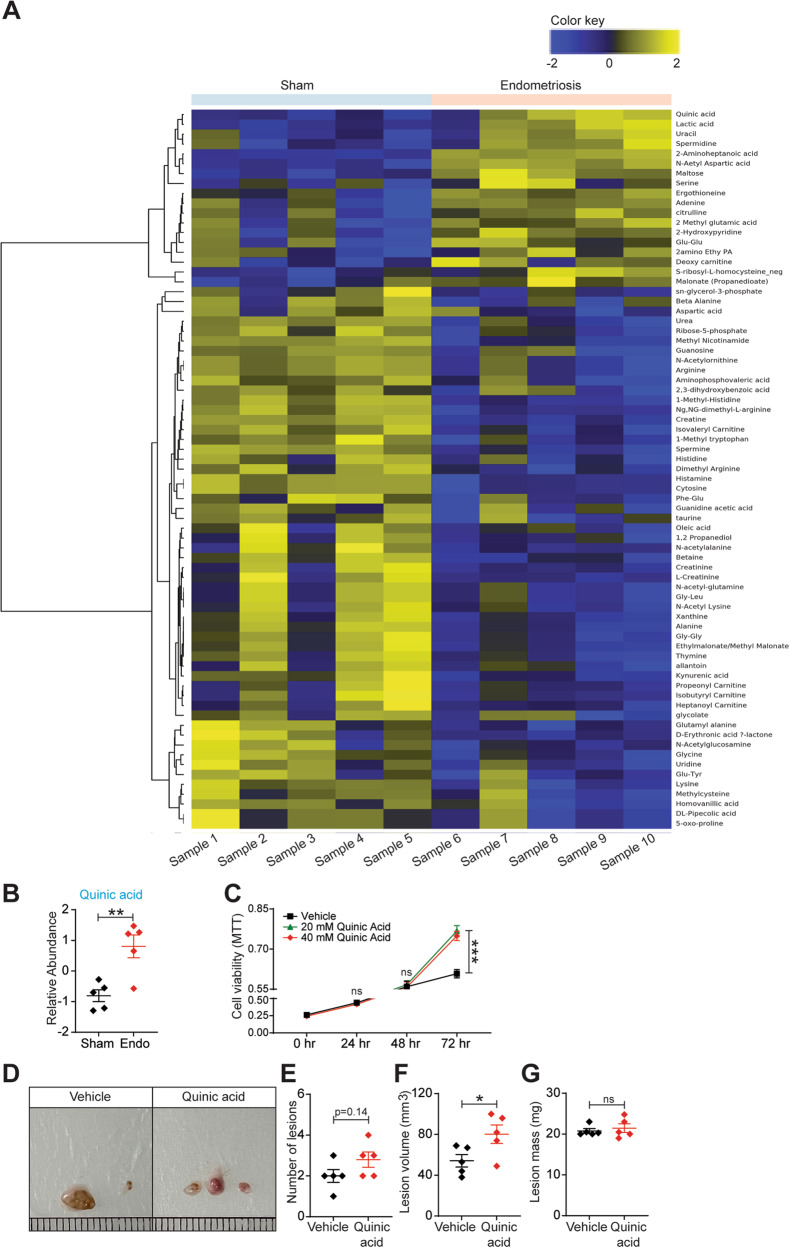
Endometriosis is a pathological condition of the female reproductive tract characterized by the existence of endometrium-like tissue at ectopic sites, affecting 10% of women between the age 15 and 49 in the USA. However, currently there is no reliable non-invasive method to detect the presence of endometriosis without surgery and many women find hormonal therapy and surgery as ineffective in avoiding the recurrences. There is a lack of knowledge on the etiology and the factors that contribute to the development of endometriosis. A growing body of recent evidence suggests an association between gut microbiota and endometriosis pathophysiology. However, the direct impact of microbiota and microbiota-derived metabolites on the endometriosis disease progression is largely unknown. To understand the causal role of gut microbiota and endometriosis, we have implemented a novel model using antibiotic-induced microbiota-depleted (MD) mice to investigate the endometriosis disease progression. Interestingly, we found that MD mice showed reduced endometriotic lesion growth and, the transplantation of gut microbiota by oral gavage of feces from mice with endometriosis rescued the endometriotic lesion growth. Additionally, using germ-free donor mice, we indicated that the uterine microbiota is dispensable for endometriotic lesion growth in mice. Furthermore, we showed that gut microbiota modulates immune cell populations in the peritoneum of lesions-bearing mice. Finally, we found a novel signature of microbiota-derived metabolites that were significantly altered in feces of mice with endometriosis. Finally, we found one the altered metabolite, quinic acid promoted the survival of endometriotic epithelial cells in vitro and lesion growth in vivo, suggesting the disease-promoting potential of microbiota-derived metabolites. In summary, these data suggest that gut microbiota and microbiota-derived metabolome contribute to lesion growth in mice, possibly through immune cell adaptations. Of translational significance, these findings will aid in designing non-invasive diagnostics using stool metabolites for endometriosis.


Conflict of interest statement

The authors declare no competing interests.

Figures

Fig. 1
Fig. 1. Generation of microbiota-depleted mice using antibiotics.
A Schematic of experimental timeline and procedures. B Quantification of relative abundances of Bacteroidetes, Firmicutes, and Gamma-proteobacteria in feces from vehicle and Microbiota-depleted (MD) mice. C-D Wet weights of C spleen and D caecum in indicated treatment groups at a sacrifice. E The number of Peyer’s patches from indicated treatment groups. F Representative images of Hematoxylin and Eosin-stained uterine cross-sections from the indicated treatment groups. LE, Luminal Epithelium; GE, Glandular Epithelium; S, Stroma. Yellow arrows indicate the gland. G-H Mouse G body weight and H water consumption at indicated time points in vehicle and MD mice. I-J Relative level of I 17β-Estradiol in serum and J IL-1β in peritoneal fluid of indicated treatment groups. Data are presented as mean ± SE (n = 5 mice per group). **P < 0.01, ***P < 0.001, and ns, non-significant.
Fig. 2
Fig. 2. Gut microbiota promotes endometriosis disease progression in mice.
A, J Ectopic endometriotic lesion representative images from A suture model J injection model. B, K The endometriotic lesion volumes from B suture model and K injection model. C, L The endometriotic lesion masses from C suture model and L injection model from the indicated groups 21 days postinduction of endometriosis. M Number of lesions per mouse in injection model from the indicated groups 21 days post-induction of endometriosis. D, N Representative images of ectopic lesions from D suture model and N injection model from the indicated treatment groups stained with Hematoxylin & Eosin (H&E). E, O Representative images of ectopic lesions from E suture model and O injection model from the indicated treatment groups stained with anti-Ki-67 antibody. F, P Percentages of Ki-67-positive cells in endometriotic lesion epithelium, F suture model and P injection model; G, Q stroma, G suture model and Q injection model. Representative images of ectopic lesions stained with H, R anti-CD31 in H suture model and R injection model; I, S anti-F4/80 I suture model and S injection model from the indicated treatment groups. White arrows indicate positive cells. Data are presented as mean ± SE (n = 5), *P < 0.05, **P < 0.01 and ***P < 0.001.
Fig. 3
Fig. 3. Gut microbiota is required for endometriotic lesion growth in mice.
A, E Schematic of experimental timeline and procedures for A suture model and E injection model for the fecal microbiota transfer (FMT) experiments. Microbiota-depleted mice underwent endometriosis induction and received an oral gavage of PBS (MD + PBS), feces from mice without endometriosis (MD + NE) or feces from mice with endometriosis (MD + E). B, F Ectopic endometriotic lesion representative images from B suture model and F injection model. C, G Ectopic lesion volumes from C suture model and G injection model. D, H Ectopic lesion masses from D suture model and H injection model from the indicated groups 21 days after the induction of endometriosis. I Number of lesions per mouse in injection model from the indicated groups 21 days post-induction of endometriosis. All the indicated data is 21 days after induction of endometriosis. Data are presented as mean ± SE (n = 5), *P < 0.05, **P < 0.01, ***P < 0.001, and ns, nonsignificant.
Fig. 4
Fig. 4. Uterine microbiota might be dispensable for endometriotic lesion growth in mice.
A, F, K Schematic of experimental timeline and procedures. Ectopic endometriotic lesion B, G, O representative images, C, H, L number of lesions per mouse, D, I, M volumes and E, J, N masses from the indicated groups 21 days after induction of endometriosis. Data are presented as mean ± SE (n = 5), *P < 0.05, **P < 0.01, ***P < 0.001 and ns nonsignificant.
Fig. 5
Fig. 5. Gut microbiota depletion affects the macrophage and B cell population in the peritoneal fluid of mice with endometriosis.
The endometriosis was induced in the vehicle and MD mice as shown in Fig. S1B and flow cytometric analysis was carried out on the peritoneal fluid from mice with endometriosis. A Flowchart of flow cytometric plots for the cell sorting using the Cytek Aurora. B The relative number of total microphages per mL and C flow cytometric plots in the peritoneal fluid from vehicle and MD mice. D The relative number of CD206+ M2-like macrophages (M2-like mac) per mL and E flow cytometric plots in the peritoneal fluid from vehicle and MD mice. F, G Mean fluorescence intensity of F CD206+ CD11b+ F4/80 hi mac (M2 like Macrophage) and G CD86+ CD11b+ F4/80 hi mac (M1like macrophage) in the peritoneal fluid from vehicle and MD mice. H The relative number of CD19+ B-cells per mL and I flow cytometric plots in the peritoneal fluid from vehicle and MD mice. All the indicated data is 21 days after the induction of endometriosis. Data are presented as mean ± SE (n = 4), *P < 0.05 and ns nonsignificant.
Fig. 6
Fig. 6. Gut microbiota depletion affects total, CD4+ and CD8+ T cell population in the peritoneal fluid from mice with endometriosis.
A Flowchart of Flow cytometric plots for the cell sorting using Cytek. B-D The relative number of B total T cells per mL, C CD4+ T cells per mL and D CD8+ cells per mL in the peritoneal fluid from vehicle and MD mice. The endometriosis was induced in the vehicle and MD mice as shown in Fig. S1B and flow cytometric analysis was carried out on the peritoneal fluid 21 days after the induction of endometriosis. Data are presented as mean ± SE (n = 4), *P < 0.05.
Fig. 7
Fig. 7. Fecal metabolites differ between mice with and without endometriosis.
A The heat map depicting the metabolites that were differentially present between mice with and without endometriosis with cutoff of FDR < 0.25. B Quinic acid is present at higher level in feces of mice with endometriosis. Stool samples from mice with and without endometriosis were subjected to LC-MS (in the Baylor College of Medicine Metabolomics Core) to detect ~150 water-soluble metabolites. Each row is a metabolite, and each column is a stool sample from an individual mouse; (n = 5 mice per group). Data are presented as mean ± SE. C MTT cell viability assays of iHEECs/Luc treated with 20 mM and 40 mM Quinic acid for indicated time points. Results are shown as mean ± SE (n = 3) and experiment repeated three times. Ectopic endometriotic lesion D representative images, E number of lesions per mouse, F volumes and G masses from the vehicle and QA treated groups,14 days after induction of endometriosis. Data are presented as mean ± SE (n = 5), *P < 0.05, **P < 0.01, ***P < 0.001 and ns, non-significant.

References

    1. Zondervan KT, Becker CM, Koga K, Missmer SA, Taylor RN, Vigano P. Endometriosis. Nat Rev Dis Prim. 2018;4:9. doi: 10.1038/s41572-018-0008-5. - DOI - PubMed
    1. Rogers PA, D’Hooghe TM, Fazleabas A, Gargett CE, Giudice LC, Montgomery GW, et al. Priorities for endometriosis research: recommendations from an international consensus workshop. Reprod Sci. 2009;16:335–46. doi: 10.1177/1933719108330568. - DOI - PMC - PubMed
    1. Kvaskoff M, Mahamat-Saleh Y, Farland LV, Shigesi N, Terry KL, Harris HR, et al. Endometriosis and cancer: a systematic review and meta-analysis. Hum Reprod Update. 2020;27:393–420. - PubMed
    1. Chen I, Veth VB, Choudhry AJ, Murji A, Zakhari A, Black AY, et al. Pre- and postsurgical medical therapy for endometriosis surgery. Cochrane Database Syst Rev. 2020;11:CD003678. - PMC - PubMed
    1. Kvaskoff M, Mahamat-Saleh Y, Farland LV, Shigesi N, Terry KL, Harris HR, et al. Endometriosis and cancer: a systematic review and meta-analysis. Hum Reprod Update. 2021;27:393–420. doi: 10.1093/humupd/dmaa045. - DOI - PubMed