

Connectivity-based parcellation of the amygdala and identification of its main white matter connections
- PMID: 36693904
- PMCID: PMC9873600
- DOI: 10.1038/s41598-023-28100-6
Connectivity-based parcellation of the amygdala and identification of its main white matter connections
Erratum in
-
Author Correction: Connectivity-based parcellation of the amygdala and identification of its main white matter connections.Sci Rep. 2023 Apr 4;13(1):5495. doi: 10.1038/s41598-023-32200-8. Sci Rep. 2023. PMID: 37015960 Free PMC article. No abstract available.
Abstract
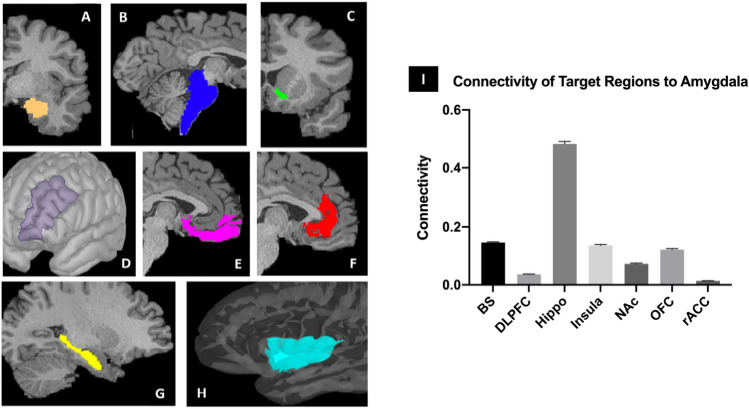
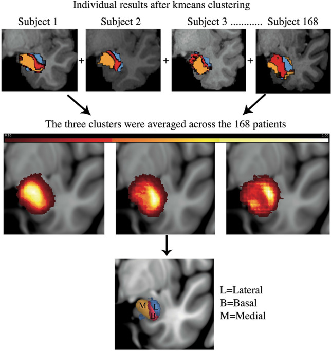
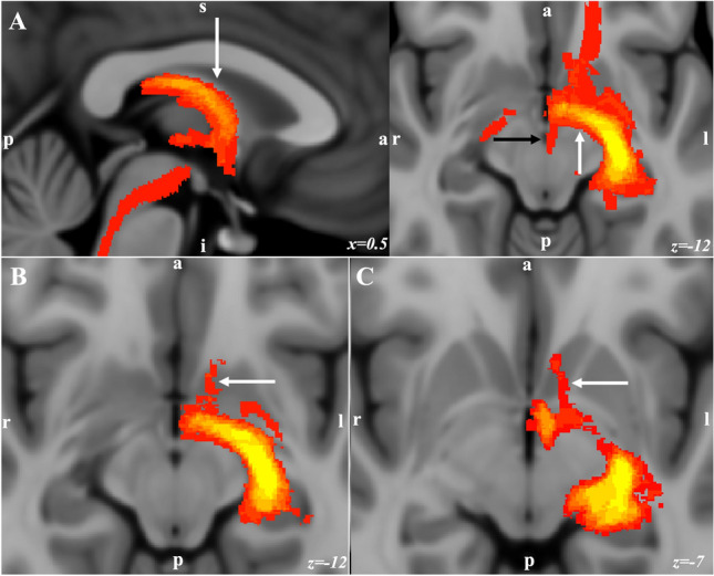
The amygdala plays a role in emotion, learning, and memory and has been implicated in behavioral disorders. Better understanding of the amygdala circuitry is crucial to develop new therapies for these disorders. We used data from 200 healthy-subjects from the human connectome project. Using probabilistic tractography, we created population statistical maps of amygdala connectivity to brain regions involved in limbic, associative, memory, and reward circuits. Based on the amygdala connectivity with these regions, we applied k-means clustering to parcellate the amygdala into three clusters. The resultant clusters were averaged across all subjects and the main white-matter pathways of the amygdala from each averaged cluster were generated. Amygdala parcellation into three clusters showed a medial-to-lateral pattern. The medial cluster corresponded with the centromedial and cortical nuclei, the basal cluster with the basal nuclei and the lateral cluster with the lateral nuclei. The connectivity analysis revealed different white-matter pathways consistent with the anatomy of the amygdala circuit. This in vivo connectivity-based parcellation of the amygdala delineates three clusters of the amygdala in a mediolateral pattern based on its connectivity with brain areas involved in cognition, memory, emotion, and reward. The human amygdala circuit presented in this work provides the first step for personalized amygdala circuit mapping for patients with behavioral disorders.
© 2023. The Author(s).
Conflict of interest statement
The authors declare no competing interests.
Figures


Similar articles
-
Deconstructing white matter connectivity of human amygdala nuclei with thalamus and cortex subdivisions in vivo.Hum Brain Mapp. 2017 Aug;38(8):3927-3940. doi: 10.1002/hbm.23639. Epub 2017 May 17. Hum Brain Mapp. 2017. PMID: 28512761 Free PMC article.
-
Functional connectivity-based parcellation of amygdala using self-organized mapping: a data driven approach.Hum Brain Mapp. 2014 Apr;35(4):1247-60. doi: 10.1002/hbm.22249. Epub 2013 Feb 18. Hum Brain Mapp. 2014. PMID: 23418140 Free PMC article.
-
Track-Weighted Dynamic Functional Connectivity Profiles and Topographic Organization of the Human Pulvinar.Hum Brain Mapp. 2024 Dec 1;45(17):e70062. doi: 10.1002/hbm.70062. Hum Brain Mapp. 2024. PMID: 39639553 Free PMC article.
-
Asymmetries in the human brain.Handb Clin Neurol. 2025;208:15-36. doi: 10.1016/B978-0-443-15646-5.00030-0. Handb Clin Neurol. 2025. PMID: 40074393 Review.
-
The Original Social Network: White Matter and Social Cognition.Trends Cogn Sci. 2018 Jun;22(6):504-516. doi: 10.1016/j.tics.2018.03.005. Epub 2018 Apr 5. Trends Cogn Sci. 2018. PMID: 29628441 Free PMC article. Review.
Cited by
-
Brainwide mesoscale functional networks revealed by focal infrared neural stimulation of the amygdala.Natl Sci Rev. 2024 Dec 24;12(4):nwae473. doi: 10.1093/nsr/nwae473. eCollection 2025 Apr. Natl Sci Rev. 2024. PMID: 40170996 Free PMC article.
-
Functional and structural connectivity of the subregions of the amygdala in ADHD children with or without ODD.BMC Psychiatry. 2025 Jan 24;25(1):74. doi: 10.1186/s12888-025-06500-4. BMC Psychiatry. 2025. PMID: 39856610 Free PMC article.
-
Direct parieto-occipital connectivity of the amygdala via the parahippocampal segment of the cingulum bundle.Neuroradiol J. 2025 May 14:19714009251339083. doi: 10.1177/19714009251339083. Online ahead of print. Neuroradiol J. 2025. PMID: 40366130 Free PMC article.
-
Neurotensin and Alcohol Use Disorders: Towards a Pharmacological Treatment.Int J Mol Sci. 2023 May 12;24(10):8656. doi: 10.3390/ijms24108656. Int J Mol Sci. 2023. PMID: 37240004 Free PMC article. Review.
