. 2023 Jan 24;13(1):1317.
doi: 10.1038/s41598-023-28363-z.

Pre-analytical sample handling effects on tear fluid protein levels

Affiliations

Pre-analytical sample handling effects on tear fluid protein levels

Marlies Gijs et al. Sci Rep. .

Abstract

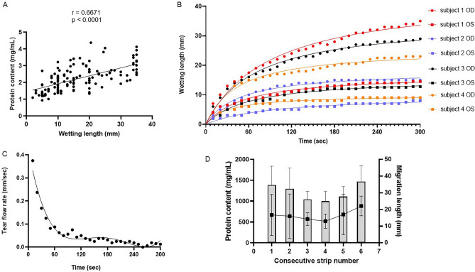
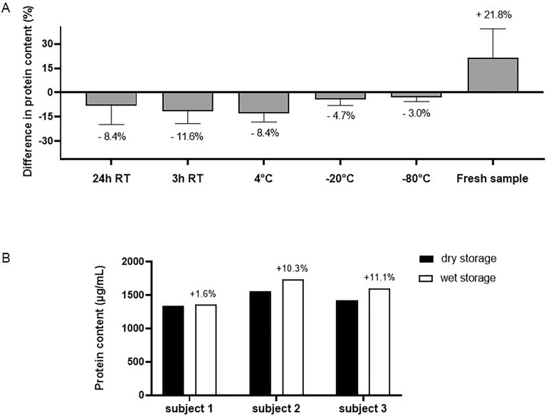
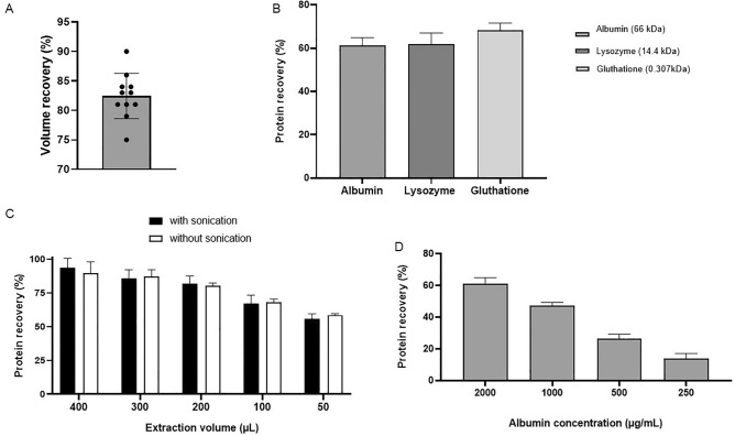
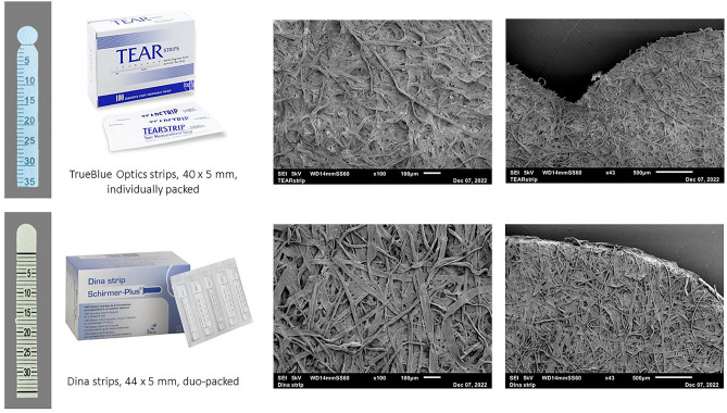
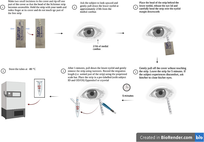
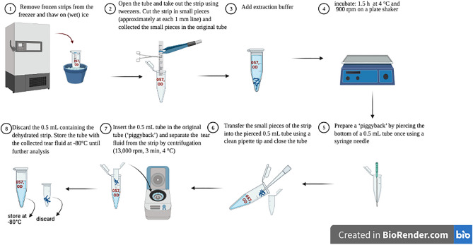
Tear fluid is emerging as a source of non-invasive biomarkers, both for ocular and systemic conditions. Accurate quantification of tear proteins can be improved by standardizing methods to collect and process tear fluid. The aim of this study was to determine sample handling factors that may influence the tear protein biomarker profile. Tear fluid was collected using Schirmer's strips. Tear proteins were extracted by elution through centrifugation. Total protein content was determined using the bicinchoninic acid assay. Key concepts that apply to the entire sample processing cycle are tear sampling, tear storage, protein extraction and data normalization. Differences in wetting or migration length were observed between Schirmer's strips from different manufacturers, and between protein-free and protein-rich solutions. One unit of migration length (mm) did not correspond to one unit of volume (µL). A positive correlation (r = 0.6671, p < 0.0001) was observed between migration length and total tear protein content. The most beneficial storage conditions were strips that were not stored (+ 21.8%), or underwent 'wet' storage (+ 11.1%). Protein recovery was the highest in 400 µL extraction buffer and independent of protein molecular weight. This study helps to explain inter- and intra-variability that is often seen with tear biomarker research. This information is critical to ensure accuracy of test results, as tear biomarkers will be used for patient management and in clinical trials in the near future. This study also highlights the need for standardization of Schirmer's strip manufacturing, tear fluid processing and analyte concentration normalization.


Conflict of interest statement

The authors declare no competing interests.

Figures

Figure 1
Figure 1
Factors influencing tear fluid sampling. (A) Migration length of fixed volumes of PBS or plasma on Schirmer’s strips (TrueBlue and Dina) from different manufacturers. Bars indicate mean ± SD. n = 3 for PBS and plasma, n = 1 for capillary tears, (B) differences in wetting length as compared to the theoretical/assumed values. Bars indicate mean ± SD. n = 3 for PBS and plasma, n = 1 for capillary tears.
Figure 2
Figure 2
Factors influencing tear fluid sampling. (A) Scatter plot of total protein content versus wetting length, n = 115, (B) wetting length as measured each 10 s after insertion, n = 8, (C) average tear flow rate plotted versus time, (D) total protein content of consecutive sampled tears. Consecutive strip numbers correspond to the time of strip collection from baseline: strip 1 (0 min), strip 2 (5 min), strip 3 (15 min), strip 4 (35 min), strip 5 (60 min) and strip 6 (90 min). Bars and squares indicate mean ± SD. n = 4. Differences between time points were not significant (p = 0.3769).
Figure 3
Figure 3
Factors influencing tear fluid storage. (A) Protein content of strips stored at different storage conditions, bars represent mean % ± SD change in protein content from control (= − 80 °C), n = 3, (B) Protein content of strips stored in wet versus dry storage conditions, bars represent individual values, labels indicate % change in protein content from control (= dry storage), n = 3 subjects. Differences between storage temperatures were not significant (p = 0.0611).
Figure 4
Figure 4
Factors influencing tear protein extraction. (A) Volume recovery (%) after application of a fixed volume PBS to an empty strip, n = 11, (B) protein recovery (%) of three proteins with different molecular weight, bars represent mean ± SD, n = 3, (C) protein recovery (%) of extraction performed with different extraction volumes and with and without sonication, bars represent mean ± SD, n = 3, (D) protein recovery (%) of extraction performed in samples with a different initial albumin concentrations, bars represent mean ± SD, n = 3.
Figure 5
Figure 5
Schirmer’s strips (TrueBlue TEAR strips and Dina strips) Illustration and SEM images of the Schirmer’s strips from different manufacturers that were used in this study.
Figure 6
Figure 6
Tear fluid sampling protocol. Tear fluid was collected from the left and right eye without topical anesthesia. Care was taken not to touch the strip with fingertips. The migration length was read after 5 min from the preprinted scale bar on the strips. Immediately after sampling, samples were stored at − 80 °C, unless noted otherwise. Image created in Biorender.com.
Figure 7
Figure 7
Tear fluid extraction protocol. Tear fluid was extracted from the Schirmer’s strip by agitating small cut pieces of these strips in a volume of extraction buffer at 4 °C for 1.5 h. Tear fluid was then eluted by centrifugation (‘piggyback method’) and stored at − 80 °C until further use. Image created in Biorender.com. The label “057 OD” illustrates an example patient ID and right eye.

References

    1. Zhou L, Beuerman RW. The power of tears: how tear proteomics research could revolutionize the clinic. Expert Rev. Proteom. 2017;14:189–191. doi: 10.1080/14789450.2017.1285703. - DOI - PubMed
    1. Hagan S, Martin E, Enríquez-de-Salamanca A. Tear fluid biomarkers in ocular and systemic disease: potential use for predictive, preventive and personalised medicine. Epma J. 2016;7:1–20. doi: 10.1186/s13167-016-0065-3. - DOI - PMC - PubMed
    1. You J, et al. Tear fluid protein biomarkers. Adv. Clin. Chem. 2013;62:151–196. doi: 10.1016/B978-0-12-800096-0.00004-4. - DOI - PubMed
    1. Akkurt Arslan M, et al. Proteomic analysis of tears and conjunctival cells collected with schirmer strips using timsTOF Pro: Preanalytical considerations. Metabolites. 2021 doi: 10.3390/metabo12010002. - DOI - PMC - PubMed
    1. Rentka A, et al. Evaluation of commonly used tear sampling methods and their relevance in subsequent biochemical analysis. Ann. Clin. Biochem. 2017;54:521–529. doi: 10.1177/0004563217695843. - DOI - PubMed