. 2023 Jan 24;80(2):50.
doi: 10.1007/s00018-023-04699-7.

Circ-sh3rf3/GATA-4/miR-29a regulatory axis in fibroblast-myofibroblast differentiation and myocardial fibrosis

Affiliations

Circ-sh3rf3/GATA-4/miR-29a regulatory axis in fibroblast-myofibroblast differentiation and myocardial fibrosis

Cai-Xia Ma et al. Cell Mol Life Sci. .

Abstract

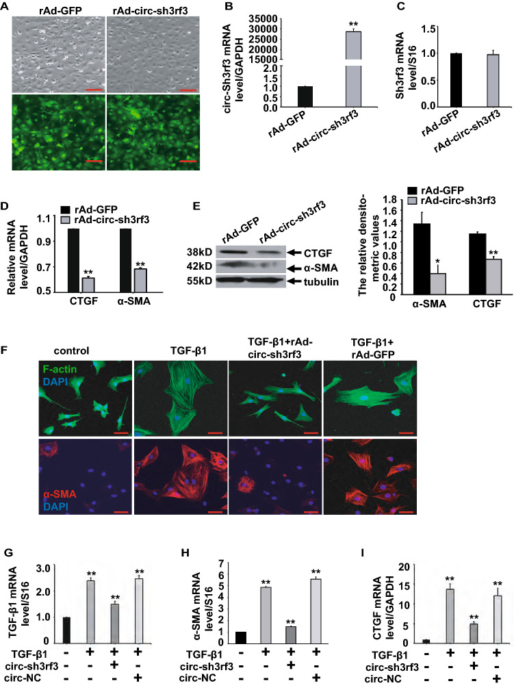
The transdifferentiation from cardiac fibroblasts to myofibroblasts is an important event in the initiation of cardiac fibrosis. However, the underlying mechanism is not fully understood. Circ-sh3rf3 (circular RNA SH3 domain containing Ring Finger 3) is a novel circular RNA which was induced in hypertrophied ventricles by isoproterenol hydrochloride, and our work has established that it is a potential regulator in cardiac hypertrophy, but whether circ-sh3rf3 plays a role in cardiac fibrosis remains unclear, especially in the conversion of cardiac fibroblasts into myofibroblasts. Here, we found that circ-sh3rf3 was down-regulated in isoproterenol-treated rat cardiac fibroblasts and cardiomyocytes as well as during fibroblast differentiation into myofibroblasts. We further confirmed that circ-sh3rf3 could interact with GATA-4 proteins and reduce the expression of GATA-4, which in turn abolishes GATA-4 repression of miR-29a expression and thus up-regulates miR-29a expression, thereby inhibiting fibroblast-myofibroblast differentiation and myocardial fibrosis. Our work has established a novel Circ-sh3rf3/GATA-4/miR-29a regulatory cascade in fibroblast-myofibroblast differentiation and myocardial fibrosis, which provides a new therapeutic target for myocardial fibrosis.

Keywords: Circular RNA; Fibroblast–myofibroblast differentiation; Myocardial fibrosis; microRNAs.


Conflict of interest statement

The authors declare no competing interest.

Figures

Fig. 1
Fig. 1
Decreased circ-sh3rf3 expression in isoproterenol (ISO)-induced cardiac fibrosis. (A, B) qPCR for circRNA_Sh3rf3 and Sh3rf3 in isoproterenol-treated rat cardiomyocytes (A) and cardiac fibroblasts (B). (C) qPCR assays for atrial natriuretic factor (ANF) and B-type natriuretic peptide (BNP) were performed in isoproterenol-treated rat cardiomyocytes. (D) The expression of transforming growth factor-β1 (TGF-β1), connective tissue growth factor (CTGF), matrix metalloproteinase-2 (MMP-2), matrix metalloproteinase-9 (MMP-9), collagen type I alpha 1 (COL1A1), and α-smooth muscle actin (α-SMA) was determined by qPCR in isoproterenol-treated rat cardiac fibroblasts. (E) Masson trichrome staining of left ventricular sections from mice infused with isoproterenol or saline (control) for 2 weeks (left), and fibrotic areas were quantitated by with Image J software (right). Scale bars: 50 μm. Iso isoproterenol. The data shown are the means ± SEMs from three independent experiments. *P < 0.05, **P < 0.01 compared with the control, and rat cardiomyocytes and cardiac fibroblasts treated with PBS (AD) served as the control
Fig. 2
Fig. 2
circ-sh3rf3 expression decreases during fibroblast differentiation into myofibroblasts. (A) Representative images of cardiac fibroblasts cultured in 1% or 10% serum using immunofluorescence. Cardiac fibroblasts spontaneously differentiated into myofibroblasts in 10% serum with marked changes in cell morphology and the presence of stress fibers by staining with F-actin. (BC) α-SMA expression was determined by Western blot (B) and qPCR (C). The protein bands were quantitated by densitometry (B, lower). (D) qPCR assay for circ-sh3rf3 in cardiac fibroblasts maintained in 10% serum. (E) Cardiac fibroblasts were starved for 24 h, followed by treatment with different concentrations of TGF-β1 (5, 10, 20 ng/ml) for 24 or 48 h. The mRNA level of α-SMA was analyzed by qPCR. (F) Representative images of fibroblasts treated with 5 ng/ml TGF-β1 for 48 h using immunofluorescence. F-actin staining (upper panel) shows marked changes in cell morphology, and immunofluorescence (lower panel) shows increased expression of α-SMA in cardiac fibroblasts stimulated with TGF-β1. (GI) qPCR for α-SMA, CTGF, and TGF-β1 in cardiac fibroblasts treated with TGF-β1. (J) Western blotting for α-SMA, CTGF, and TGF-β1 in TGF-β1-treated cardiac fibroblasts (left), and the protein bands were quantitated by densitometry (right). (K) qPCR analysis of circ-sh3rf3 in cardiac fibroblasts treated with TGF-β1. The data shown are the means ± SEMs from three independent experiments, **P < 0.01 compared with the control, and cardiac fibroblasts cultured in 1% serum (BD) and treated with PBS (E, GK) served as the control. Scale bars: 50 μm
Fig. 3
Fig. 3
circ-sh3rf3 inhibits fibroblast-to-myofibroblast differentiation. (A) Adenovirus-mediated overexpression of circ-sh3rf3 in cardiac fibroblasts cultured in medium with 10% serum. Recombinant circ-sh3rf3 adenovirus or empty adenovirus vector with the coexpression of green fluorescent protein (GFP) was constructed and then used to infect cardiac fibroblasts, and the infection efficiency of recombinant circ-sh3rf3 adenovirus (rAd-circ-sh3rf3) was reflected by the expression of GFP. rAd-GFP, empty adenovirus vector. Scale bars: 100 μm. (BC) qPCR of circ-sh3rf3 (B) and sh3rf3 (C) in cardiac fibroblasts infected with rAd-circ-sh3rf3 or rAd-GFP (control). (DE) Expression of CTGF and α-SMA was determined by qPCR (D) and Western blotting (E, left) in adenovirus-infected cardiac fibroblasts, rAd-GFP served as the control. and the protein bands were quantitated by densitometry (E, right). (F) Immunofluorescence analysis of F-actin (upper panel) and α-SMA (lower panel) in cardiac fibroblasts treated with TGF-β1 and/or circ-Sh3rf3. (GI) Expression of TGF-β1, α-SMA and CTGF in cardiac fibroblasts treated with TGF-β1 and/or circ-sh3rf3 was detected by qPCR, and cardiac fibroblasts treated with PBS served as the control. The data shown are the means ± SEMs from three independent experiments, **P < 0.01 compared with the control. Scale bars: 50 μm. circ-NC: circular RNA negative control
Fig. 4
Fig. 4
circ-sh3rf3 attenuates fibroblast-to-myofibroblast differentiation by upregulating miR-29a. (A) qPCR assays for miR-29a in cardiac fibroblasts infected with rAd-circ-sh3rf3. The empty vector served as the control. (B) Masson trichrome staining of left ventricular sections from mice infused with Iso and/or miR-29a agomir or agomir negative control. (CF) mRNA levels of CTGF, α-SMA, collagen type III alpha 1 (COL3A1), and MMP-2 were determined by qPCR in the left ventricles of mice treated with Iso and/or miR-29a agomir or agomir negative control, and treatment with saline served as the control. (G) The expression of miR-29a in isoproterenol-treated cardiac myocytes(cMs) and fibroblasts (cFs) was determined by qPCR, and treatment with PBS served as the control. (H) qPCR for miR-29a in cardiac fibroblasts transfected with miR-29a agomir or agomir negative control, and PBS treatment served as the control. (IL) qPCR for CTGF, TGF-β1, α-SMA, and MMP-2 in rat cardiac fibroblasts treated with Iso and/or miR-29a agomir or agomir negative control, and PBS treatment served as the control. (M) Western blotting for CTGF, TGF-β1, α-SMA, MMP-2, TGFβ-receptor1 (TGFβ-R1), and Smad3 phosphorylation (p-Smad3) in rat cardiac fibroblasts treated with Iso and/or miR-29a agomir or agomir negative control (upper). PBS treatment served as the control. The protein bands were quantitated by densitometry (lower). (N) F-actin and α-SMA staining using immunofluorescence analysis in cardiac fibroblasts treated with 5 ng/ml TGF-β1 for 48 h and/or miR-29a agomir. (O) qPCR for CTGF, α-SMA and TGF-β1 in cardiac fibroblasts overexpressed with circ-Sh3rf3 and/or transfected with miR-29a antagomir (left), and the expression of miR-29a was determined by qPCR (right). Cardiac fibroblasts overexpressed with antagomir negative control (right) and circ-Sh3rf3 (left) served as the control. The data shown are the means ± SEMs from three independent experiments, *P < 0.05, **P < 0.01 compared with the control. Scale bars: 50 μm. Ctr: control, c-sh3rf3: circular sh3rf3. Iso: isoproterenol, 29a: miR-29a agomir, NC: miR-29a agomir negative control, anti-29a: miR-29a antagomir, anti-NC: antagomir negative control
Fig. 5
Fig. 5
circ-sh3rf3 upregulates the expression of miR-29a by inhibiting GATA-4 expression. (A) Bioinformatic analysis of potential GATA-4 binding sites on the circ-sh3rf3 sequence using RBPmap. (B) Cell lysates from cardiac fibroblasts infected with recombinant circ-sh3rf3 adenovirus were mixed with biotinylated circ-sh3rf3 or random probe, incubated with streptavidin beads and finally subjected to Western blotting using anti-GATA-4 antibody. (CD) Cell lysates from H9C2 cells (C) or cardiac fibroblasts (D) infected with recombinant circ-sh3rf3 adenovirus were subjected to immunoprecipitation with antibodies against rabbit IgG (control) or GATA-4, followed by qPCR. (E) The mRNA level of GATA-4 in cardiac fibroblasts transfected with circ-sh3rf3 was determined by q-PCR (Left), and the protein level of GATA-4 and GATA-6 were detected by Western blotting and quantitated by densitometry (right). (F) Expression of miR-29a (upper) and GATA-4 (lower) in cardiac fibroblasts transfected with RNAi GATA-4 by qPCR. (G) mRNA levels of CTGF, TGF-β1, and α-SMA in cardiac fibroblasts transfected with RNAi GATA-4 by qPCR. The empty vector served as the control (EG). (HI) qPCR for CTGF and α-SMA in cardiac fibroblasts treated with TGF-β1 and Ri-GATA4. (J) Cardiac fibroblasts were treated with TGF-β1 and/or transfected with Ri-GATA4, and F-actin (upper panel) and α-SMA (lower panel) staining was determined by immunofluorescence. PBS treatment served as the control (HJ). The data shown are the means ± SEMs from three independent experiments,**P < 0.01 compared with the control. Scale bars: 50 μm. rAd-circ-sh3rf3 Recombinant circ-sh3rf3 adenovirus, CFs cardiac fibroblasts, ctr control, c-sh3rf3 circular sh3rf3, Ri-G4 RNAi-GATA-4, TGF-β1
Fig. 6
Fig. 6
Model depicting the role of the circ-sh3rf3-GATA-4-miR-29a regulatory axis in fibroblast–myofibroblast differentiation and myocardial fibrosis. This regulatory axis involves circ-sh3rf3 interacting with GATA-4 and inhibiting GATA-4 expression, which subsequently elevated miR-29a expression by abolishing the suppression of miR-29a expression mediated by GATA-4, thus further inhibiting fibroblast-myofibroblast differentiation and myocardial fibrosis

References

    1. Bevilacqua A, Willis MS, Bultman SJ. SWI/SNF chromatin-remodeling complexes in cardiovascular development and disease. Cardiovasc Pathol. 2014;23:85–91. doi: 10.1016/j.carpath.2013.09.003. - DOI - PMC - PubMed
    1. Caja L, Dituri F, Mancarella S, Caballero-Diaz D, Moustakas A, Giannelli G, Fabregat I. TGF-beta and the tissue microenvironment: relevance in fibrosis and cancer. Int J Mol Sci. 2018;19:1294. doi: 10.3390/ijms19051294. - DOI - PMC - PubMed
    1. Chothani S, Schafer S, Adami E, Viswanathan S, Widjaja AA, Langley SR, Tan J, Wang M, Quaife NM, Jian Pua C, D'Agostino G, Guna Shekeran S, George BL, Lim S, Yiqun Cao E, van Heesch S, Witte F, Felkin LE, Christodoulou EG, Dong J, Blachut S, Patone G, Barton PJR, Hubner N, Cook SA, Rackham OJL. Widespread translational control of fibrosis in the human heart by RNA-binding proteins. Circulation. 2019;140:937–951. doi: 10.1161/CIRCULATIONAHA.119.039596. - DOI - PMC - PubMed
    1. de Salvi GF, de Moraes WM, Bozi LH, Souza PR, Antonio EL, Bocalini DS, Tucci PJ, Ribeiro DA, Brum PC, Medeiros A. Dexamethasone-induced cardiac deterioration is associated with both calcium handling abnormalities and calcineurin signaling pathway activation. Mol Cell Biochem. 2017;424:87–98. doi: 10.1007/s11010-016-2846-3. - DOI - PubMed
    1. Dey S, Kwon JJ, Liu S, Hodge GA, Taleb S, Zimmers TA, Wan J, Kota J. miR-29a is repressed by MYC in pancreatic cancer and its restoration drives tumor-suppressive effects via downregulation of LOXL2. Mol Cancer Res MCR. 2020;18:311–323. doi: 10.1158/1541-7786.MCR-19-0594. - DOI - PMC - PubMed