

Effects of sustained hyperprolactinemia in late gestation on the mammary parenchymal tissue transcriptome of gilts
- PMID: 36694114
- PMCID: PMC9875420
- DOI: 10.1186/s12864-023-09136-4
Effects of sustained hyperprolactinemia in late gestation on the mammary parenchymal tissue transcriptome of gilts
Abstract
Background: Gilts experiencing sustained hyperprolactinemia from d 90 to 109 of gestation showed an early onset of lactogenesis coupled with premature mammary involution. To better understand the molecular mechanisms underlying the premature mammary involution observed in these gilts, a transcriptomic analysis was undertaken. Therefore, this study aimed to explore the effect of hyperprolactinemia on the global transcriptome in the mammary tissue of late gestating gilts and identify the molecular pathways involved in triggering premature mammary involution.
Methods: On d 90 of gestation, gilts received daily injections of (1) canola oil until d 109 ± 1 of gestation (CTL, n = 18); (2) domperidone (to induce hyperprolactinemia) until d 96 ± 1 of gestation (T7, n = 17) or; (3) domperidone (until d 109 ± 1 of gestation (T20, n = 17). Mammary tissue was collected on d 110 of gestation and total RNA was isolated from six CTL and six T20 gilts for microarray analysis. The GeneChip® Porcine Gene 1.0 ST Array was used for hybridization. Functional enrichment analyses were performed to explore the biological significance of differentially expressed genes, using the DAVID bioinformatics resource.
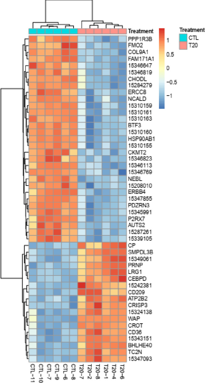
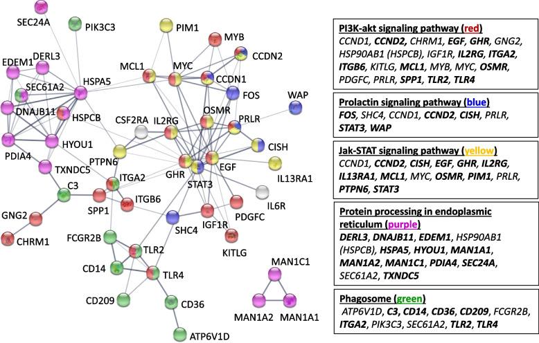
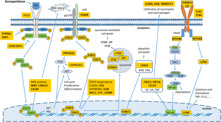
Results: The expression of 335 genes was up-regulated and that of 505 genes down-regulated in the mammary tissue of T20 vs CTL gilts. Biological process GO terms and KEGG pathways enriched in T20 vs CTL gilts reflected the concurrent premature lactogenesis and mammary involution. When looking at individual genes, it appears that mammary cells from T20 gilts can simultaneously upregulate the transcription of milk proteins such as WAP, CSN1S2 and LALBA, and genes triggering mammary involution such as STAT3, OSMR and IL6R. The down-regulation of PRLR expression and up-regulation of genes known to inactivate the JAK-STAT5 pathway (CISH, PTPN6) suggest the presence of a negative feedback loop trying to counteract the effects of hyperprolactinemia.
Conclusions: Genes and pathways identified in this study suggest that sustained hyperprolactinemia during late-pregnancy, in the absence of suckling piglets, sends conflicting pro-survival and cell death signals to mammary epithelial cells. Reception of these signals results in a mammary gland that can simultaneously synthesize milk proteins and initiate mammary involution.
Keywords: Domperidone; Gestation; Gilt; Mammary gland; Prolactin; Transcriptomic.
© 2023. Crown.
Conflict of interest statement
The authors declare that they have no competing interest.
Figures
Similar articles
-
Effects of sustained hyperprolactinemia in late gestation on mammary development of gilts.Domest Anim Endocrinol. 2020 Jul;72:106408. doi: 10.1016/j.domaniend.2019.106408. Epub 2019 Nov 7. Domest Anim Endocrinol. 2020. PMID: 32007676
-
Hyperprolactinemia using domperidone in prepubertal gilts: Effects on hormonal status, mammary development and mammary and pituitary gene expression.Domest Anim Endocrinol. 2021 Jul;76:106630. doi: 10.1016/j.domaniend.2021.106630. Epub 2021 Apr 14. Domest Anim Endocrinol. 2021. PMID: 33979716
-
Using domperidone to induce and sustain hyperprolactinemia in late-pregnant gilts.Domest Anim Endocrinol. 2019 Jan;66:14-20. doi: 10.1016/j.domaniend.2018.05.004. Epub 2018 May 17. Domest Anim Endocrinol. 2019. PMID: 30205268
-
Review: Mammary gland development in swine: embryo to early lactation.Animal. 2019 Jul;13(S1):s11-s19. doi: 10.1017/S1751731119000521. Animal. 2019. PMID: 31280748 Review.
-
Suckling effects in sows: importance for mammary development and productivity.Animal. 2013 Dec;7(12):1964-8. doi: 10.1017/S1751731113001201. Epub 2013 Jul 10. Animal. 2013. PMID: 23842122 Review.
Cited by
-
A long-lasting prolactin stimulates galactopoiesis in mice.iScience. 2025 Jul 15;28(8):113112. doi: 10.1016/j.isci.2025.113112. eCollection 2025 Aug 15. iScience. 2025. PMID: 40792031 Free PMC article.
-
Prolactin Regulates Ovine Ovarian Granulosa Cell Apoptosis by Affecting the Expression of MAPK12 Gene.Int J Mol Sci. 2023 Jun 17;24(12):10269. doi: 10.3390/ijms241210269. Int J Mol Sci. 2023. PMID: 37373417 Free PMC article.
References
-
- King RH, Pettigrew JE, McNamara JP, McMurtry JP, Henderson TL, Hathaway MR, et al. The effect of exogenous prolactin on lactation performance of first-litter sows given protein-deficient diets during the first pregnancy. Anim Reprod Sci. 1996;41:37–50. doi: 10.1016/0378-4320(95)01438-1. - DOI
MeSH terms
Substances
LinkOut - more resources
Full Text Sources
Research Materials
Miscellaneous
