

Magnetofection of miR-21 promoted by electromagnetic field and iron oxide nanoparticles via the p38 MAPK pathway contributes to osteogenesis and angiogenesis for intervertebral fusion
- PMID: 36694219
- PMCID: PMC9875474
- DOI: 10.1186/s12951-023-01789-3
Magnetofection of miR-21 promoted by electromagnetic field and iron oxide nanoparticles via the p38 MAPK pathway contributes to osteogenesis and angiogenesis for intervertebral fusion
Abstract
Background: Magnetofection-mediated gene delivery shows great therapeutic potential through the regulation of the direction and degree of differentiation. Lumbar degenerative disc disease (DDD) is a serious global orthopaedic problem. However, even though intervertebral fusion is the gold standard for the treatment of DDD, its therapeutic effect is unsatisfactory. Here, we described a novel magnetofection system for delivering therapeutic miRNAs to promote osteogenesis and angiogenesis in patients with lumbar DDD.
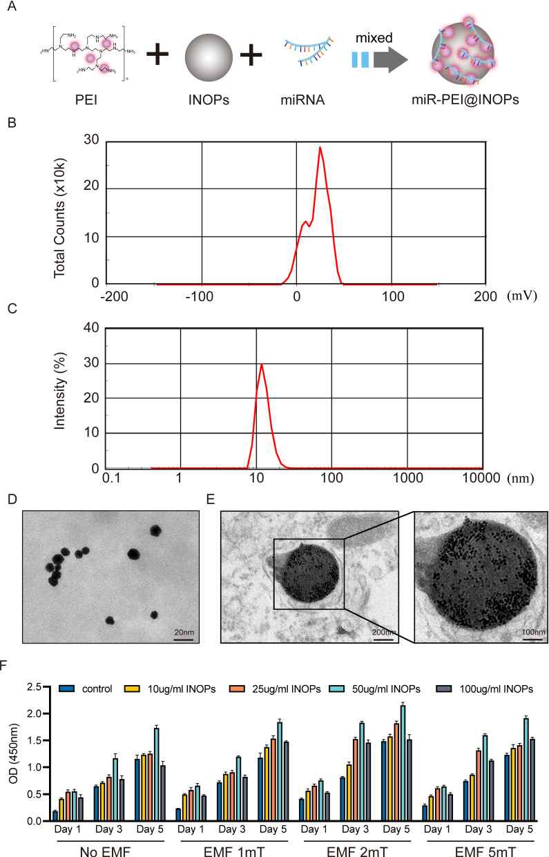
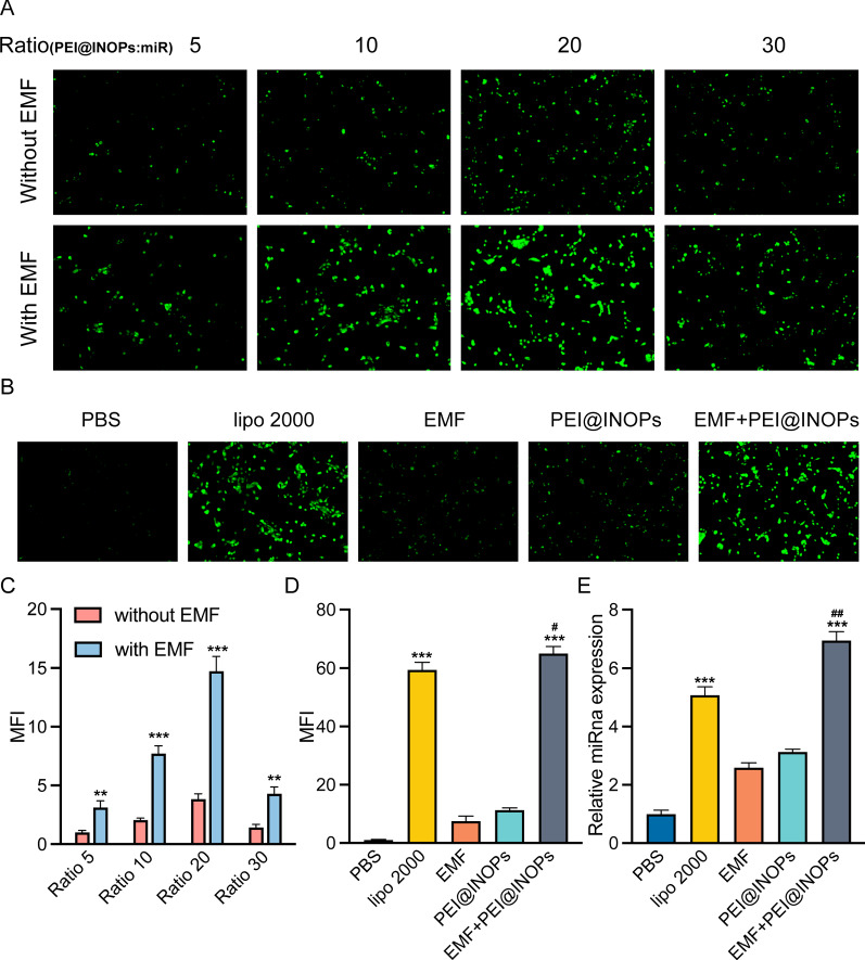
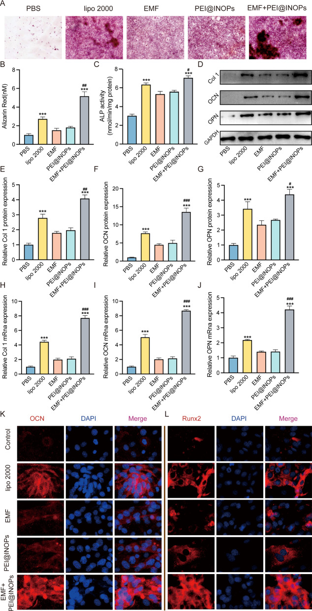
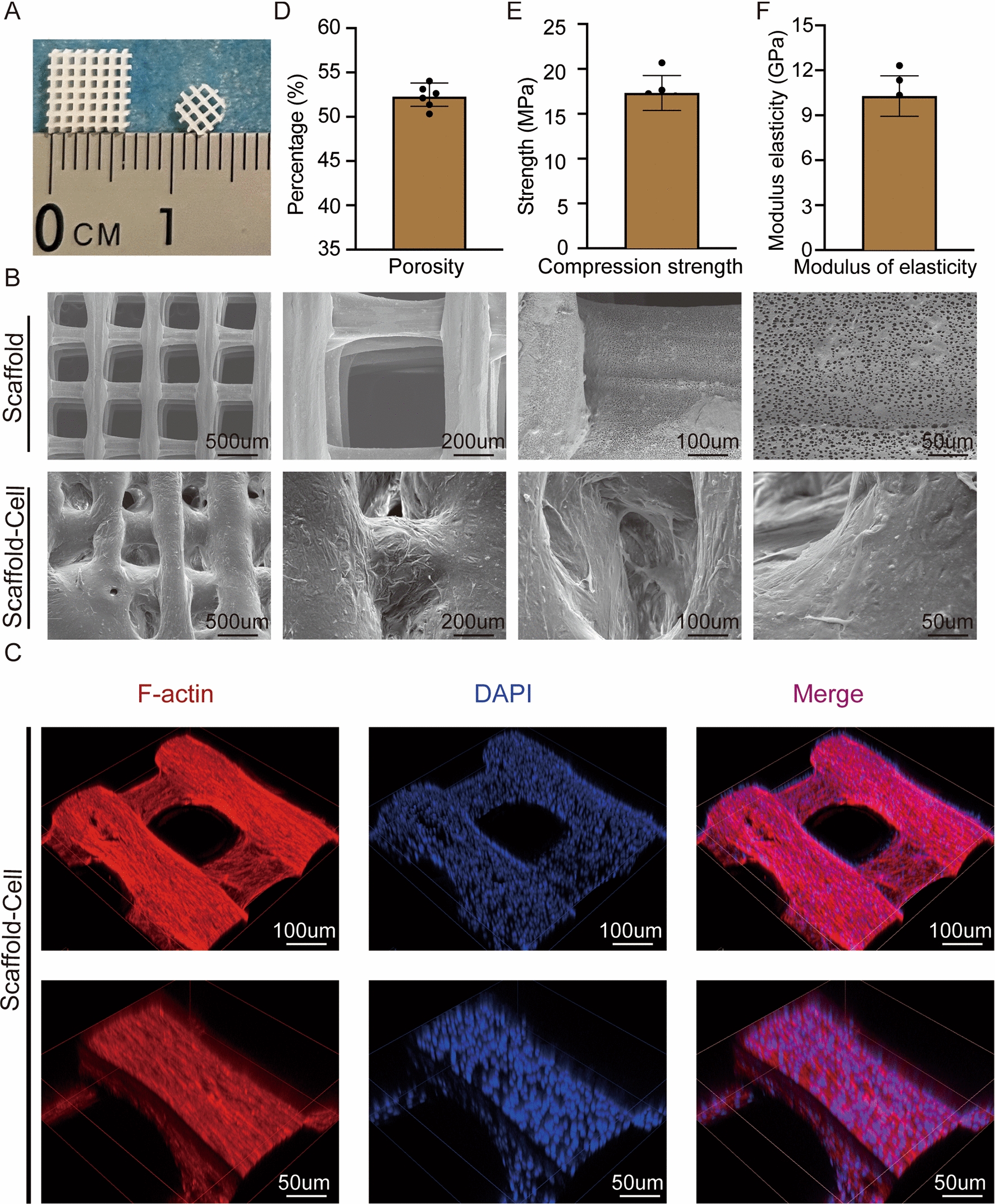
Results: Co-stimulation with electromagnetic field (EMF) and iron oxide nanoparticles (IONPs) enhanced magnetofection efficiency significantly. Moreover, in vitro, magnetofection of miR-21 into bone marrow mesenchymal stem cells (BMSCs) and human umbilical endothelial cells (HUVECs) influenced their cellular behaviour and promoted osteogenesis and angiogenesis. Then, gene-edited seed cells were planted onto polycaprolactone (PCL) and hydroxyapatite (HA) scaffolds (PCL/HA scaffolds) and evolved into the ideal tissue-engineered bone to promote intervertebral fusion. Finally, our results showed that EMF and polyethyleneimine (PEI)@IONPs were enhancing transfection efficiency by activating the p38 MAPK pathway.
Conclusion: Our findings illustrate that a magnetofection system for delivering miR-21 into BMSCs and HUVECs promoted osteogenesis and angiogenesis in vitro and in vivo and that magnetofection transfection efficiency improved significantly under the co-stimulation of EMF and IONPs. Moreover, it relied on the activation of p38 MAPK pathway. This magnetofection system could be a promising therapeutic approach for various orthopaedic diseases.
Keywords: Bone tissue engineering; Electromagnetic field; Gene therapy; Iron oxide nanoparticles; Magnetofection.
© 2023. The Author(s).
Conflict of interest statement
The authors have no financial disclosures or conflicts of interest with the research presented.
Figures


Similar articles
-
Low-frequency electromagnetic fields combined with tissue engineering techniques accelerate intervertebral fusion.Stem Cell Res Ther. 2021 Feb 17;12(1):143. doi: 10.1186/s13287-021-02207-x. Stem Cell Res Ther. 2021. PMID: 33597006 Free PMC article.
-
Sinusoidal electromagnetic fields accelerate bone regeneration by boosting the multifunctionality of bone marrow mesenchymal stem cells.Stem Cell Res Ther. 2021 Apr 13;12(1):234. doi: 10.1186/s13287-021-02302-z. Stem Cell Res Ther. 2021. PMID: 33849651 Free PMC article.
-
The combinatory effect of sinusoidal electromagnetic field and VEGF promotes osteogenesis and angiogenesis of mesenchymal stem cell-laden PCL/HA implants in a rat subcritical cranial defect.Stem Cell Res Ther. 2019 Dec 16;10(1):379. doi: 10.1186/s13287-019-1464-x. Stem Cell Res Ther. 2019. PMID: 31842985 Free PMC article.
-
New Insights into Biocompatible Iron Oxide Nanoparticles: A Potential Booster of Gene Delivery to Stem Cells.Small. 2020 Sep;16(37):e2001588. doi: 10.1002/smll.202001588. Epub 2020 Jul 28. Small. 2020. PMID: 32725792 Review.
-
Stem cells in preclinical spine studies.Spine J. 2014 Mar 1;14(3):542-51. doi: 10.1016/j.spinee.2013.08.031. Epub 2013 Nov 15. Spine J. 2014. PMID: 24246748 Review.
Cited by
-
Recent advances of nanoparticles on bone tissue engineering and bone cells.Nanoscale Adv. 2024 Feb 12;6(8):1957-1973. doi: 10.1039/d3na00851g. eCollection 2024 Apr 16. Nanoscale Adv. 2024. PMID: 38633036 Free PMC article. Review.
-
How to enhance the ability of mesenchymal stem cells to alleviate intervertebral disc degeneration.World J Stem Cells. 2023 Nov 26;15(11):989-998. doi: 10.4252/wjsc.v15.i11.989. World J Stem Cells. 2023. PMID: 38058958 Free PMC article. Review.
-
Impact of Long-Lasting Environmental Factors on Regulation Mediated by the miR-34 Family.Biomedicines. 2024 Feb 12;12(2):424. doi: 10.3390/biomedicines12020424. Biomedicines. 2024. PMID: 38398026 Free PMC article. Review.
-
Energizing Healing with Electromagnetic Field Therapy in Musculoskeletal Disorders.J Orthop Sports Med. 2024;6(2):89-106. doi: 10.26502/josm.511500147. Epub 2024 May 17. J Orthop Sports Med. 2024. PMID: 39036742 Free PMC article.
-
Effective delivery of miR-150-5p with nucleus pulposus cell-specific nanoparticles attenuates intervertebral disc degeneration.J Nanobiotechnology. 2024 May 27;22(1):292. doi: 10.1186/s12951-024-02561-x. J Nanobiotechnology. 2024. PMID: 38802882 Free PMC article.
References
-
- Cheung KM, Karppinen J, Chan D, Ho DW, Song YQ, Sham P, Cheah KS, Leong JC, Luk KD. Prevalence and pattern of lumbar magnetic resonance imaging changes in a population study of one thousand forty-three individuals. Spine (Phila Pa 1976) 2009;34(9):934–40. doi: 10.1097/BRS.0b013e3181a01b3f. - DOI - PubMed
-
- Global regional. National incidence, prevalence, and years lived with disability for 354 diseases and injuries for 195 countries and territories, 1990–2017: a systematic analysis for the global burden of Disease Study 2017. Lancet. 2018;392(10159):1789–858. doi: 10.1016/S0140-6736(18)32279-7. - DOI - PMC - PubMed
MeSH terms
Substances
Grants and funding
LinkOut - more resources
Full Text Sources
Medical
Research Materials
