. 2023 Jun 7;31(6):1636-1646.
doi: 10.1016/j.ymthe.2023.01.019. Epub 2023 Jan 23.

A trans-amplifying RNA simplified to essential elements is highly replicative and robustly immunogenic in mice

Affiliations

A trans-amplifying RNA simplified to essential elements is highly replicative and robustly immunogenic in mice

Mario Perkovic et al. Mol Ther. .

Erratum in

Abstract

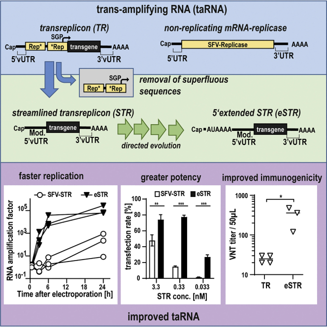
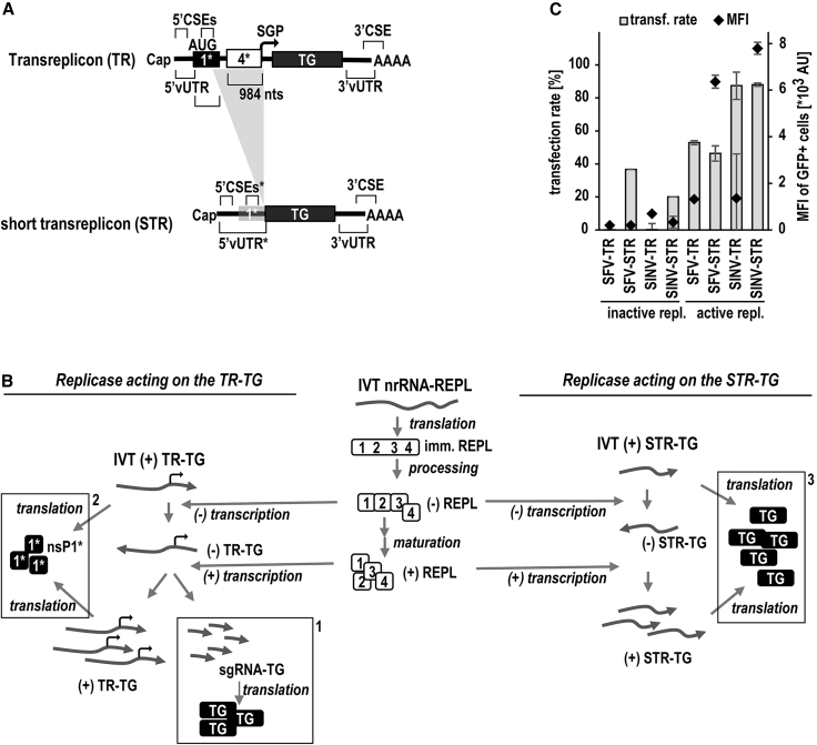
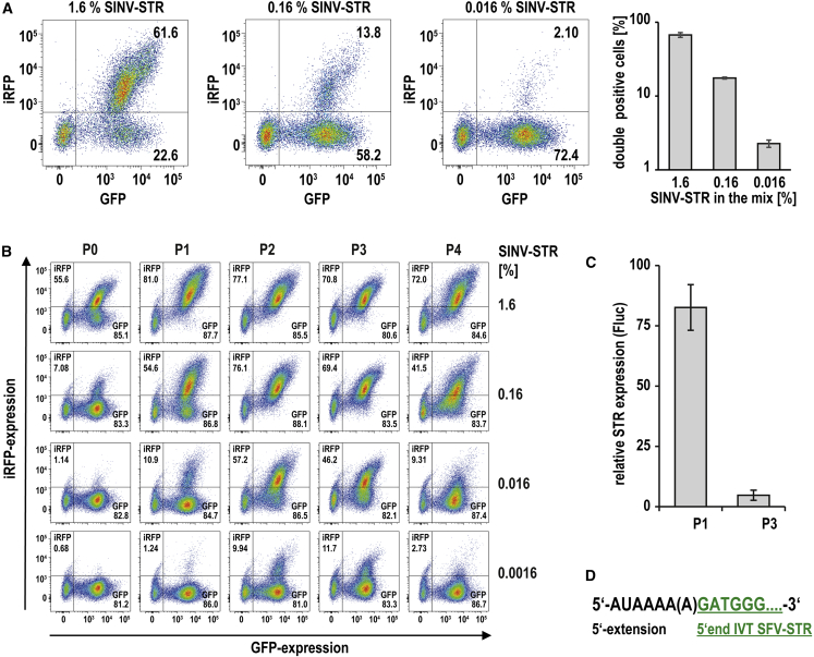
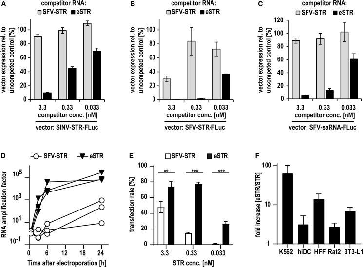
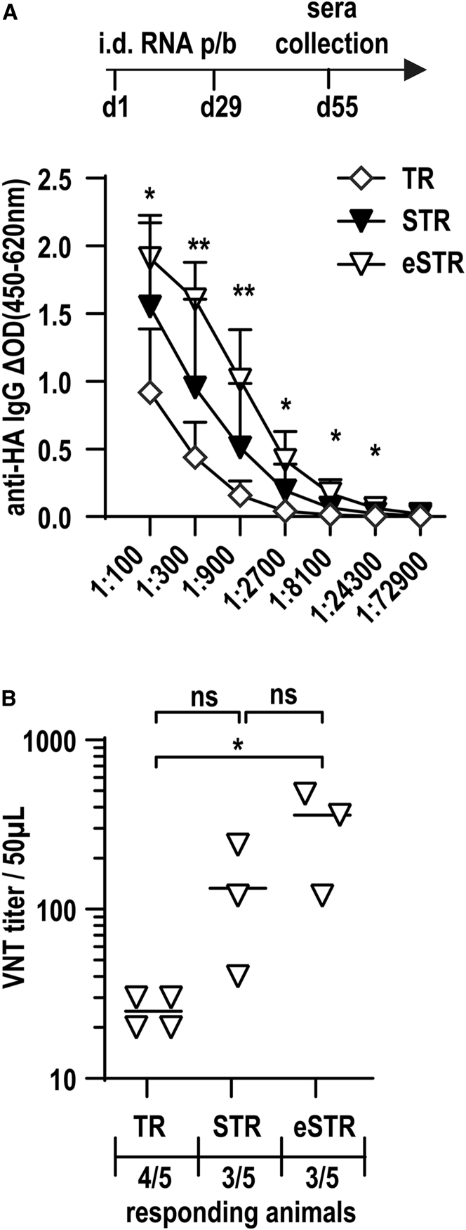
Trans-amplifying RNA (taRNA) is a split-vector derivative of self-amplifying RNA (saRNA) and a promising vaccine platform. taRNA combines a non-replicating mRNA encoding an alphaviral replicase and a transreplicon (TR) RNA coding for the antigen. Upon translation, the replicase amplifies the antigen-coding TR, thereby requiring minimal amounts of TR for immunization. TR amplification by the replicase follows a complex mechanism orchestrated by genomic and subgenomic promoters (SGPs) and generates genomic and subgenomic amplicons whereby only the latter are translated into therapeutic proteins. This complexity merits simplification to improve the platform. Here, we eliminated the SGP and redesigned the 5' untranslated region to shorten the TR (STR), thereby enabling translation of the remaining genomic amplicon. We then applied a directed evolution approach to select for faster replicating STRs. The resulting evolved STR (eSTR) had acquired A-rich 5' extensions, which improved taRNA expression thanks to accelerated replication. Consequently, we reduced the minimal required TR amount by more than 10-fold without losing taRNA expression in vitro. Accordingly, eSTR-immunized mice developed greater antibody titers to taRNA-encoded influenza HA than TR-immunized mice. In summary, this work points the way for further optimization of taRNA by combining rational design and directed evolution.

Keywords: alphaviral vector; directed evolution; trans-amplifying RNA; transreplicon; vaccine.


Conflict of interest statement

Declaration of interests U.S., T.B., and M.P. are inventors on patents and patent applications, which cover parts of this article. U.S. is employee at BioNTech Corporation (Mainz, Germany), a privately owned company developing therapeutic RNA.

Figures

None
Graphical abstract
Figure 1
Figure 1
Transreplicon simplification and mechanism of trans-replication (A) Design of simplified and shortened transreplicons. Conserved sequence elements (CSEs) serving as replicase recognition signals are located within the 5′ UTR of viral origin (5′ vUTR), at the beginning of nsP1, at the end of nsP4 (subgenomic promoter [SGP]), and at the end of the 3′ vUTR extending into the poly(A) tail (reviewed in Pietilä et al.11). In the first generation of transreplicons (TRs, top), the replicase gene was largely deleted, but all CSEs were conserved. Consequently, 221 nucleotides of nsP1 (1∗) and 984 nucleotides of nsP4 (4∗) comprising the 5′ CSE or the SGP, respectively, remained in the TR. To trim and simplify the TR, we removed the nsP4-SGP (indicated by the gray triangular background) and mutated all AUG triplets within the 5′ CSE/nsP1 (gray shaded 1∗), resulting in the start codon of the transgene becoming the most upstream one. Thus, the short TR (STR; bottom) closely resembles an mRNA and can be translated without replication if capped during in vitro transcription. (B) Mechanism of TR and STR trans-replication. Full-length immature replicase (imm. REPL, center) is translated from in-vitro-transcribed (IVT) nrRNA-REPL and then processed by intramolecular proteolysis mediated by nsP2. First, the negative-strand-specific replicase complex made of nsP123 and nsP4 is formed, followed by further cleavages and release of nsP1, nsP2, and nsP3, eventually forming the mature positive-strand-specific replicase complex ((+)REPL). Common replication steps of TR (left) and an STR (right) include the synthesis of a negatively sensed copy of the positively sensed transfected IVT RNA vectors in full length, followed by transcription of novel full-length positive-sensed copies ((+)TR-TG or (+)STR-TG, respectively). These steps mirror the replication of genomic RNA of alphaviruses. In the case of the TR-TG, the (+)REPL transcribes, in addition, a subgenomic RNA (sgRNA-TG) identical to that found in the parental saRNA, which is translated into the transgene (left, dashed box no. 1). Due to the presence of the original replicase start codon, a truncated nsP1 peptide (nsP1∗; 1∗) may be translated from the TR transgene (left, dashed box no. 2). The replication of the STR, however, no longer supports subgenomic transcription, and the AUG codon mutations prevent a putative nsP1-peptide translation (right). Thus, STR replication is limited to genomic-like replication, and all positive-stranded copies are translated exclusively to the transgene (right, dashed box no. 3). (C) STRs improve taRNA performance compared with TRs. K562 cells were co-electroporated with 1 μg nrRNA encoding either inactive or active SFV replicase and 0.2 μg TR or STRs of SFV or SINV. The GFP expression mediated by TR and STRs was measured by flow cytometry to investigate transfection rates and level of expression in individual cells. Data are shown as mean ± SD of three independent experiments.
Figure 2
Figure 2
The SINV-STR is preferentially amplified by the SFV replicase, but it is outcompeted by a 5′-modified SFV-STR upon directed evolution (A) Preferential expression of SINV-STR. GFP-SecNLuc-encoding uncapped STRs of 6 alphaviruses (FMV, AURAV, HJV, MADV, CHIKV, SFV) were mixed in equal amounts and spiked with a minor amount of an uncapped SINV-STR encoding a GFP-iRFP fusion reporter. SINV-STR abundance ranged from 1.6% to 0.016%. This blend of STRs was mixed with capped nrRNA-SFV replicase and electroporated into K562 cells. Dot plots show the expression of both fluorescent reporters of a representative experiment the day after electroporation (left). Data are also shown as mean and SD of 3 independent experiments (right). (B) Evolution of iRFP expression upon serial RNA transfer. K562 cells were co-electroporated with SINV-spiked uncapped STR mixtures and capped nrRNA-replicase, with SINV-STR abundance ranging from 1.6% to 0.0016%. After 16 h, GFP and iRFP expression was measured before the total RNA of the transfected cells was extracted, supplemented with IVT nrRNA-replicase, and used to electroporate K562 cells again. This serial RNA transfer was done four times, iRFP expression indicates the abundance of the SINV-STR after each passage (P0 = cells transfected with IVT RNA; P1 to Px = cells transfected with cellular RNA). (C) Competitive inhibition of a SINV-STR by cellular RNA of serially transfected cells. Total cellular RNA of mock electroporated K562 cells or total cellular RNA of passages P1 and P3 were mixed with very low amount (250 pg) of uncapped IVT SINV-STR coding for luciferase. Upon co-electroporation with SFV-replicase nrRNA into K562 cells, luciferase expression was measured after 24 h. Data are shown as the expression relative to the mock control. (D) Identification of a 5′-extended SFV-STR enriched upon serial transfer. Cellular RNA was extracted from cells that were serially transfected 4 times with the STR mixture containing 0.0016% SINV-STR. The RNA was reverse transcribed, and the resulting cDNA was used to amplify the 5′ end of STRs. 5′ end PCR products were subcloned and sequenced. SFV-STRs extended at the 5′ end by AUAAAA or AUAAAAA were found in 59% of the clones.
Figure 3
Figure 3
eSTRs competitively inhibit co-transfected STRs and saRNA, replicate faster, and increase expression of taRNA (A and B) Expression of a SINV- or an SFV-STR in the presence of eSTRs. K562 cells were co-electroporated with 3.3 nM capped SFV-replicase nrRNA and 0.033 to 3.3 nM capped wild-type (WT) SFV-STRs or capped eSTRs (5′ extension AUAAAA) expressing GFP-SecNLuc. Capped WT SINV-STR (A) or capped SFV-STR (B) expressing firefly luciferase (0.33 nM) were added, and luciferase expression was measured 24 h after transfer and normalized to the expression of the uncompeted control. (C) Inhibition of SFV saRNA by eSTRs. K562 cells were co-electroporated with 3.3 nM capped SFV saRNA encoding firefly luciferase and 0.033 to 3.3 nM capped WT SFV-STR or capped eSTRs. Controls were electroporated with saRNA alone and used to normalize the luciferase expression data measured 24 h after transfection. Data are shown as mean and SD of 3 independent experiments (A–C). (D) Replication rate of eSTR compared with STR. BHK21 cells were co-electroporated with 3.3 nM capped SFV-replicase nrRNA and 0.33 nM capped WT SFV-STR or capped eSTR encoding firefly luciferase. RNA was extracted at indicated time points, and STR was quantified by qRT-PCR. Controls were co-electroporated with inactive replicase and levels of STRs found in these cells used to calculate the factor of amplification at each time point. Data of three independent experiments are shown as individual line graphs. (E) Expression of taRNA with STR and eSTR. K562 cells were co-electroporated with 3.3 nM capped SFV-replicase nrRNA and 0.033 to 3.3 nM capped WT SFV-STR or capped eSTR. GFP expression resulting from STRs was assessed by flow cytometry 24 h after transfer. Data from three independent experiments are shown (mean ± SD). Unpaired Student’s t test was performed (∗p < 0.05; ∗∗p < 0.01; ∗∗∗p < 0.001). (F) Improvement of taRNA expression by eSTR in various cell types and species. Human immature dendritic cells (hiDCs), human foreskin fibroblast (HFFs), Rattus norvegicus fibroblasts (Rat2), and Mus musculus fibroblasts (3T3-L1) were seeded into 96-well plates lipofected with 20 ng taRNA encoding firefly luciferase per well (19.6 ng capped SFV-replicase nrRNA and 0.4 ng capped SFV-STR or eSTR), using 80 nL MessengerMax per well. 24 h after transfection, luciferase expression levels were measured and the fold increase in expression by eSTR over STR calculated for each cell type. Relative expression (eSTR/STR) of three independent experiments are shown (mean ± SD).
Figure 4
Figure 4
eSTRs improve vaccination with SFV taRNA BALB/c mice (group sizes n = 5) were immunized at days 1 and 29 by intradermal injection of 50 ng uncapped SFV-TR, SFV-STR, or eSTR encoding the influenza A virus hemagglutinin (HA) together with 20 μg capped nrRNA encoding SFV replicase. Negative controls received 20 μL buffer without RNA. Sera for the quantification of IgG titer were sampled on day 55. (A) Total anti-HA IgG antibody in sera of responding mice were determined by ELISA. Unpaired Student’s t test was performed for each dilution step (TR vs. eSTR: ∗p < 0.05, ∗∗p < 0.01; TR vs. STR: all non-significant). (B) Virus neutralization test (VNT) titer of each responding animal is displayed, with the limit of detection of 10. All animals receiving PBS alone were non-responding, and therefore data are not shown. For all other groups, the number of responding over total number of animals is given below the x axis. One-way ANOVA was performed (∗p < 0.05; ns p > 0.05).

Comment in

Similar articles

Cited by

References

    1. Kumar A., Blum J., Thanh Le T., Havelange N., Magini D., Yoon I.-K. The mRNA vaccine development landscape for infectious diseases. Nat. Rev. Drug Discov. 2022;21:333–334. - PubMed
    1. Polack F.P., Thomas S.J., Kitchin N., Absalon J., Gurtman A., Lockhart S., Perez J.L., Pérez Marc G., Moreira E.D., Zerbini C., et al. Safety and efficacy of the BNT162b2 mRNA covid-19 vaccine. N. Engl. J. Med. 2020;383:2603–2615. - PMC - PubMed
    1. Simon-Loriere E., Schwartz O. Towards SARS-CoV-2 serotypes? Nat. Rev. Microbiol. 2022;20:187–188. - PMC - PubMed
    1. Bloom K., van den Berg F., Arbuthnot P. Self-amplifying RNA vaccines for infectious diseases. Gene Ther. 2021;28:117–129. - PMC - PubMed
    1. Kis Z., Kontoravdi C., Shattock R., Shah N. Resources, production scales and time required for producing RNA vaccines for the global pandemic demand. Vaccines. 2020;9:3. - PMC - PubMed