

A Tumor Treated With Antibiotics: A Rare Case
- PMID: 36694498
- PMCID: PMC9863745
- DOI: 10.7759/cureus.32819
A Tumor Treated With Antibiotics: A Rare Case
Abstract
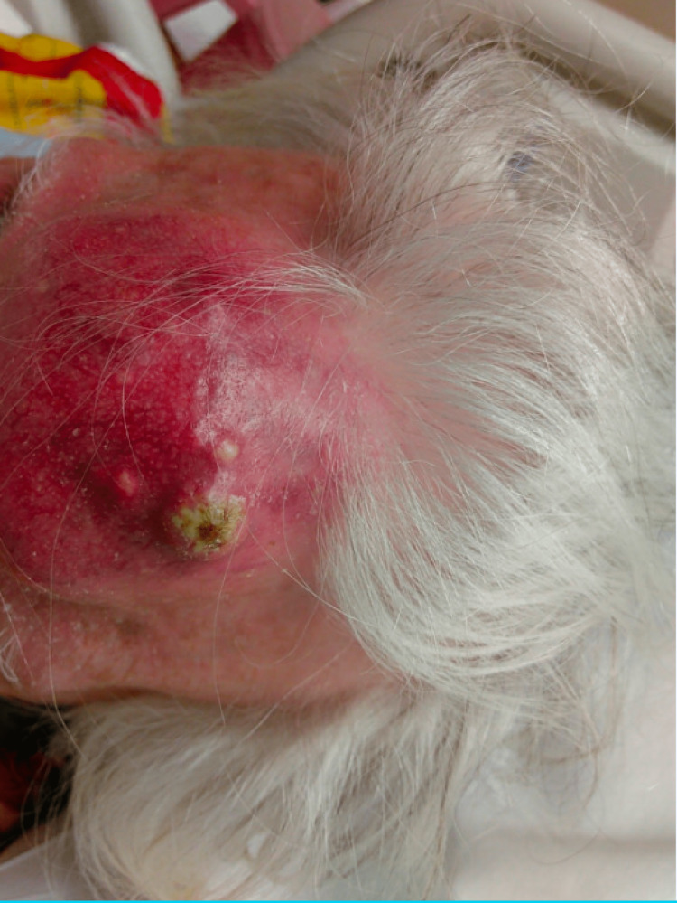
Pott's puffy tumor, a rare condition, is an osteomyelitis involving the frontal bone with accompanying subperiosteal abscess. Patients typically present with swelling of the scalp and forehead, headache, fever, tenderness of the frontal sinus, and nasal discharge. MRI is the modality of choice for diagnosis and assessment. The standard of care is incision and drainage with long-course antibiotics. The prognosis is excellent; however, complications from a hematogenous spread can lead to meningitis or epidural spaces if not treated properly.
Keywords: anaerobe; antibiotic usage; frontal sinus; gram-positive bacteria; incision and drainage of abscess; omfs; osteomyelitis; pott's puffy tumor; recurrent sinusitis.
Copyright © 2022, Mahmoud et al.
Conflict of interest statement
The authors have declared that no competing interests exist.
Figures


Similar articles
-
Pott's Puffy Tumor Associated with Subperiosteal Empyema and Osteomyelitis in a Pediatric Patient.Eur J Case Rep Intern Med. 2025 Feb 12;12(3):005185. doi: 10.12890/2025_005185. eCollection 2025. Eur J Case Rep Intern Med. 2025. PMID: 40051739 Free PMC article.
-
Pott's Puffy Tumour: A Rare Complication of Sinusitis.Eur J Case Rep Intern Med. 2024 Mar 11;11(4):004287. doi: 10.12890/2024_004287. eCollection 2024. Eur J Case Rep Intern Med. 2024. PMID: 38584903 Free PMC article.
-
Patient presenting with frontal subperiosteal abscess and headache: a case of Pott's puffy tumour.Br J Neurosurg. 2019 Jun;33(3):275-277. doi: 10.1080/02688697.2017.1330944. Epub 2017 May 22. Br J Neurosurg. 2019. PMID: 28532175
-
[Pott's Puffy tumor: a forgotten complication of sinusitis].Radiologia. 2011 Mar-Apr;53(2):175-8. doi: 10.1016/j.rx.2010.05.007. Epub 2010 Jul 24. Radiologia. 2011. PMID: 20656307 Review. Spanish.
-
Pott's puffy tumor in a 23-month-old: Youngest known case of a rare disease.Auris Nasus Larynx. 2022 Aug;49(4):713-716. doi: 10.1016/j.anl.2020.12.004. Epub 2021 Jan 6. Auris Nasus Larynx. 2022. PMID: 33422370 Review.
Cited by
-
Streptococcus intermedius: From a Normal Oral Commensal to a Life-Threatening Organism.Cureus. 2023 Dec 18;15(12):e50708. doi: 10.7759/cureus.50708. eCollection 2023 Dec. Cureus. 2023. PMID: 38234954 Free PMC article.
References
-
- Evaluation of adult Pott's puffy tumor: our five cases and 27 literature cases. Akiyama K, Karaki M, Mori N. Laryngoscope. 2012;122:2382–2388. - PubMed
-
- Intracranial complications of frontal sinusitis. Remmler D, Boles R. Laryngoscope. 1980;90:1814–1824. - PubMed
-
- The Pott puffy tumor revisited: neurosurgical implications of this unforgotten entity. Case report and review of the literature. Kombogiorgas D, Solanki GA. J Neurosurg. 2006;105:143–149. - PubMed
-
- Complications of frontal sinusitis and their management. Goldberg AN, Oroszlan G, Anderson TD. Otolaryngol Clin North Am. 2001;34:211–225. - PubMed
-
- Pott's puffy tumor and intranasal cocaine abuse. Pansini A, Copelli C, Manfuso A, d'Ecclesia A, Califano L, Cocchi R. J Craniofac Surg. 2020;31:0–20. - PubMed
Publication types
LinkOut - more resources
Full Text Sources