. 2023 Jan 11;18(1):20220613.
doi: 10.1515/med-2022-0613. eCollection 2023.

circ-IARS depletion inhibits the progression of non-small-cell lung cancer by circ-IARS/miR-1252-5p/HDGF ceRNA pathway

Affiliations

circ-IARS depletion inhibits the progression of non-small-cell lung cancer by circ-IARS/miR-1252-5p/HDGF ceRNA pathway

Jinhua Yang et al. Open Med (Wars). .

Abstract

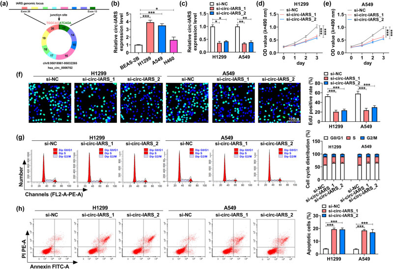
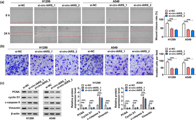
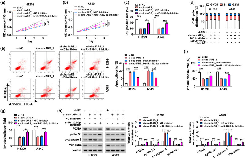
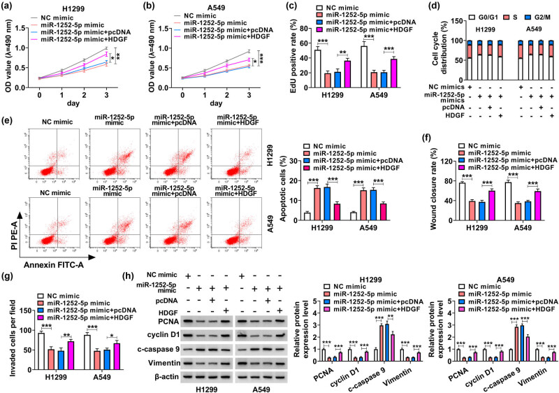
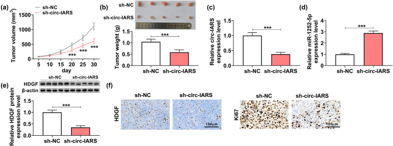
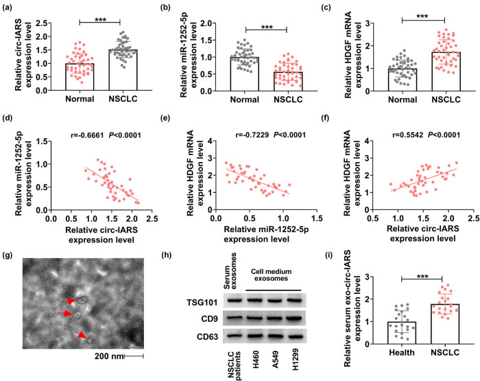
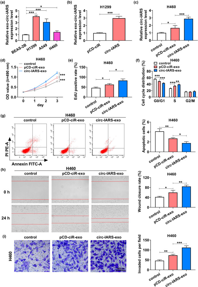
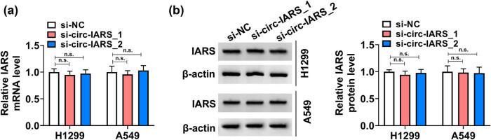
This study aims to explore the role and mechanism of circ-IARS in non-small-cell lung cancer (NSCLC) progression. Expression of circ-IARS, microRNA (miR)-1252-5p, and hepatoma-derived growth factor (HDGF) was measured by real-time quantitative PCR and western blotting. The interactions among circ-IARS, miR-1252-5p, and HDGF were determined by dual-luciferase reporter assay and RNA immunoprecipitation. Cell behaviors were measured by 3-(4,5-dimethylthiazol-2-yl)-2,5-diphenyltetrazolium bromide (MTT), 5-ethynyl-2'-deoxyuridine (EdU) assay, flow cytometry, scratch wound assay, and transwell assay, and validated in in vivo xenograft model. Exosomes were isolated using commercial kit, and the expression and functions of exosomal circ-IARS (exo-circ-IARS) were analyzed as described above. Results showed that the expression of circ-IARS was upregulated in NSCLC cells, NSCLC tissues, and serum exosomes from NSCLC patients. circ-IARS exhaustion antagonized cell proliferation, cell cycle progression, migration, and invasion and promoted apoptosis in NSCLC. Molecularly, circ-IARS could sponge miR-1252-5p to modulate the expression of the downstream gene HDGF. In addition, miR-1252-5p downregulation attenuated circ-IARS exhaustion-mediated effects in H1299 and A549 cells. MiR-1252-5p mimic-induced effects were relieved by increasing HDGF expression in H1299 and A549 cells. Exo-circ-IARS promoted H460 cell proliferation, migration, and invasion and inhibited cell apoptosis. Silencing circ-IARS retarded tumor growth of NSCLC cells in vivo. Thus, circ-IARS, secreted by exosomes, was a novel oncogene in NSCLC and regulated the malignant development of NSCLC cells via circ-IARS/miR-1252-5p/HDGF competing endogenous RNA regulatory axis.

Keywords: HDGF; NSCLC; circ-IARS; exosome; miR-1252-5p.


Conflict of interest statement

Conflict of interest: The authors declare that they have no competing interest.

Figures

Figure 1
Figure 1
Exhausting circ-IARS could antagonize NSCLC cell growth. (a) Schematic diagram showing the back-splicing of exons 13–20 of the IARS gene generating the circRNA hsa_circ_0006702. (b and c) RT-qPCR is used to measure the relative circ-IARS expression level in (b) H1299, A549, H460, and BEAS-2B cells, and in (c) H1299 and A549 cells transfected with si-circ-IARS_1, si-circ-IARS_2, or si-NC. In transfected H1299 (d) and A549 cells (e), MTT assay monitored the OD value at 490 nm, (f) EdU assay determined the EdU-positive rate, (g and h) FCM method analyzed the cell cycle distribution (%) in G0/G1, S, and G2/M phases, and apoptotic cells (%). *P < 0.05, **P < 0.01, and ***P < 0.001.
Figure 2
Figure 2
Exhausting circ-IARS could antagonize cell migration and invasion of NSCLC cells. (a) Scratch wound assay measured wound closure rate in H1299 and A549 cells after transfection. (b) Transwell assay examined the invaded cells per field (100×) after transfection. (c) Western blotting detected the relative protein expression levels of PCNA, cyclin D1, c-caspase 9, and vimentin in transfected cells, normalized to β-actin. ***P < 0.001.
Figure 3
Figure 3
Circ-IARS served as a ceRNA for miR-1252-5p to regulate HDGF in NSCLC cells. (a) Circinteractome website and StarBase website together presented 12 miRNAs that were complementary to circ-IARS. (b and c) RT-qPCR detected the relative expression levels of 8 miRNAs in H1299 and A549 cells transfected with si-circ-IARS_1 or si-NC. (d) Schematic diagram showing the predicted binding sites between WT-circ-IARS and miR-1252-5p sequences. (e) Dual-luciferase reporter assay measured the relative luciferase activity of WT-circ-IARS reporter vector and MUT-circ-IARS reporter vector in HEK293T cells transfected with miR-1252-5p mimic or NC mimic. (f and g) RIP assay evaluated the relative enrichment of circ-IARS and miR-1252-5p by AGO2 or IgG in H1299 and A549 cells, normalized to the input of cells. (h and i) RT-qPCR measured the relative miR-1252-5p expression level in (h) BEAS-2B, H1299, A549, and H460 cells, and (i) H1299 and A549 cells transfected with si-NC, si-circ-IARS_1, si-circ-IARS_1 + miR-1252-5p inhibitor, or si-circ-IARS_1 + NC inhibitor. (j) Schematic diagram showing the predicted binding sites between WT-HDGF 3′UTR and miR-1252-5p sequences. (k) Dual-luciferase reporter assay measured the relative luciferase activity of WT-HDGF 3′UTR reporter vector and MUT-HDGF 3′UTR reporter vector in HEK293T cells transfected with miR-1252-5p mimic or NC mimic. (l and m) RIP assay evaluated the relative enrichment of HDGF and miR-1252-5p by AGO2 or IgG in H1299 and A549 cells, normalized to the input of cells. (n) RT-qPCR measured the relative miR-1252-5p expression level in H1299 and A549 cells transfected with miR-1252-5p mimic, miR-1252-5p inhibitor, NC mimic, or NC inhibitor. (o–r) Western blotting detected the relative HDGF protein expression in (o and p) H1299 and A549 cells transfected with HDGF vector, pcDNA vector, miR-1252-5p mimic, NC mimic, miR-1252-5p mimic + HDGF vector, or miR-1252-5p mimic + pcDNA vector, and (q) BEAS-2B, H1299, A549, and H460 cells, as well as (r) H1299 and A549 cells transfected with si-NC, si-circ-IARS_1, si-circ-IARS_1 + miR-1252-5p inhibitor, or si-circ-IARS_1 + NC inhibitor. *P < 0.05, **P < 0.01, and ***P < 0.001.
Figure 4
Figure 4
Downregulation of miR-1252-5p counteracted the effects of circ-IARS exhaustion in NSCLC cells. H1299 and A549 cells transfected with si-NC, si-circ-IARS_1, si-circ-IARS_1 + miR-1252-5p inhibitor, or si-circ-IARS_1 + NC inhibitor. (a and b) MTT assay monitored the OD value at 490 nm. (c) EdU assay determined the EdU-positive rate. (d) FCM method analyzed the cell cycle distribution (%) in G0/G1, S, and G2/M phases. (e) FCM method analyzed the apoptotic cells (%). (f) Scratch wound assay measured the wound closure rate. (g) Transwell assay examined the invaded cells per field (100×). (h) Western blotting detected the relative protein expression level of PCNA, cyclin D1, c-caspase 9, and vimentin in transfected cells, normalized to β-actin. *P < 0.05, **P < 0.01, and ***P < 0.001.
Figure 5
Figure 5
Overexpressing miR-1252-5p suppressed the development of NSCLC cells through HDGF. H1299 and A549 cells transfected with NC mimic, miR-1252-5p mimic, miR-1252-5p mimic + HDGF vector, or miR-1252-5p mimic + pcDNA vector. (a and b) MTT assay monitored the OD value at 490 nm. (c) EdU assay determined the EdU positive rate. (d and e) FCM method analyzed the cell cycle distribution (%) and apoptotic cells (%). (f) Scratch wound assay measured the percentage of wound closure rate. (g) Transwell assay examined the invaded cells per field (100×). (h) Western blotting detected the relative protein expression levels of PCNA, cyclin D1, c-caspase 9, and vimentin in transfected cells, normalized to β-actin. *P < 0.05, **P < 0.01, and ***P < 0.001.
Figure 6
Figure 6
Silencing circ-IARS retarded the tumor growth of NSCLC in vivo. (a) Tumor volume was monitored every 5 days after inoculation of sh-circ-IARS or sh-NC-transfected H1299 cells into nude mice (N = 5). (b) Tumor weight was examined at the end day of the xenograft experiment. (c and d) RT-qPCR detected the relative circ-IARS and miR-1252-5p expression, and (e) western blotting measured the relative HDGF protein expression in tumor tissues from nude mice. (f) IHC examined the HDGF and Ki67 expression in paraffin-embedded xenograft tumor tissues. ***P < 0.001.
Figure 7
Figure 7
Circ-IARS was upregulated in NSCLC patients. (a–c) RT-qPCR detected the relative expression of circ-IARS, miR-1252-5p, and HDGF mRNA in NSCLC tumor tissues (NSCLC; N = 44), normalized to that in normal para-carcinoma tissues (Normal; N = 44). (d–f) Spearman’s rank correlation analysis determined the linear correlation among circ-IARS, miR-1252-5p, and HDGF mRNA expression in NSCLC tumors. (g) TEM showed the exosomes isolated from the serum of NSCLC patients. (h) Western blotting measured the expression of TSG101, CD9, and CD63 in isolated exosomes from NSCLC serum and NSCLC cell medium. (i) RT-qPCR detected the relative exo-circ-IARS expression in the serum of NSCLC patients (NSCLC; N = 22), normalized to serum from healthy people (Healthy; N = 22). ***P < 0.001.
Figure 8
Figure 8
Ex-circ-IARS contributed to the growth, migration, and invasion of NSCLC cells in vitro. (a and b) RT-qPCR detected the relative exo-circ-IARS expression level in (a) BEAS-2B, H1299, A549, and H460 cells, and (b) H1299 cells transfected with circ-IARS or pCD-ciR vector. (c–i) H460 cells were co-cultured with H1299 cells-based exosomes after circ-IARS or pCD-ciR vector transfection (pCD-ciR-exo or circ-IARS-exo), normalized to control cells (without exosome treatment). (c) RT-qPCR detected the relative circ-IARS expression level. (d) MTT assay monitored the OD value at 490 nm. (e) EdU assay determined the EdU-positive rate. (f and g) FCM method analyzed the cell cycle distribution (%) and apoptotic cells (%). (h) Scratch wound assay measured the wound closure rate. (i) Transwell assay examined the number of invaded cells per field (100×). *P < 0.05, **P < 0.01, and ***P < 0.001.
Figure A1
Figure A1
The effects of si-circ-IARS_1 and si-circ-IARS_2 on IARS mRNA and protein expression were analyzed by RT-qPCR (A) and Western blotting (B) in H1299 and A549 cells. ns: no significant difference.

Similar articles

Cited by

References

    1. Siegel RL, Miller KD, Jemal A. Cancer statistics, 2018. CA Cancer J Clin. 2018;68(1):7–30. - PubMed
    1. Chen W, Zheng R, Baade PD, Zhang S, Zeng H, Bray F, et al. Cancer statistics in China, 2015. CA Cancer J Clin. 2016;66(2):115–32. - PubMed
    1. Blandin Knight S, Crosbie PA, Balata H, Chudziak J, Hussell T, Dive C. Progress and prospects of early detection in lung cancer. Open Biol. 2017;7(9):170070. - PMC - PubMed
    1. Molina JR, Yang P, Cassivi SD, Schild SE, Adjei AA. Non-small cell lung cancer: epidemiology, risk factors, treatment, and survivorship. Mayo Clin Proc. 2008;83(5):584–94. - PMC - PubMed
    1. Mesko S, Gomez D. Proton therapy in non-small cell lung cancer. Curr Treat Options Oncol. 2018;19(12):76. - PubMed

LinkOut - more resources