

Refractory Hydroa Vacciniforme-like Lymphoma: Biological Insights from Morphoproteomic Analysis
- PMID: 36694700
- PMCID: PMC9831872
Refractory Hydroa Vacciniforme-like Lymphoma: Biological Insights from Morphoproteomic Analysis
Abstract
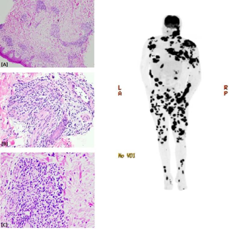
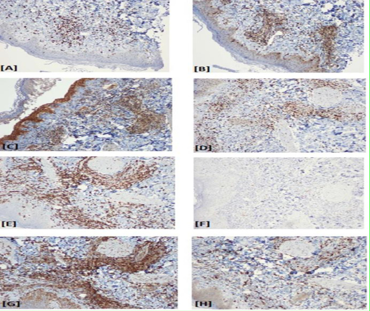
T-cell/natural killer cell lymphoproliferative disorders are rare, associated with poor overall survival, and have limited treatment options. We report a case of a patient who developed hydroa vacciniforme-like lymphoma (HVLL, an EBV-peripheral T-cell lymphoma), refractory to multiple lines of systemic therapy including methotrexate, mycophenolate mofetil, dapsone, thalidomide, prednisone, and romidepsin. We conducted morphoproteomic analysis of the patient's tumor which provided important biological insights. Histopathology showed primarily lymphohistiocytic infiltrates strongly positive EBV expression with a Ki-67 of >50% in the pretreatment biopsy and approximately 90% in the post-treatment biopsy, strong expression of Enhancer of Zester Homolog 2 (EZH2), a constitutively active mTOR pathway, 50% cytoplasmic BCL-2 expression; largely negative PD-1 positive CD8 T-cells. Based on this morphoproteomic analysis and published literature, we postulated that novel agents, including venetoclax, tazemetostat, and other agents may provide a targeted approach for treating HVLL. This case illustrates the use of morphoproteomic analysis to better understand the biology of tumors.
Keywords: Epstein-Barr virus; Hydroa vacciniforme-like lymphoma (HVLL); Morphoproteomics; Resistance mechanisms; T-cell lymphoproliferative disorders.
Copyright © 2022 Tehran University of Medical Sciences.
Figures
Similar articles
-
Hydroa vacciniforme-like lymphoma: a chronic EBV+ lymphoproliferative disorder with risk to develop a systemic lymphoma.Blood. 2013 Oct 31;122(18):3101-10. doi: 10.1182/blood-2013-05-502203. Epub 2013 Aug 27. Blood. 2013. PMID: 23982171
-
Hydroa Vacciniforme and Hydroa Vacciniforme-Like Lymphoproliferative Disorder: A Spectrum of Disease Phenotypes Associated with Ultraviolet Irradiation and Chronic Epstein-Barr Virus Infection.Int J Mol Sci. 2020 Dec 7;21(23):9314. doi: 10.3390/ijms21239314. Int J Mol Sci. 2020. PMID: 33297336 Free PMC article. Review.
-
Epstein-Barr virus-associated hydroa vacciniforme-like cutaneous lymphoma in seven Chinese children.Pediatr Dermatol. 2010 Sep-Oct;27(5):463-9. doi: 10.1111/j.1525-1470.2010.01094.x. Pediatr Dermatol. 2010. PMID: 20497358
-
Systemic lymphoma arising from hydroa vacciniforme-like lymphoma: report of two cases with review of literature.Int J Clin Exp Pathol. 2014 Aug 15;7(9):6403-8. eCollection 2014. Int J Clin Exp Pathol. 2014. PMID: 25337300 Free PMC article. Review.
-
Adult-onset hydroa vacciniforme-like lymphoma in a long-term resident of the United States.JAAD Case Rep. 2018 Mar 31;4(4):314-317. doi: 10.1016/j.jdcr.2017.10.023. eCollection 2018 May. JAAD Case Rep. 2018. PMID: 29693057 Free PMC article. No abstract available.
References
-
- Carbone A, Gloghini A, Dotti G. EBV-associated lymphoproliferative disorders: classification and treatment. Oncologist. 2008;13(5):577–85. - PubMed
-
- Gupta G, Man I, Kemmett D. Hydroa vacciniforme: A clinical and follow-up study of 17 cases. J Am Acad Dermatol. 2000;42(2 Pt 1):208–13. - PubMed
-
- Magaña M, Sangüeza P, Gil-Beristain J, et al. Angiocentric cutaneous T-cell lymphoma of childhood (hydroa-like lymphoma): a distinctive type of cutaneous T-cell lymphoma. J Am Acad Dermatol. 1998;38(4):574–9. - PubMed
-
- Barrionuevo C, Anderson VM, Zevallos-Giampietri E, et al. Hydroa-like cutaneous T-cell lymphoma: a clinicopathologic and molecular genetic study of 16 pediatric cases from Peru. Appl Immunohistochem Mol Morphol. 2002;10(1):7–14. - PubMed
-
- Doeden K, Molina-Kirsch H, Perez E, et al. Hydroa-like lymphoma with CD56 expression. J Cutan Pathol. 2008;35(5):488–94. - PubMed
Publication types
LinkOut - more resources
Full Text Sources
Research Materials
Miscellaneous