Occurrence and Phylogenetic Description of Cystic Echinococcosis Isolate from Egyptian Camel (Camelus Dromedarius)
- PMID: 36694828
- PMCID: PMC9831517
- DOI: 10.2478/helm-2022-0026
Occurrence and Phylogenetic Description of Cystic Echinococcosis Isolate from Egyptian Camel (Camelus Dromedarius)
Abstract
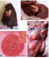
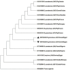
Cystic echinococcosis is one of the most significant cyclo-zoonotic diseases of major economic and public health significance worldwide. The current study was carried out to determine the epidemiological profile of cystic echinococcosis as well as to investigate its molecular and phylogenic status from one-humped camel (Camelus dromedarius) in the southern region of Egypt. In the present work, 110 camels freshly slaughtered at Daraw abattoirs, Aswan governorate were inspected for the presence of Hydatid cysts (HCs) visually and manually by palpation and incision, over a period of one year (June, 2018 - May, 2019). Furthermore, fourteen fertile hydatid cyst samples were collected from lungs of slaughtered camels. DNA extraction from two fertile samples was successfully achieved followed by phylogenetic analysis on two mitochondrial genes (cox1and nad1). Out of 110 camels slaughtered 11 (10 %) were found harboring hydatid cysts. The infection was found to prevail throughout the year, with the highest peak encountered in winter (45.5 %). The lungs were the most frequently infected organs (72.7 %) with liver cysts occurring at a significantly lower rate (27.3 %). The mean value of total protein, glucose, urea, cholesterol, magnesium, potassium, copper and creatinine was higher in cystic fluid from camels as compared to cattle. Blast and phylogenetic analysis on sequenced genes showed the presence of Echinococcus intermedius, originally the pig genotype (G7) in camels for the first time in Egypt. To the best of our knowledge, the current research provides a description of the current epidemiological and molecular situation of camel hydatidosis in the southern region of Egypt. Furthermore, the current results may have significant implications for hydatid disease control in the studied region.
Keywords: Camelus dromedarius; Cystic echinococcosis; Egypt; Epidemiological status; Molecular characterization.
© 2022 I. S. Elshahawy, M. A. El-Seify, Z. K. Ahamed, M. M. Fawaz, published by Sciendo.
Conflict of interest statement
Conflict of Interest Authors state no conflict of interest.
Figures


Similar articles
-
A Novel Designed Sandwich ELISA for the Detection of Echinococcus granulosus Antigen in Camels for Diagnosis of Cystic Echinococcosis.Trop Med Infect Dis. 2023 Aug 6;8(8):400. doi: 10.3390/tropicalmed8080400. Trop Med Infect Dis. 2023. PMID: 37624338 Free PMC article.
-
Molecular Studies on Cystic Echinococcosis of Camel (Camelus dromedarius) and Report of Echinococcus ortleppi in Iran.Iran J Parasitol. 2017 Jul-Sep;12(3):323-331. Iran J Parasitol. 2017. PMID: 28979341 Free PMC article.
-
Epidemiological, Morphometric, and Molecular Investigation of Cystic Echinococcosis in Camel and Cattle From Upper Egypt: Current Status and Zoonotic Implications.Front Vet Sci. 2021 Oct 4;8:750640. doi: 10.3389/fvets.2021.750640. eCollection 2021. Front Vet Sci. 2021. PMID: 34671663 Free PMC article.
-
Prevalence and bacterial isolation from hydatid cysts in dromedary camels (Camelus dromedarius) slaughtered at Sharkia abattoirs, Egypt.J Parasit Dis. 2021 Mar;45(1):236-243. doi: 10.1007/s12639-020-01300-x. Epub 2020 Nov 3. J Parasit Dis. 2021. PMID: 33746409 Free PMC article.
-
The global status and genetic characterization of hydatidosis in camels (Camelus dromedarius): a systematic literature review with meta-analysis based on published papers.Parasitology. 2021 Mar;148(3):259-273. doi: 10.1017/S0031182020001705. Epub 2020 Sep 17. Parasitology. 2021. PMID: 32940199 Free PMC article.
Cited by
-
A Novel Designed Sandwich ELISA for the Detection of Echinococcus granulosus Antigen in Camels for Diagnosis of Cystic Echinococcosis.Trop Med Infect Dis. 2023 Aug 6;8(8):400. doi: 10.3390/tropicalmed8080400. Trop Med Infect Dis. 2023. PMID: 37624338 Free PMC article.
-
A bibliometric analysis of six decades of camel research in North Africa: Trends, collaboration, and emerging themes.Open Vet J. 2024 Dec;14(12):3505-3524. doi: 10.5455/OVJ.2024.v14.i12.34. Epub 2024 Dec 31. Open Vet J. 2024. PMID: 39927358 Free PMC article.
References
-
- Ahmed Z.K. Studies on internal helminths infecting camels. Ph.D. Thesis. Faculty of Veterinary Medicine, South Valley University; 2021.
-
- Abdel-Aziz A.R., El-Meghanawy R.A.. Molecular characterization of hydatid cyst from Egyptian one humped camels (Camelus dromedaries) PSM Vet Res. 2016;1(1):13–16.
LinkOut - more resources
Full Text Sources
Research Materials
