. 2022 Oct-Dec;14(4):20-37.
doi: 10.32607/actanaturae.11820.

Flow-Seq Method: Features and Application in Bacterial Translation Studies

Affiliations

Flow-Seq Method: Features and Application in Bacterial Translation Studies

E S Komarova et al. Acta Naturae. 2022 Oct-Dec.

Abstract

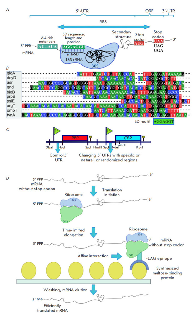
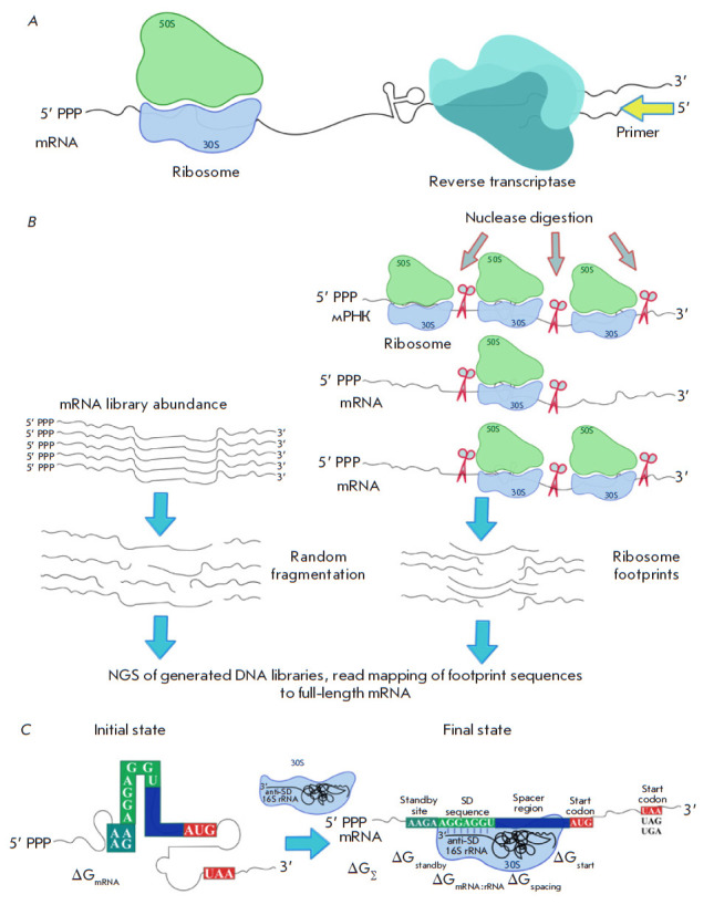
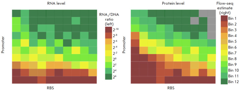
The Flow-seq method is based on using reporter construct libraries, where a certain element regulating the gene expression of fluorescent reporter proteins is represented in many thousands of variants. Reporter construct libraries are introduced into cells, sorted according to their fluorescence level, and then subjected to next-generation sequencing. Therefore, it turns out to be possible to identify patterns that determine the expression efficiency, based on tens and hundreds of thousands of reporter constructs in one experiment. This method has become common in evaluating the efficiency of protein synthesis simultaneously by multiple mRNA variants. However, its potential is not confined to this area. In the presented review, a comparative analysis of the Flow-seq method and other alternative approaches used for translation efficiency evaluation of mRNA was carried out; the features of its application and the results obtained by Flow-seq were also considered.

Keywords: Flow-seq; NGS; bacteria; flow cytometry; high-throughput sequencing; translation.


Figures

Fig. 1
Fig. 1
(A) – Structural features of mRNA in bacteria. 5’ and 3’ UTR – the 5’ and 3’ untranslated regions, respectively. RBS – the ribosome binding site on mRNA. ORF – the open reading frame containing the protein-coding sequence. SD and anti-SD – Shine–Dalgarno and anti-Shine–Dalgarno sequences, respectively. (B) – An example of 5’ UTR mRNA sequence alignment used in a large-scale analysis of untranslated gene regions with the SD motif highlighted. (C) – An example of a dual-reporter construct with control 5’ UTR upstream of the RFP fluorescent protein gene and a variable 5’ UTR upstream of the second CER fluorescent sensor protein gene to assess the effect of the features of the variable region on the translation efficiency. (D) – The scheme of affinity isolation of ribosomes with efficiently translated mRNA. Selection was carried out by limiting the time of in vitro translation. The mRNA contains 5’ UTR, the coding region that includes the region encoding the FLAG epitope interacting with the synthesized maltose-binding protein and TolA allowing the epitope to exit the ribosome tunnel and fold properly. There is no stop codon in the mRNA construct, so the ribosome remains on it. The drawing was executed in the Inkscape software
Fig. 2
Fig. 2
(A) – The principle of the toeprinting technique. Stable ribosome complexes stop reverse transcriptase at a specific mRNA position, thus generating short cDNA products of a specific length. Primers for reverse transcriptase can be radioactively or fluorescently labeled. (B) – The scheme of the ribosome profiling method (Ribo-seq). After translation initiation, mRNA is cut by a specific nuclease at the sites where it is not protected by ribosomes. In parallel, the original mRNA library is prepared for sequencing by random fragmentation. It will be used as a reference sequence. All obtained ribosome footprints are used to prepare a DNA library, which is further deeply sequenced. Based on the NGS results, footprint sequence reads are mapped to full-length mRNA. (C) – The thermodynamic model of bacterial translation initiation. Changes in free energy during the initiation stage depend on the five types of molecular interactions defining the initial and the final states of the system. The drawing was executed in the Inkscape software
Fig. 3
Fig. 3
The scheme of the Flow-seq method (as exemplified by working with randomized 5’ UTR upstream of the CER protein gene and control 5’ UTR upstream of the RFP protein gene). The stages of plasmid library construction, transformation, sorting, and sequencing are presented. (A) – Cloning of a randomized DNA fragment into a reporter vector upstream of the CER protein gene. A constant 5’ UTR is retained upstream of the RFP protein gene. (B) – Electroporation of the entire plasmid library into E. coli cells. (C) – Cell separation based on the CER/RFP fluorescence intensity ratio by a cell sorter. (D) – Cell fraction collection (e.g., F1–F6) according to the CER/RFP ratio. (E) – DNA isolation and randomized region amplification followed by high-throughput sequencing (NGS). The drawing was executed in the Inkscape software
Fig. 4
Fig. 4
A schematic image of the exemplary representative maps of RNA and protein synthesis efficiency levels. RNA (left) and protein (right) levels for a small set of constructs are gridded according to the identity of the promoters (the Y axis) and ribosome binding sites (RBS, the X axis). Promoters and RBSs are sorted in ascending order of the average efficiency of RNA and protein synthesis, respectively. Gray cells indicate constructs corresponding to levels below an empirically defined threshold. Scales of RNA levels (the RNA to DNA ratios) and protein levels (ratios of GFP (green) to RFP (red) fluorescence proteins) are shown to the right of their respective maps. The drawing was executed based on the source [67] in the Inkscape software

Similar articles

Cited by

References

    1. Saier M.H. Jr.. Journal of Bacteriology. 2019;201(15):e00091–e119. - PMC - PubMed
    1. Evfratov S.A., Osterman I.A., Komarova E.S., Pogorelskaya A.M., Rubtsova M.P., Zatsepin T.S., Semashko T.A., Kostryukova E.S., Mironov A.A., Burnaev E.. Nucleic Acids Research. 2017;45(6):3487–3502. - PMC - PubMed
    1. Brenneis M., Soppa J.. PLoS One. 2009;4(2):e4484. - PMC - PubMed
    1. Shine J., Dalgarno L.. Nature. 1975;254:34–38. - PubMed
    1. Shine J., Dalgarno L.. Proc. Natl. Acad. Sci. USA. 1974;71(4):1342–1346. - PMC - PubMed

LinkOut - more resources