. 2023 Feb 1;129(2):455-464.
doi: 10.1152/jn.00345.2022. Epub 2023 Jan 25.

BDNF-induced phrenic motor facilitation shifts from PKCθ to ERK dependence with mild systemic inflammation

Affiliations

BDNF-induced phrenic motor facilitation shifts from PKCθ to ERK dependence with mild systemic inflammation

Ibis M Agosto-Marlin et al. J Neurophysiol. .

Abstract

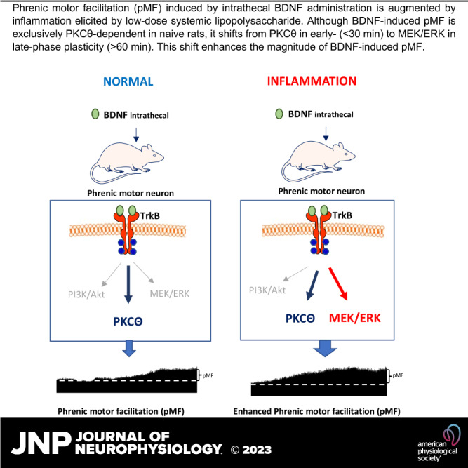
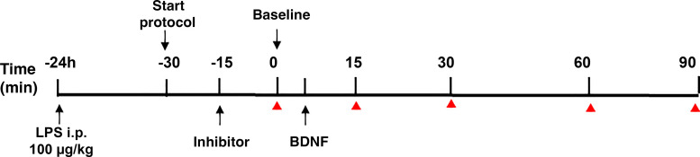
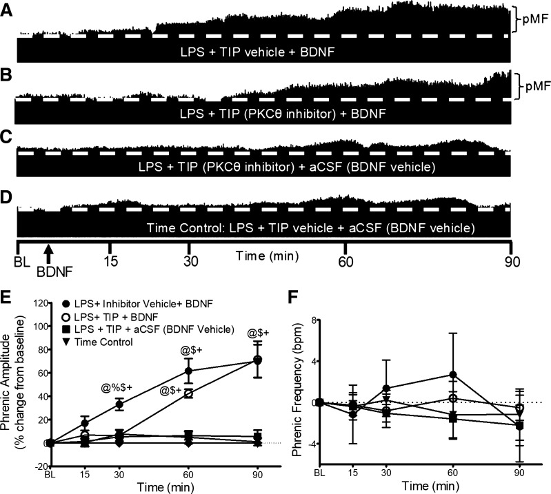
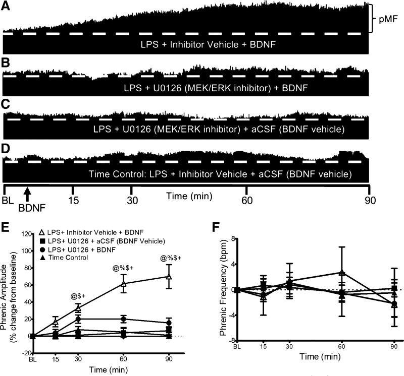
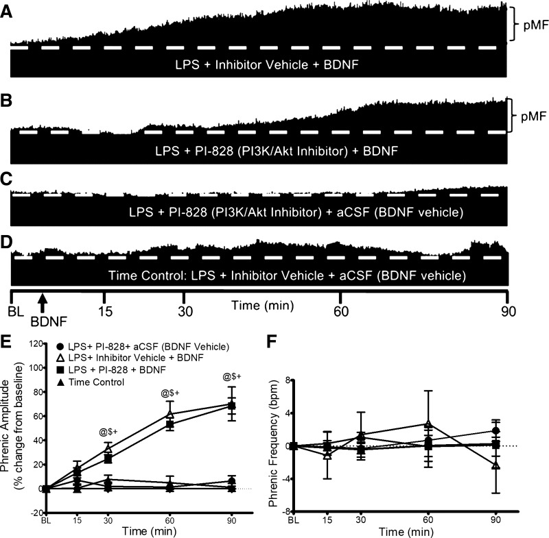
Moderate acute intermittent hypoxia (mAIH) elicits a form of phrenic motor plasticity known as phrenic long-term facilitation (pLTF), which requires spinal 5-HT2 receptor activation, ERK/MAP kinase signaling, and new brain-derived neurotrophic factor (BDNF) synthesis. New BDNF protein activates TrkB receptors that normally signal through PKCθ to elicit pLTF. Phrenic motor plasticity elicited by spinal drug administration (e.g., BDNF) is referred to by a more general term: phrenic motor facilitation (pMF). Although mild systemic inflammation elicited by a low lipopolysaccharide (LPS) dose (100 µg/kg; 24 h prior) undermines mAIH-induced pLTF upstream from BDNF protein synthesis, it augments pMF induced by spinal BDNF administration through unknown mechanisms. Here, we tested the hypothesis that mild inflammation shifts BDNF/TrkB signaling from PKCθ to alternative pathways that enhance pMF. We examined the role of three known signaling pathways associated with TrkB (MEK/ERK MAP kinase, PI3 kinase/Akt, and PKCθ) in BDNF-induced pMF in anesthetized, paralyzed, and ventilated Sprague Dawley rats 24 h post-LPS. Spinal PKCθ inhibitor (TIP) attenuated early BDNF-induced pMF (≤30 min), with minimal effect 60-90 min post-BDNF injection. In contrast, MEK inhibition (U0126) abolished BDNF-induced pMF at 60 and 90 min. PI3K/Akt inhibition (PI-828) had no effect on BDNF-induced pMF at any time. Thus, whereas BDNF-induced pMF is exclusively PKCθ-dependent in normal rats, MEK/ERK is recruited by neuroinflammation to sustain, and even augment downstream plasticity. Because AIH is being developed as a therapeutic modality to restore breathing in people living with multiple neurological disorders, it is important to understand how inflammation, a common comorbidity in many traumatic or degenerative central nervous system disorders, impacts phrenic motor plasticity.NEW & NOTEWORTHY We demonstrate that even mild systemic inflammation shifts signaling mechanisms giving rise to BDNF-induced phrenic motor plasticity. This finding has important experimental, biological, and translational implications, particularly since BDNF-dependent spinal plasticity is being translated to restore breathing and nonrespiratory movements in diverse clinical disorders, such as spinal cord injury (SCI) and amyotrophic lateral sclerosis (ALS).

Keywords: brain-derived neurotrophic factor; cell signaling; inflammation; phrenic motor neuron; plasticity.


Conflict of interest statement

No conflicts of interest, financial or otherwise, are declared by the authors.

Figures

None
Graphical abstract
Figure 1.
Figure 1.
Experimental protocol. Rats were injected with LPS (100 µg/kg, ip) 24 h before neurophysiology experiments. Rats were anesthetized and prepared for phrenic nerve recordings (start protocol). Blood gases were measured before and after stable nerve recordings were established. Kinase inhibitors were administered, and 20 min allowed before establishing baseline (BL) blood gas values just before intrathecal BDNF injection. Blood samples and phrenic nerve recordings were taken at baseline 15, 30, 60, and 90 min post-BDNF (red triangle). BDNF, brain-derived neurotrophic factor.
Figure 2.
Figure 2.
PKCθ inhibitor (TIP) and BDNF-induced pMF. Representative traces of compressed integrated phrenic neurograms (A–D). BDNF-induced pMF after LPS is PKCθ-dependent at 30 min, but not 60 or 90 min. A: post-LPS and intrathecal TIP vehicle (100% aCSF, 12 µL), BDNF elicits robust pMF. B: post-LPS, intrathecal TIP (100 mM/12 µL) 20 min before BDNF impaired pMF at 30 min. At 60 and 90 min post-BDNF, phrenic nerve amplitude is increased. C: with TIP (100 mM/12 µL) plus BDNF vehicle (0.1% BSA and aCSF), pMF is not observed. D: with intrathecal inhibitor vehicle (100% aCSF, 12 µL) plus BDNF vehicle (0.1% BSA and aCSF), pMF is not observed. E: group data for phrenic burst amplitude, expressed as percent change from baseline. LPS + TIP vehicle + BDNF (black circle, n = 8), LPS + PKCθ inhibitor + BDNF (white circles, n = 5), LPS + PKCθ inhibitor + aCSF (squares, n = 4), and LPS + inhibitor vehicle + aCSF (Time Control; black triangles, n = 5) were compared to compare groups at similar time post-BDNF/vehicle injection, and within each group vs. baseline. Phrenic amplitude after LPS + TIP vehicle + BDNF is significantly elevated above the other 3 groups at 30 min only, and above LPS + TIP + aCSF and time control at 60 and 90 min. LPS + TIP vehicle+ BDNF and LPS + PKCθ inhibitor + BDNF were not significantly different at 60 and 90 min post-BDNF. LPS + PKCθ inhibitor + aCSF and time control were not significantly different at any time. Significance is P ≤ 0.05; @significant difference from baseline; %significant difference from LPS + TIP+ BDNF; $significant difference from LPS + TIP + aCSF; and +significant difference from time control. F: phrenic burst frequency, expressed as a change from baseline (burst/min). Phrenic frequency was scaled to equal phrenic amplitude for direct comparison of magnitudes among groups; there were no significant differences between any group at any time; P > 0.05. aCSF, artificial cerebrospinal fluid; BDNF, brain-derived neurotrophic factor; LPS, lipopolysaccharide; pMF, phrenic motor facilitation; TIP, theta inhibitory peptide.
Figure 3.
Figure 3.
MEK/ERK inhibitor (U0126) and BDNF-induced pMF. Representative traces of compressed integrated phrenic neurograms (A–D). After LPS (100 µg/kg, 24 h post), BDNF-induced pMF is MEK/ERK dependent since pMF at later times (60–90 min) was blocked by U0126. A: after U0126 vehicle (20% DMSO/80% saline), BDNF elicits robust pMF 24 h post-LPS. B: after U0126 (100 mM/12 µL) given 20 min before BDNF in LPS-treated rats, pMF is impaired. C: after U0126 (100 mM/12 µL) plus BDNF vehicle (0.1% BSA and aCSF), pMF is not observed. D: after intrathecal inhibitor vehicle (20% DMSO/80% saline) plus BDNF vehicle (0.1% BSA and aCSF), pMF is not observed. E: group data for phrenic burst amplitude, expressed as a percent increase from baseline. LPS + inhibitor vehicle + BDNF (white triangles, n = 8), LPS + MEK/ERK inhibitor + BDNF (circles, n = 6), LPS + MEK/ERK inhibitor + BDNF vehicle (squares, n = 7), and LPS + inhibitor vehicle + BDNF vehicle (Time Control; black triangles, n = 4) were compared to determine significance between groups, and each group vs. baseline. Rats that received LPS + inhibitor vehicle + BDNF showed elevated phrenic amplitude vs. the other 3 groups at 30, 60 and 90 min. LPS + MEK/ERK inhibitor + BDNF, LPS + MEK/ERK inhibitor + BDNF vehicle and LPS + inhibitor vehicle + BDNF vehicle were not significantly different at any time. Significance is P ≤ 0.05; @significant difference from baseline; %significant difference from LPS + MEK/ERK inhibitor + BDNF; $significant difference from LPS + MEK/ERK inhibitor + BDNF vehicle; and +significant difference from time control. F: phrenic burst frequency, expressed as a change from baseline in burst/min. Phrenic frequency was scaled equal to phrenic amplitude for direct comparison of magnitudes in the groups. There were no significant differences between any group at any time (all P > 0.05). aCSF, artificial cerebrospinal fluid; BDNF, brain-derived neurotrophic factor; LPS, lipopolysaccharide; pMF, phrenic motor facilitation.
Figure 4.
Figure 4.
PI3K/Akt inhibitor (PI-828) and BDNF-induced pMF. Representative traces of compressed integrated phrenic neurograms (A–D). After LPS (100 µg/kg; 24 h post), BDNF-induced pMF remains independent of PI3K/Akt signaling. A: intrathecal inhibitor vehicle (20% DMSO/80% saline) 20 min before intrathecal BDNF exhibits pMF. B: after LPS, BDNF still elicits pMF after intrathecal PI3K/Akt inhibitor (100 mM/12 µL; 20 min before). C: PI3K/Akt inhibitor (PI-828; 100 mM/12 µL; 20 min before) with BDNF vehicle (0.1% BSA and aCSF) does not elicit pMF. D: inhibitor vehicle (20% DMSO/80% saline; 20 min pre) with BDNF vehicle (0.1% BSA and aCSF) does not elicit pMF. E: group data for phrenic burst amplitude, expressed as percent increase from baseline. LPS + inhibitor vehicle + BDNF (white triangles, n = 8), LPS + PI3K/Akt inhibitor + BDNF (squares, n = 5), LPS + PI3K/Akt inhibitor + BDNF vehicle (circles, n = 5), and LPS + inhibitor vehicle + BDNF vehicle (Time Control; black triangles, n = 5) were compared to determine significant differences between groups and vs. baseline. @Significant difference from baseline; $significant difference from LPS + inhibitor vehicle + BDNF; and +significant difference from time control. F: phrenic burst frequency, expressed as a change from baseline in burst/min. Phrenic frequency was scaled equal to phrenic amplitude for direct comparison of magnitudes in the groups. There were no significant differences between any group at any time; P > 0.05. aCSF, artificial cerebrospinal fluid; BDNF, brain-derived neurotrophic factor; LPS, lipopolysaccharide; pMF, phrenic motor facilitation.

Similar articles

Cited by

References

    1. Mancini A, de Iure A, Picconi B. Basic mechanisms of plasticity and learning. Handb Clin Neurol 184: 21–34, 2022. doi:10.1016/B978-0-12-819410-2.00002-3. - DOI - PubMed
    1. Wang Y, Fu AKY, Ip NY. Instructive roles of astrocytes in hippocampal synaptic plasticity: neuronal activity-dependent regulatory mechanisms. FEBS J 289: 2202–2218, 2021. doi:10.1111/febs.15878. - DOI - PMC - PubMed
    1. Feldman JL, Mitchell GS, Nattie EE. Breathing: rhythmicity, plasticity, chemosensitivity. Annu Rev Neurosci 26: 239–266, 2003. doi:10.1146/annurev.neuro.26.041002.131103. - DOI - PMC - PubMed
    1. Powell FL, Milsom WK, Mitchell GS. Time domains of the hypoxic ventilatory response. Respir Physiol 112: 123–134, 1998. doi:10.1016/s0034-5687(98)00026-7. - DOI - PubMed
    1. Mitchell GS, Johnson SM. Neuroplasticity in respiratory motor control. J Appl Physiol (1985) 94: 358–374, 2003. doi:10.1152/japplphysiol.00523.2002. - DOI - PubMed

Publication types

MeSH terms

Substances