. 2023 Feb 14;11(1):e0411622.
doi: 10.1128/spectrum.04116-22. Epub 2023 Jan 25.

Mutation of Phenylalanine 23 of Newcastle Disease Virus Matrix Protein Inhibits Virus Release by Disrupting the Interaction between the FPIV L-Domain and Charged Multivesicular Body Protein 4B

Affiliations

Mutation of Phenylalanine 23 of Newcastle Disease Virus Matrix Protein Inhibits Virus Release by Disrupting the Interaction between the FPIV L-Domain and Charged Multivesicular Body Protein 4B

Yu Pei et al. Microbiol Spectr. .

Abstract

The matrix (M) protein FPIV L-domain is conserved among multiple paramyxoviruses; however, its function and the associated mechanism remain unclear. In this study, the paramyxovirus Newcastle disease virus (NDV) was employed to study the FPIV L-domain. Two recombinant NDV strains, each carrying a single amino acid mutation at the Phe (F23) or Pro (P24) site of 23FPIV/I26 L-domain, were rescued. Growth defects were observed in only the recombinant SG10-F23A (rSG10-F23A) strain. Subsequent studies focused on rSG10-F23A revealed that the virulence, pathogenicity, and replication ability of this strain were all weaker than those of wild-type strain rSG10 and that a budding deficiency contributed to those weaknesses. To uncover the molecular mechanism underlying the rSG10-F23A budding deficiency, the bridging proteins between the FPIV L-domain and endosomal sorting complex required for transported (ESCRT) machinery were explored. Among 17 candidate proteins, only the charged multivesicular body protein 4 (CHMP4) paralogues were found to interact more strongly with the NDV wild-type M protein (M-WT) than with the mutated M protein (M-F23A). Overexpression of M-WT, but not of M-F23A, changed the CHMP4 subcellular location to the NDV budding site. Furthermore, a knockdown of CHMP4B, the most abundant CHMP4 protein, inhibited the release of rSG10 but not that of rSG10-F23A. From these findings, we can reasonably infer that the F23A mutation of the FPIV L-domain blocks the interaction between the NDV M protein and CHMP4B and that this contributes to the budding deficiency and consequent growth defects of rSG10-F23A. This work lays the foundation for further study of the FPIV L-domain in NDV and other paramyxoviruses. IMPORTANCE Multiple viruses utilize a conserved motif, termed the L-domain, to act as a cellular adaptor for recruiting host ESCRT machinery to their budding site. Despite the FPIV type L-domain having been identified in some paramyxoviruses 2 decades ago, its function in virus life cycles and its method of recruiting the ESCRT machinery are poorly understood. In this study, a single amino acid mutation at the F23 site of the 23FPIV26 L-domain was found to block NDV budding at the late stage. Furthermore, CHMP4B, a core component of the ESCRT-III complex, was identified as a main factor that links the FPIV L-domain and ESCRT machinery together. These results extend previous understanding of the FPIV L-domain and, therefore, not only provide a new approach for attenuating NDV and other paramyxoviruses but also lay the foundation for further study of the FPIV L-domain.

Keywords: CHMP4B; ESCRT machinery; FPIV L-domain; M protein; Newcastle disease virus; virus release.


Conflict of interest statement

The authors declare no conflict of interest.

Figures

FIG 1
FIG 1
Rescue and confirmation of the rNDVs. (A) Amino acid sequence alignment of the M proteins from the family Paramyxoviridae. The conserved FPIV-like sequence is outlined by a black dotted line. (B) Schematic diagram of rSG10. The mutant sites of rSG10-F23A and rSG10-P24A are marked by a black underline. (C and D) One-step (MOI = 1) and multistep (MOI = 0.01) growth kinetics of rSG10, rSG10-F23A, and rSG10-P24A in Vero cells. Cell supernatants from infected cells were collected at the indicated time points, and the viral titers were determined by titration on corresponding cells. P values were calculated by two-way ANOVA.
FIG 2
FIG 2
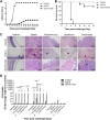
Pathogenicity and replication ability of rNDVs in vivo. Three-week-old SPF chickens were inoculated with PBS (uninfected control group) or rNDVs (rSG10 or rSG10-F23A). (A) Clinical symptoms of 10 birds per group were scored daily during the 14-day observation according to the following standard: healthy, 0; sick, 1; wing drop, torticollis, or lack of coordination, 2; prostration, 3; and death, 4. (B) Survival of the control uninfected, rSG10-infected, and rSG10-F23A-infected groups, determined from 10 birds per group. (C) Tissue histopathology analysis at 5 dpc of 3-week-old SPF chickens that had been inoculated with PBS or rNDVs. Tissues were fixed, embedded in paraffin wax, sectioned into slices, and stained in eosin. The lesions of tissues were observed under light microscopy. Black arrowheads indicate the pathological changes observed in tissues. Scale bars: 200 μm. (D) Birds from different groups were euthanized at 3, 5, or 7 dpc, and the viral loads in different tissues were evaluated by RT-qPCR. The comparative threshold cycle (ΔΔCT) method was applied, and the 2−ΔΔCT value of each tissue from the uninfected control group was set to 1. The 2−ΔΔCT value of each tissue from the rSG10- and rSG10-F23A-infected groups was normalized to the corresponding value of the uninfected control group. The significance of each difference was analyzed by two-way ANOVA.
FIG 3
FIG 3
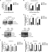
Analysis of the budding efficiency of rNDVs. (A and B) Vero cells were infected with rSG10, rSG10-F23A, or rSG10-P24A at an MOI of 1 (A) or 0.01 (B). At the indicated time points, the extracellular virial titers were determined by titration on the corresponding cells. The significance of each difference was calculated with one-way or two-way ANOVA. (C) Virions were purified via ultracentrifugation. Equal amounts of virions and WCLs were subjected to Western blotting. The HN and M proteins of NDV were detected using specific antibodies. (D) The budding index of rNDVs was defined as the grayscale value of the HN or M protein bands in virions divided by their values in WCLs. The budding efficiency of rSG10 was set to 1, and the budding efficiencies of rSG10-F23A and rSG10-P24A were obtained via comparison with rSG10. The significance of each difference was calculated with two-way ANOVA. The values ± SD from triplicate experiments are shown. (E to G) Virions were purified via ultracentrifugation at the indicated time points. The F, HN, and M proteins of NDV were detected using specific antibodies. The budding efficiency of rNDVs was calculated as described for panel D. The values ± SD from triplicate experiments are shown.
FIG 4
FIG 4
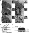
Analysis of the budding efficiency of NDV VLPs. (A) TEM images of thin-sectioned Vero cells that had been infected with an rNDV at an MOI of 0.01. Typical paramyxovirus particles (diameters, 100 to 200 nm) were observed in the plasma membrane. Scale bars = 1 μm. (B) Vero cells were transfected with plasmids expressing Flag-M-WT or Flag-M-F23A. At 36 hpt, the VLPs were purified. Equal amounts of cell lysates and VLPs were subjected to Western blotting (IB). The budding efficiency was calculated as described for Fig. 3D. The assays were performed three times. (C) Collected VLPs and cell lysates were treated with different concentrations (1, 5, or 20 μg/mL) of proteinase K for 30 min in the presence of 1% Triton X-100 or were left untreated. The resulting digestions were subjected to Western blotting.
FIG 5
FIG 5
Detection of interaction between ESCRT proteins and the FPIV L-domain. HA-tagged ALIX and Flag-tagged M protein (M-WT or M-F23A) (A) or Myc-tagged M protein (M-WT or M-F23A) and Flag-tagged ESCRT protein (TSG101, EAP20, EAP30, EAP45, CHMP1A, CHMP1B, CHMP2A, CHMP2B, CHMP3, CHMP4A, CHMP4B, CHMP4C, CHMP5, CHMP6, CHMP7, or IST1) (B to F) were cotransfected into HEK293T cells. At 48 hpt, the cells were lysed and immunoprecipitated by using an anti-Flag M2 affinity gel. The assays were performed twice (A and B) or three times (C to F). The resulting immunoprecipitates together with cell lysates were analyzed via Western blotting. The affinity ratio between M protein (M-WT or M-F23A) and each ESCRT component was calculated by dividing the quantity of copurified HA/Myc-tagged protein by the quantity of purified Flag-tagged protein. The affinity ratio of M-WT or M-F23A was set to 1, and the relative affinity ratios of other proteins were obtained by comparison.
FIG 6
FIG 6
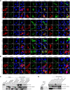
Detection of interaction between the CHMP4s and FPIV L-domain. (A and B) Vero cells were cotransfected with Myc-tagged TSG101, EAP30, EAP45, CHMP1s, CHMP2s, CHMP3, or CHMP5 (A) or CHMP4s (B) and Flag-tagged M protein (M-WT or M-F23A). At 24 hpt, cells were fixed, labeled with antibodies specific for the Myc tag or Flag tag, and incubated with Alexa Fluor 488- and 555-conjugated secondary antibodies. DAPI was used to stain the nuclei. Scale bars = 10 μm. (C and D) GST or GST-tagged CHMP4s were immobilized on glutathione Sepharose 4B and then incubated with lysates of bacteria containing His, His–M-WT, or His–M-F23A (C) or lysates of cells transfected with PIV5-M or PIV5-M-FP (D). The resulting immunoprecipitations were subjected to Western blotting conducted with antibodies specific for the GST, His, and Flag tags. The assays were run in triplicate.
FIG 7
FIG 7
Effects of endogenous CHMP4B knockdown on rSG10 virion release from cells infected at an MOI of 1 or 0.01. (A) Cell viability upon transfection with each indicated siRNA was evaluated via a CCK-8 assay. The value of the si-NC-transfected sample was set to 1. The values of si-CHMP4B- and si-CHMP4C-transfected samples were obtained by comparison with the si-NC-transfected sample. The significance of each difference was calculated by one-way ANOVA. (B) Vero cells were transfected with the indicated siRNA and harvested at 24 hpt for RNA isolation; the resulting RNA was subjected to RT-qPCR to evaluate the percentage of remaining mRNA for CHMP4B or CHMP4C after knockdown. The significance of each difference was calculated with one-way ANOVA. (C) At 48 hpt with the siRNA described above, Vero cells were harvested, and the WCLs were analyzed by Western blotting conducted using specific antibodies against endogenous CHMP4B. (D to F) After transfection with the indicated siRNAs, Vero cells were infected with the indicated rNDV at an MOI of 1 (D) or 0.01 (E and F). At 24 hpi, the extracellular viruses were collected from the infected cells and their titers were determined by titration on corresponding cells. The significance of each difference was calculated with one-way ANOVA. (G to I) The extracellular viruses described for panels D to F were also pelleted through ultracentrifugation. Equal amounts of virions and WCLs were subjected to Western blotting and the budding efficiencies were calculated as described above. The significance of each difference was calculated with one-way ANOVA. The values ± SD from triplicate experiments are shown.

Similar articles

Cited by

References

    1. Watanabe S, Shirogane Y, Sato Y, Hashiguchi T, Yanagi Y. 2019. New insights into measles virus brain infections. Trends Microbiol 27:164–175. doi:10.1016/j.tim.2018.08.010. - DOI - PubMed
    1. Johnson K, Vu M, Freiberg AN. 2021. Recent advances in combating Nipah virus. Fac Rev 10:74. doi:10.12703/r/10-74. - DOI - PMC - PubMed
    1. Bockelman C, Frawley TC, Long B, Koyfman A. 2018. Mumps: an emergency medicine-focused update. J Emerg Med 54:207–214. doi:10.1016/j.jemermed.2017.08.037. - DOI - PubMed
    1. Choi EJ, Ortega V, Aguilar HC. 2020. Feline morbillivirus, a new paramyxovirus possibly associated with feline kidney disease. Viruses 12:501. doi:10.3390/v12050501. - DOI - PMC - PubMed
    1. Pentecost M, Vashisht AA, Lester T, Voros T, Beaty SM, Park A, Wang YE, Yun TE, Freiberg AN, Wohlschlegel JA, Lee B. 2015. Evidence for ubiquitin-regulated nuclear and subnuclear trafficking among Paramyxovirinae matrix proteins. PLoS Pathog 11:e1004739. doi:10.1371/journal.ppat.1004739. - DOI - PMC - PubMed

Publication types

Substances

LinkOut - more resources