Peptidyl tRNA Hydrolase Is Required for Robust Prolyl-tRNA Turnover in Mycobacterium tuberculosis
- PMID: 36695586
- PMCID: PMC9973355
- DOI: 10.1128/mbio.03469-22
Peptidyl tRNA Hydrolase Is Required for Robust Prolyl-tRNA Turnover in Mycobacterium tuberculosis
Abstract
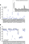
Enzymes involved in rescuing stalled ribosomes and recycling translation machinery are ubiquitous in bacteria and required for growth. Peptidyl tRNA drop-off is a type of abortive translation that results in the release of a truncated peptide that is still bound to tRNA (peptidyl tRNA) into the cytoplasm. Peptidyl tRNA hydrolase (Pth) recycles the released tRNA by cleaving off the unfinished peptide and is essential in most bacteria. We developed a sequencing-based strategy called copper sulfate-based tRNA sequencing (Cu-tRNAseq) to study the physiological role of Pth in Mycobacterium tuberculosis (Mtb). While most peptidyl tRNA species accumulated in a strain with impaired Pth expression, peptidyl prolyl-tRNA was particularly enriched, suggesting that Pth is required for robust peptidyl prolyl-tRNA turnover. Reducing Pth levels increased Mtb's susceptibility to tRNA synthetase inhibitors that are in development to treat tuberculosis (TB) and rendered this pathogen highly susceptible to macrolides, drugs that are ordinarily ineffective against Mtb. Collectively, our findings reveal the potency of Cu-tRNAseq for profiling peptidyl tRNAs and suggest that targeting Pth would open new therapeutic approaches for TB. IMPORTANCE Peptidyl tRNA hydrolase (Pth) is an enzyme that cuts unfinished peptides off tRNA that has been prematurely released from a stalled ribosome. Pth is essential in nearly all bacteria, including the pathogen Mycobacterium tuberculosis (Mtb), but it has not been clear why. We have used genetic and novel biochemical approaches to show that when Pth levels decline in Mtb, peptidyl tRNA accumulates to such an extent that usable tRNA pools drop. Thus, Pth is needed to maintain normal tRNA levels, most strikingly for prolyl-tRNAs. Many antibiotics act on protein synthesis and could be affected by altering the availability of tRNA. This is certainly true for tRNA synthetase inhibitors, several of which are drug candidates for tuberculosis. We find that their action is potentiated by Pth depletion. Furthermore, Pth depletion results in hypersensitivity to macrolides, drugs that are not active enough under ordinary circumstances to be useful for tuberculosis.
Keywords: Mycobacterium tuberculosis; antibiotic resistance; bacterial genetics; ribosomes; tRNA; tRNA sequencing; translation.
Conflict of interest statement
The authors declare no conflict of interest.
Figures


Similar articles
-
A tRNA-Acetylating Toxin and Detoxifying Enzyme in Mycobacterium tuberculosis.Microbiol Spectr. 2022 Jun 29;10(3):e0058022. doi: 10.1128/spectrum.00580-22. Epub 2022 May 31. Microbiol Spectr. 2022. PMID: 35638832 Free PMC article.
-
A physiological connection between tmRNA and peptidyl-tRNA hydrolase functions in Escherichia coli.Nucleic Acids Res. 2004 Nov 16;32(20):6028-37. doi: 10.1093/nar/gkh924. Print 2004. Nucleic Acids Res. 2004. PMID: 15547251 Free PMC article.
-
Protein synthesis factors (RF1, RF2, RF3, RRF, and tmRNA) and peptidyl-tRNA hydrolase rescue stalled ribosomes at sense codons.J Mol Biol. 2012 Apr 13;417(5):425-39. doi: 10.1016/j.jmb.2012.02.008. Epub 2012 Feb 9. J Mol Biol. 2012. PMID: 22326347
-
Structural and functional insights into peptidyl-tRNA hydrolase.Biochim Biophys Acta. 2014 Jul;1844(7):1279-88. doi: 10.1016/j.bbapap.2014.04.012. Epub 2014 Apr 21. Biochim Biophys Acta. 2014. PMID: 24768774 Review.
-
Peptidyl-tRNA hydrolase and its critical role in protein biosynthesis.Microbiology (Reading). 2006 Aug;152(Pt 8):2191-2195. doi: 10.1099/mic.0.29024-0. Microbiology (Reading). 2006. PMID: 16849786 Review.
Cited by
-
Innovative perspectives on the discovery of small molecule antibiotics.NPJ Antimicrob Resist. 2025 Mar 13;3(1):19. doi: 10.1038/s44259-025-00089-0. NPJ Antimicrob Resist. 2025. PMID: 40082593 Free PMC article. Review.
-
Nucleotidyltransferase toxin MenT extends aminoacyl acceptor ends of serine tRNAs to control Mycobacterium tuberculosis growth.Nat Commun. 2024 Nov 6;15(1):9596. doi: 10.1038/s41467-024-53931-w. Nat Commun. 2024. PMID: 39505885 Free PMC article.
-
MenT nucleotidyltransferase toxins extend tRNA acceptor stems and can be inhibited by asymmetrical antitoxin binding.Nat Commun. 2023 Aug 17;14(1):4644. doi: 10.1038/s41467-023-40264-3. Nat Commun. 2023. PMID: 37591829 Free PMC article.
-
Streptomyces rare codon UUA: from features associated with 2 adpA related locations to candidate phage regulatory translational bypassing.RNA Biol. 2023 Jan;20(1):926-942. doi: 10.1080/15476286.2023.2270812. Epub 2023 Nov 15. RNA Biol. 2023. PMID: 37968863 Free PMC article.
References
Publication types
MeSH terms
Substances
Grants and funding
- R01 AI042347/AI/NIAID NIH HHS/United States
- F31 AI167560/AI/NIAID NIH HHS/United States
- 1F31AI167560-01/HHS | NIH | National Institute of Allergy and Infectious Diseases (NIAID)
- P01AI095208/HHS | NIH | National Institute of Allergy and Infectious Diseases (NIAID)
- P01 AI095208/AI/NIAID NIH HHS/United States
LinkOut - more resources
Full Text Sources

