

Functional MRI to quantify perfusion changes of a renal allograft after embolization of an arteriovenous fistula
- PMID: 36696037
- PMCID: PMC10226906
- DOI: 10.1007/s40620-022-01539-y
Functional MRI to quantify perfusion changes of a renal allograft after embolization of an arteriovenous fistula
Abstract
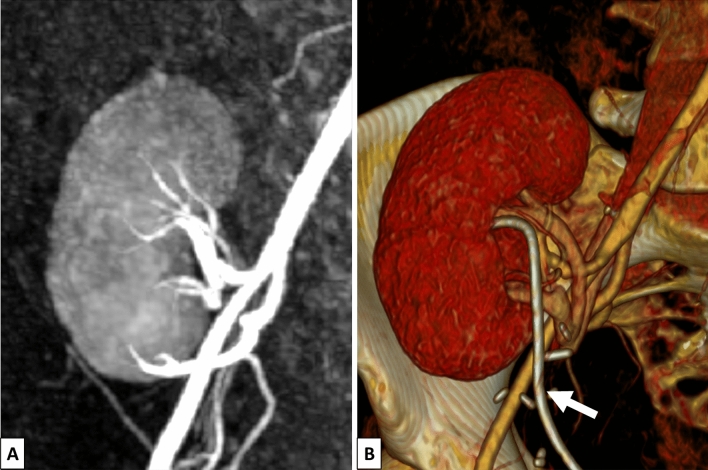
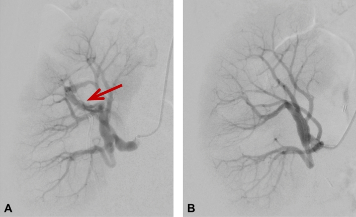
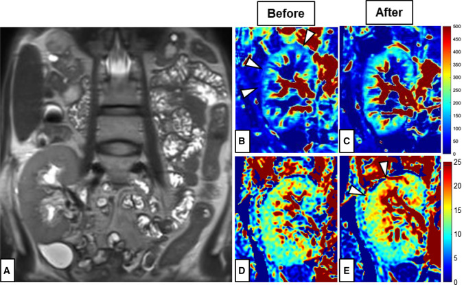
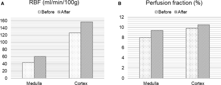
Acute allograft injury was observed in a 37-year-old woman within a few weeks after kidney transplantation. Neither renal ultrasound nor computerized tomography (CT) and magnetic resonance (MR) angiography revealed any anomaly. An MR protocol was then performed including arterial spin labeling and intravoxel incoherent motion diffusion weighted imaging. Both arterial spin labeling and the perfusion fraction in the diffusion weighted imaging showed decreased perfusion compared to reference values. The patient subsequently underwent angiography, where an arteriovenous fistula in the upper calix of the transplant kidney was detected and immediate embolization was performed. A second functional MR, performed one week later, demonstrated a 40% increase in organ perfusion. We conclude that functional MR with arterial spin labeling and intravoxel incoherent motion have the potential to provide complementary information of clinical value to conventional imaging for monitoring renal allografts.
Keywords: Arterial spin labeling; Arteriovenous fistula; Diffusion weighted imaging; Perfusion imaging; Transplant kidney.
© 2023. The Author(s).
Conflict of interest statement
The authors of this manuscript declare no relationships with any companies, whose products or services may be related to the subject matter of the article.
Figures
Similar articles
-
Evaluation of renal allografts function early after transplantation using intravoxel incoherent motion and arterial spin labeling MRI.Magn Reson Imaging. 2016 Sep;34(7):908-14. doi: 10.1016/j.mri.2016.04.022. Epub 2016 Apr 22. Magn Reson Imaging. 2016. PMID: 27114341
-
Detection of Crossed Cerebellar Diaschisis by Intravoxel Incoherent Motion MR Imaging in Subacute Ischemic Stroke.Cell Transplant. 2019 Aug;28(8):1062-1070. doi: 10.1177/0963689719856290. Epub 2019 Jun 14. Cell Transplant. 2019. PMID: 31198047 Free PMC article.
-
Multiparametric renal magnetic resonance imaging: A reproducibility study in renal allografts with stable function.NMR Biomed. 2023 Feb;36(2):e4832. doi: 10.1002/nbm.4832. Epub 2022 Oct 10. NMR Biomed. 2023. PMID: 36115029 Free PMC article.
-
Renal allograft arteriovenous fistula due to needle biopsy with late onset of symptoms--diagnosis and treatment.Nephron. 1991;59(3):482-5. doi: 10.1159/000186613. Nephron. 1991. PMID: 1758542 Review.
-
Contrecoup Injury-Induced Middle Meningeal Arteriovenous Fistula Detected by Time-of-Flight Magnetic Resonance Angiography and Magnetic Resonance Arterial Spin Labeling: Case Report and Review of the Literature.World Neurosurg. 2019 Jul;127:79-84. doi: 10.1016/j.wneu.2019.03.189. Epub 2019 Mar 28. World Neurosurg. 2019. PMID: 30928586 Review.
References
Publication types
MeSH terms
LinkOut - more resources
Full Text Sources
Medical
