

Functional MRI to quantify perfusion changes of a renal allograft after embolization of an arteriovenous fistula
- PMID: 36696037
- PMCID: PMC10226906
- DOI: 10.1007/s40620-022-01539-y
Functional MRI to quantify perfusion changes of a renal allograft after embolization of an arteriovenous fistula
Abstract
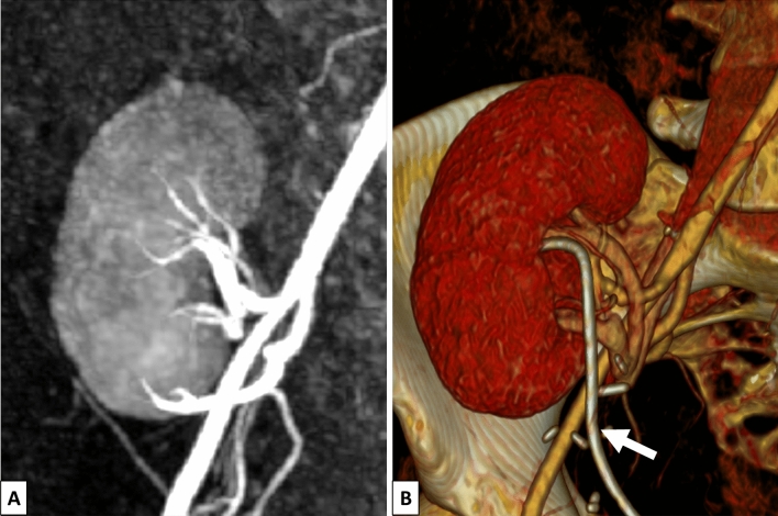
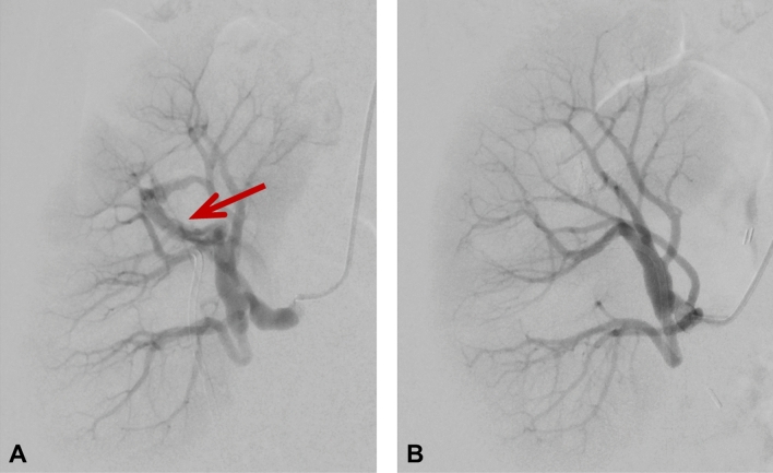
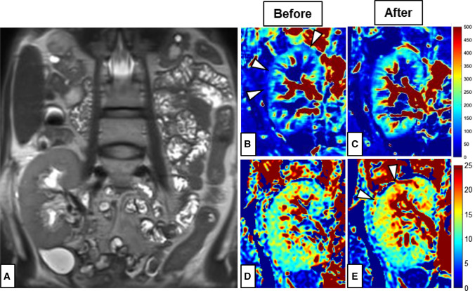
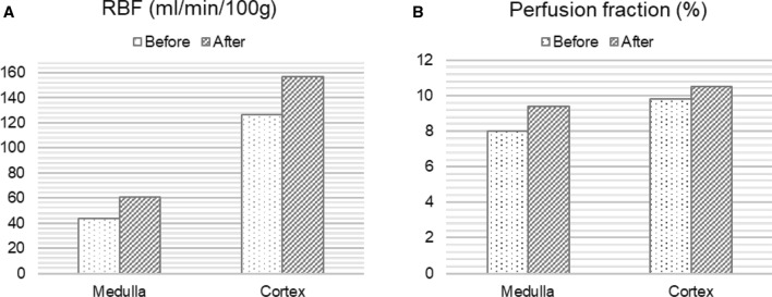
Acute allograft injury was observed in a 37-year-old woman within a few weeks after kidney transplantation. Neither renal ultrasound nor computerized tomography (CT) and magnetic resonance (MR) angiography revealed any anomaly. An MR protocol was then performed including arterial spin labeling and intravoxel incoherent motion diffusion weighted imaging. Both arterial spin labeling and the perfusion fraction in the diffusion weighted imaging showed decreased perfusion compared to reference values. The patient subsequently underwent angiography, where an arteriovenous fistula in the upper calix of the transplant kidney was detected and immediate embolization was performed. A second functional MR, performed one week later, demonstrated a 40% increase in organ perfusion. We conclude that functional MR with arterial spin labeling and intravoxel incoherent motion have the potential to provide complementary information of clinical value to conventional imaging for monitoring renal allografts.
Keywords: Arterial spin labeling; Arteriovenous fistula; Diffusion weighted imaging; Perfusion imaging; Transplant kidney.
© 2023. The Author(s).
Conflict of interest statement
The authors of this manuscript declare no relationships with any companies, whose products or services may be related to the subject matter of the article.
Figures
References
Publication types
MeSH terms
LinkOut - more resources
Full Text Sources
Medical
