

The mechanistic and structural role of von Willebrand factor in endotoxemia-enhanced deep vein thrombosis in mice
- PMID: 36696220
- PMCID: PMC11552101
- DOI: 10.1016/j.jtha.2022.11.022
The mechanistic and structural role of von Willebrand factor in endotoxemia-enhanced deep vein thrombosis in mice
Abstract
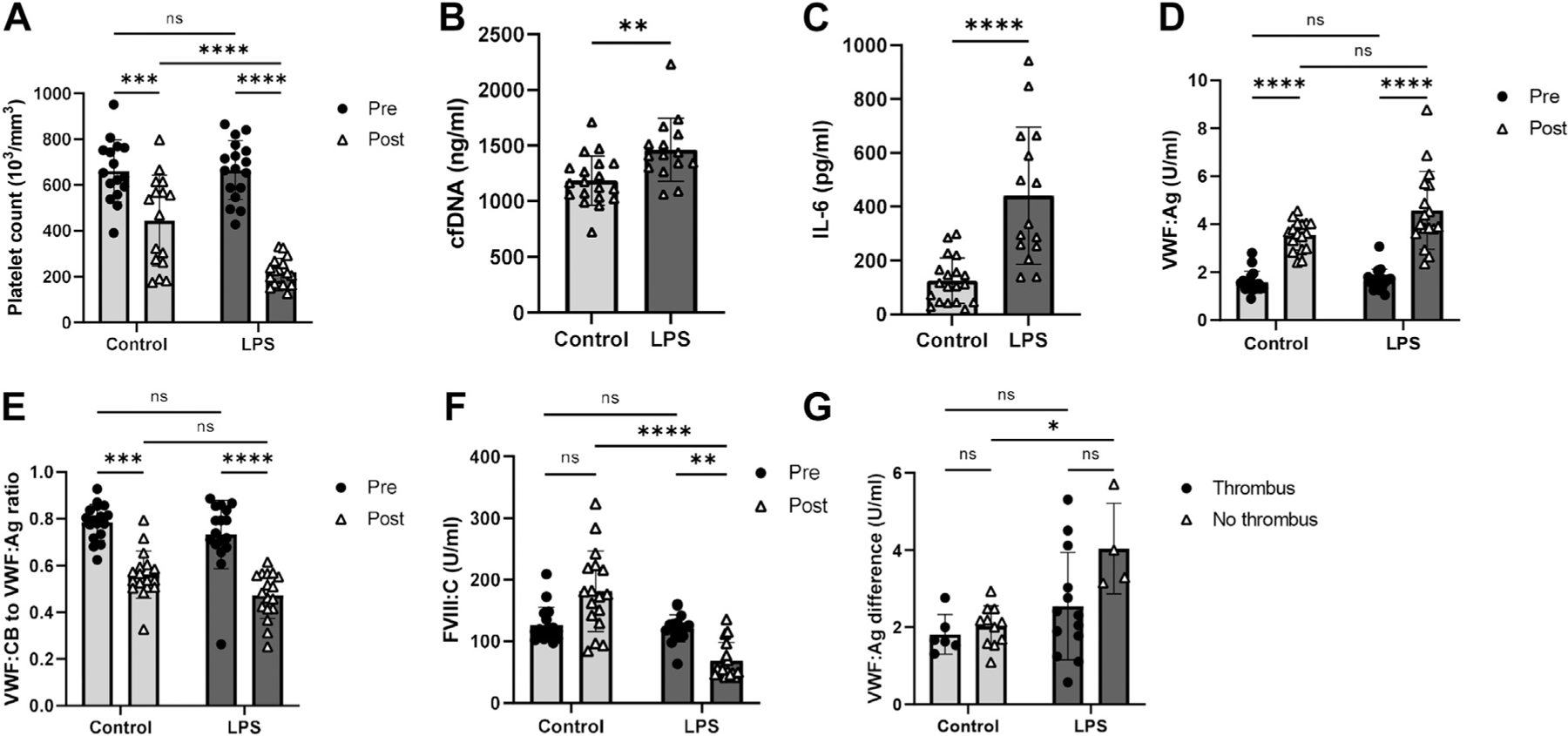
Background: Although the concept of immunothrombosis has established a link between inflammation and thrombosis, the role of inflammation in the pathogenesis of deep vein thrombosis remains to be fully elucidated. Further, although various constituents of venous thrombi have been identified, their localizations and cellular and molecular interactions are yet to be combined in a single, multiplexed analysis.
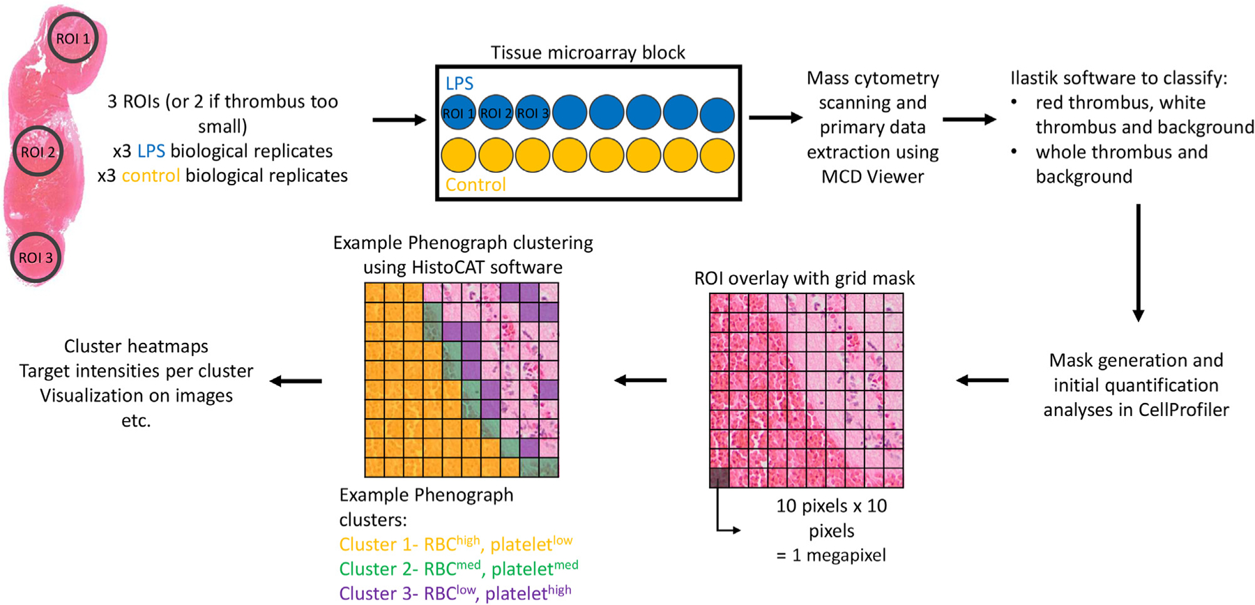
Objectives: The objective of this study was to investigate the role of the von Willebrand factor (VWF) in inflammation-associated venous thrombosis. We also performed a proof-of-concept study of imaging mass cytometry to quantitatively and simultaneously analyze the localizations and interactions of 10 venous thrombus constituents.
Methods: We combined the murine inferior vena cava stenosis model of deep vein thrombosis with the lipopolysaccharide model of endotoxemia. We also performed a proof-of-concept study of imaging mass cytometry to assess the feasibility of this approach in analyzing the structural composition of thrombi.
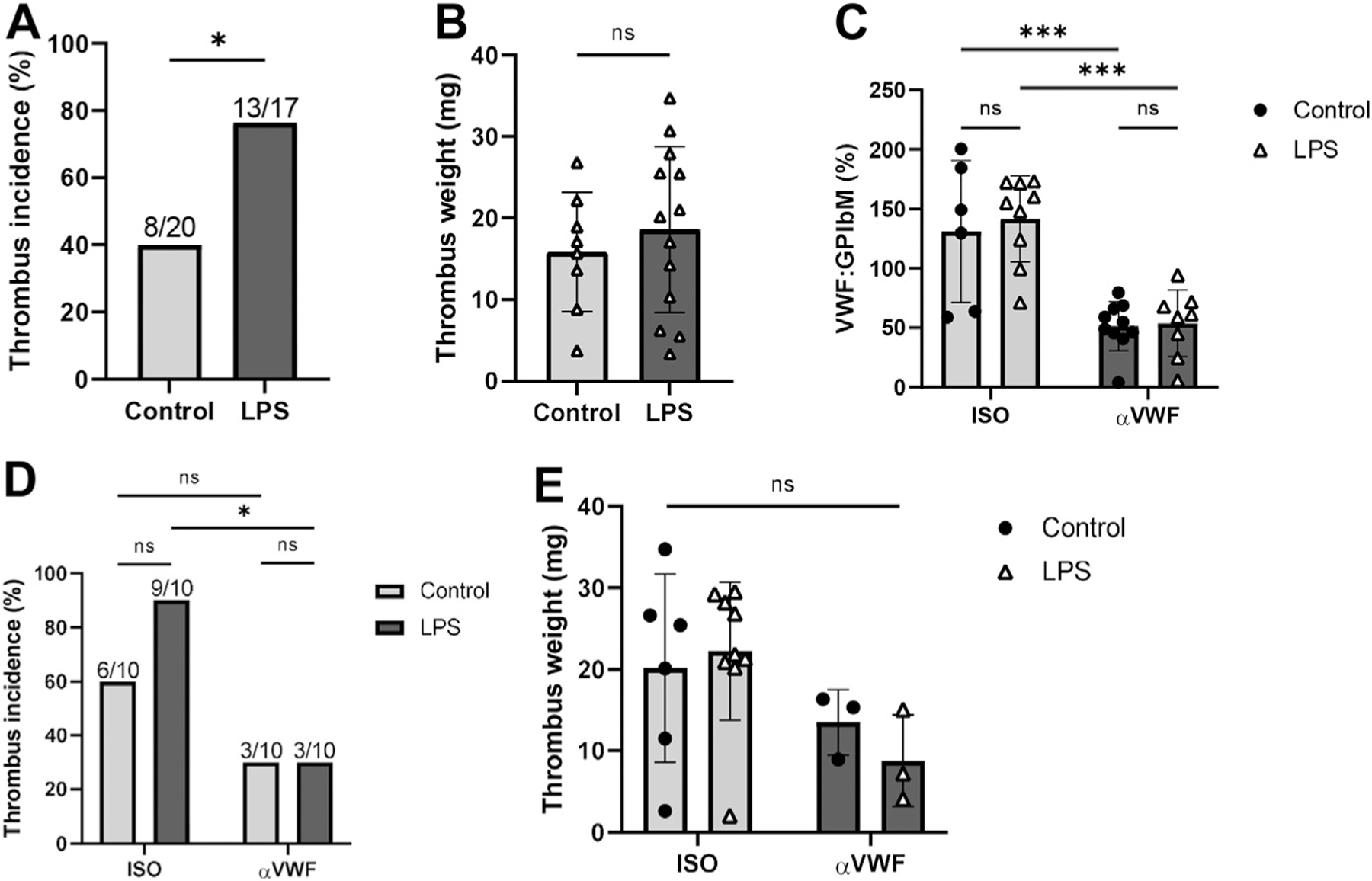
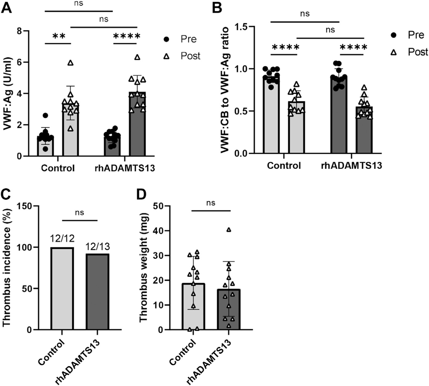
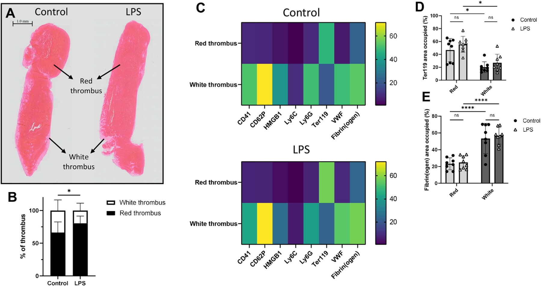
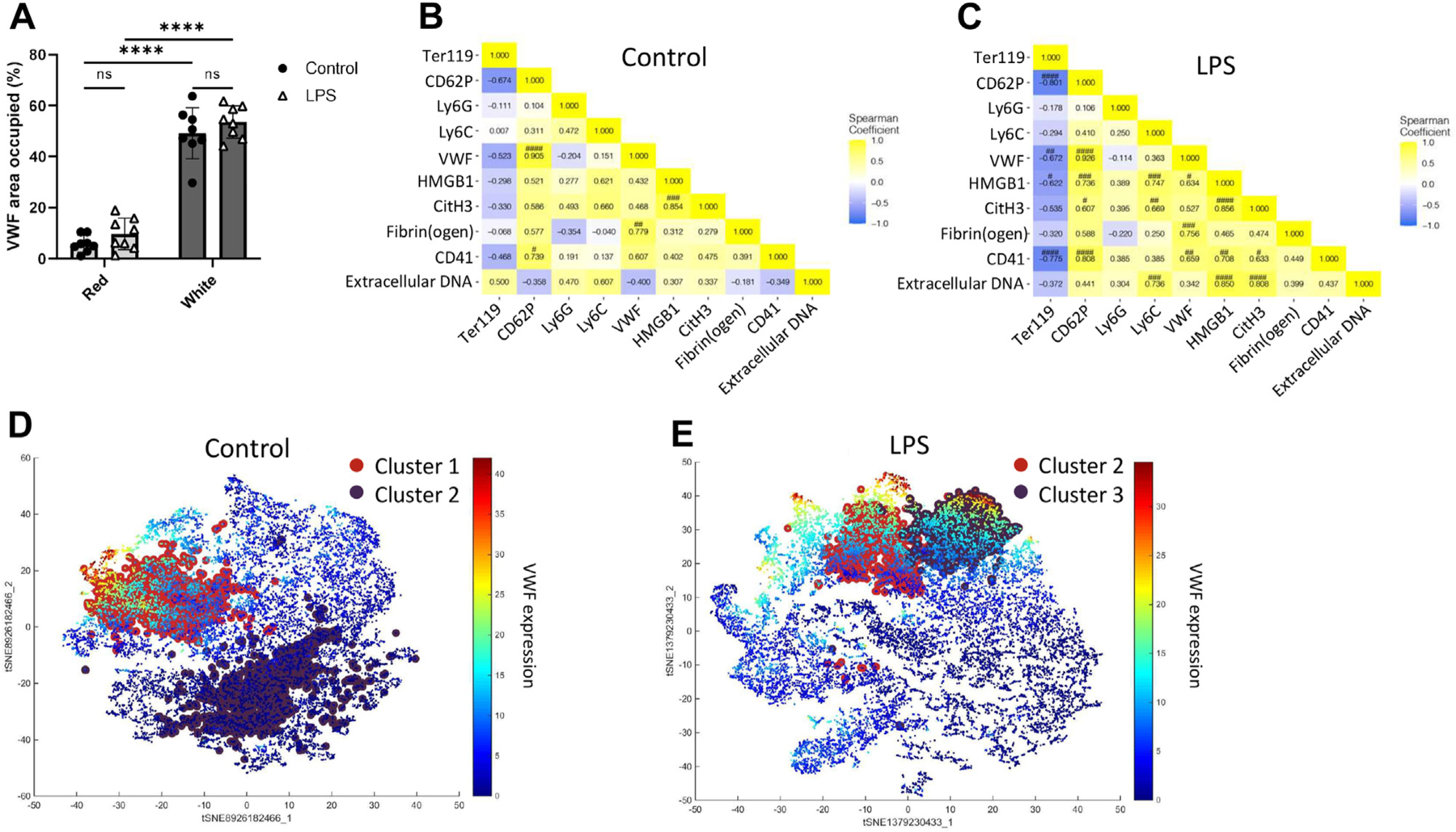
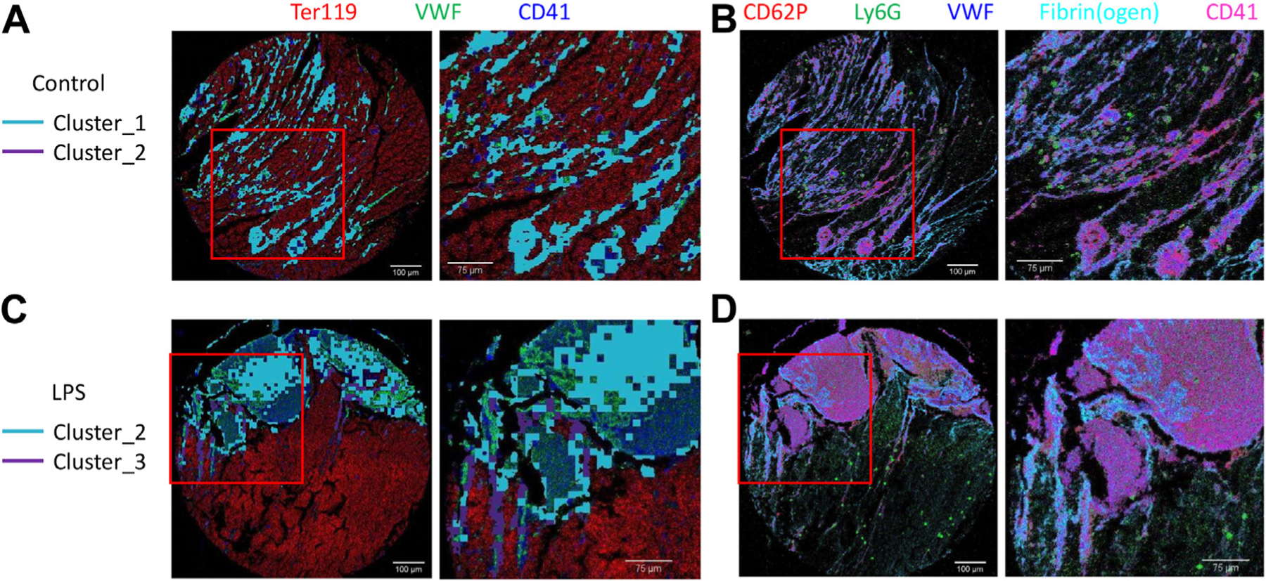
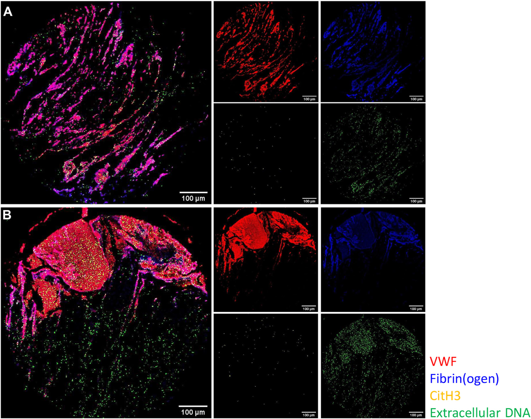
Results: We found that lipopolysaccharide-treated mice had significantly higher incidences of venous thrombosis, an effect that was mitigated when VWF was inhibited using inhibitory αVWF antibodies. Our detailed structural analysis also showed that most thrombus components are localized in the white thrombus regardless of endotoxemia. Moreover, although endotoxemia modulated the relative representation and interactions of VWF with other thrombus constituents, the scaffolding network, comprised VWF, fibrin, and neutrophil extracellular traps, remained largely unaffected.
Conclusions: We observe a key role for VWF in the pathogenesis of inflammation-associated venous thrombosis while providing a more comprehensive insight into the molecular interactions that constitute the architecture of venous thrombi.
Keywords: animal models; image cytometry; thromboinflammation; venous thrombosis; von Willebrand factor.
Crown Copyright © 2022. Published by Elsevier Inc. All rights reserved.
Conflict of interest statement
Declaration of competing interests There are no competing interests to disclose.
Figures
Comment in
-
Von Willebrand factor-inflammation crosstalk in deep vein thrombosis.J Thromb Haemost. 2023 Mar;21(3):453-455. doi: 10.1016/j.jtha.2022.11.038. J Thromb Haemost. 2023. PMID: 36858790 Free PMC article. No abstract available.
Similar articles
-
von Willebrand Factor Is a Critical Mediator of Deep Vein Thrombosis in a Mouse Model of Diet-Induced Obesity.Arterioscler Thromb Vasc Biol. 2020 Dec;40(12):2860-2874. doi: 10.1161/ATVBAHA.120.314690. Epub 2020 Sep 24. Arterioscler Thromb Vasc Biol. 2020. PMID: 32967458
-
Endothelial PTP1B Deletion Promotes VWF Exocytosis and Venous Thromboinflammation.Circ Res. 2024 May 10;134(10):e93-e111. doi: 10.1161/CIRCRESAHA.124.324214. Epub 2024 Apr 2. Circ Res. 2024. PMID: 38563147
-
Neutrophil extracellular traps promote deep vein thrombosis in mice.J Thromb Haemost. 2012 Jan;10(1):136-44. doi: 10.1111/j.1538-7836.2011.04544.x. J Thromb Haemost. 2012. PMID: 22044575 Free PMC article.
-
Insights Into Immunothrombosis: The Interplay Among Neutrophil Extracellular Trap, von Willebrand Factor, and ADAMTS13.Front Immunol. 2020 Dec 2;11:610696. doi: 10.3389/fimmu.2020.610696. eCollection 2020. Front Immunol. 2020. PMID: 33343584 Free PMC article. Review.
-
von Willebrand factor and inflammation.J Thromb Haemost. 2017 Jul;15(7):1285-1294. doi: 10.1111/jth.13696. J Thromb Haemost. 2017. PMID: 28671350 Review.
Cited by
-
Von Willebrand factor-inflammation crosstalk in deep vein thrombosis.J Thromb Haemost. 2023 Mar;21(3):453-455. doi: 10.1016/j.jtha.2022.11.038. J Thromb Haemost. 2023. PMID: 36858790 Free PMC article. No abstract available.
References
-
- Raskob GE, Angchaisuksiri P, Blanco AN, Buller H, Gallus A, Hunt BJ, Hylek EM, Kakkar A, Konstantinides SV, McCumber M, Ozaki Y, Wendelboe A, Weitz JI, ISTH Steering Committee for World Thrombosis Day. Thrombosis: a major contributor to global disease burden. Arterioscler Thromb Vasc Biol 2014;34: 2363–71. - PubMed
-
- Virani SS, Alonso A, Aparicio HJ, Benjamin EJ, Bittencourt MS, Callaway CW, Carson AP, Chamberlain AM, Cheng S, Delling FN, Elkind MSV, Evenson KR, Ferguson JF, Gupta DK, Khan SS, Kissela BM, Knutson KL, Lee CD, Lewis TT, Liu J. Heart disease and stroke statistics–2021 update: a report from the American Heart Association. Circulation. 2021;143:e254–743. - PubMed
-
- Levi M, van der Poll T. Coagulation and sepsis. Thromb Res 2017;149:38–44. - PubMed
-
- Hanify JM, Dupree LH, Johnson DW, Ferreira JA. Failure of chemical thromboprophylaxis in critically ill medical and surgical patients with sepsis. J Crit Care. 2017;37:206–10. - PubMed
Publication types
MeSH terms
Substances
Grants and funding
LinkOut - more resources
Full Text Sources
Medical
Miscellaneous
