. 2023 Jan 31;120(5):e2215575120.
doi: 10.1073/pnas.2215575120. Epub 2023 Jan 25.

Structural and functional insights into the chloroplast division site regulators PARC6 and PDV1 in the intermembrane space

Affiliations

Structural and functional insights into the chloroplast division site regulators PARC6 and PDV1 in the intermembrane space

Qingqing Sun et al. Proc Natl Acad Sci U S A. .

Abstract

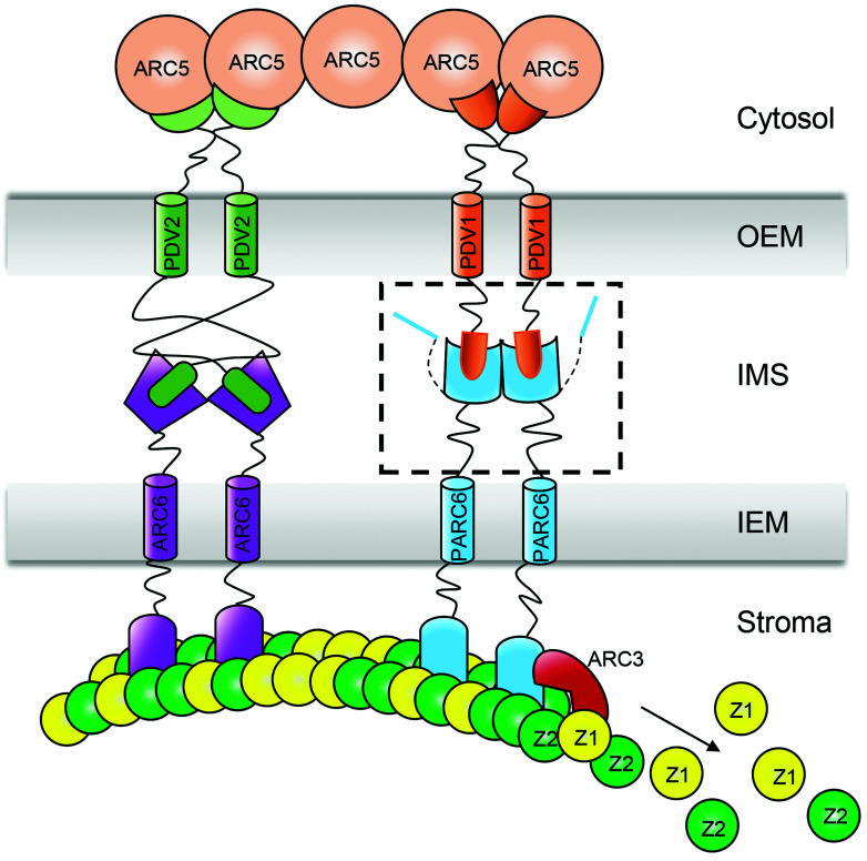
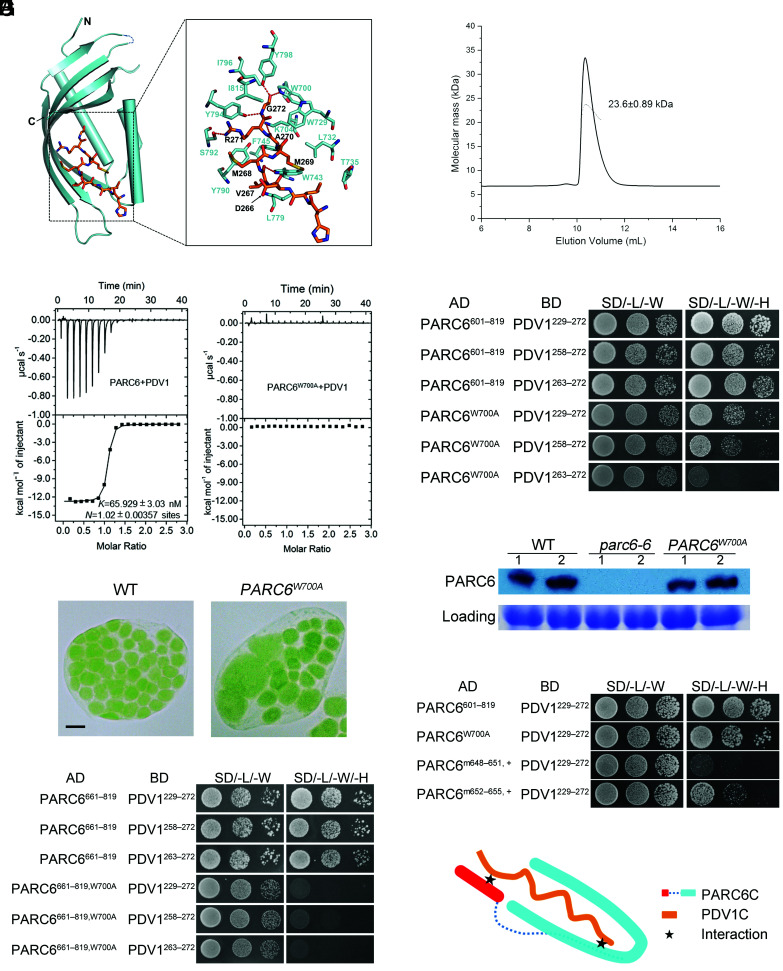
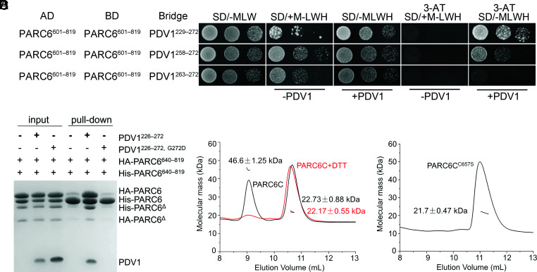
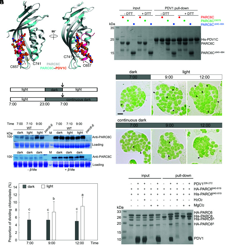
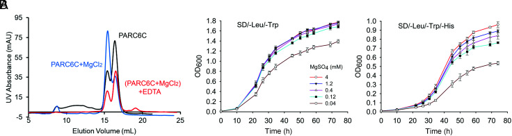
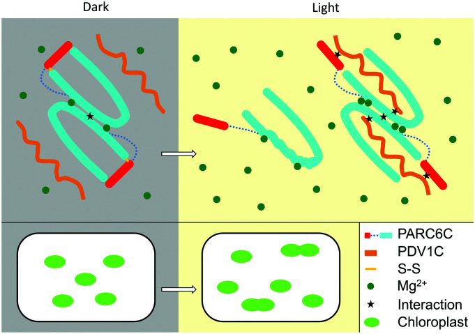
Chloroplast division involves the coordination of protein complexes from the stroma to the cytosol. The Min system of chloroplasts includes multiple stromal proteins that regulate the positioning of the division site. The outer envelope protein PLASTID DIVISION1 (PDV1) was previously reported to recruit the cytosolic chloroplast division protein ACCUMULATION AND REPLICATION OF CHLOROPLAST5 (ARC5). However, we show here that PDV1 is also important for the stability of the inner envelope chloroplast division protein PARALOG OF ARC6 (PARC6), a component of the Min system. We solved the structure of both the C-terminal domain of PARC6 and its complex with the C terminus of PDV1. The formation of an intramolecular disulfide bond within PARC6 under oxidized conditions prevents its interaction with PDV1. Interestingly, this disulfide bond can be reduced by light in planta, thus promoting PDV1-PARC6 interaction and chloroplast division. Interaction with PDV1 can induce the dimerization of PARC6, which is important for chloroplast division. Magnesium ions, whose concentration in chloroplasts increases upon light exposure, also promote the PARC6 dimerization. This study highlights the multilayer regulation of the PDV1-PARC6 interaction as well as chloroplast division.

Keywords: PARC6; PDV1; chloroplast division; crystal structure; intermembrane space.


Conflict of interest statement

The authors declare no competing interest.

Figures

Fig. 1.
Fig. 1.
A schematic diagram of the topology and interaction of chloroplast division proteins at the division site. A box with dashed lines includes the C-terminal domain of PDV1 and the C-terminal domain of PARC6 in the IMS. The mechanism of the interaction between PDV1 and PARC6 is unclear before this study. Z1, FtsZ1; Z2, FtsZ2; OEM, outer envelope membrane; IMS, inter-membrane space; IEM, inner envelope membrane.
Fig. 2.
Fig. 2.
The pdv1 mutant exhibits phenotypes similar to that of parc6. (A) A comparison of the chloroplast division phenotypes. Cells are from mesophyll. WT, wild type. Bar, 10 μm. All images are at the same magnification. (B) pdv1 and parc6 have chloroplasts with multiple division sites. Cells are from bundle sheath. Bars, 10 μm. (C) Immunofluorescence staining of FtsZ filaments. Cells are from the mesophyll of mature leaves. Bars, 10 μm.
Fig. 3.
Fig. 3.
PDV1 is important for the stability of PARC6. Immunoblot analysis of the abundance of various chloroplast division proteins in WT, pdv1, and complemented pdv1 plants (comp). Each genotype is represented by two replicates in the blots. Coomassie brilliant blue (CBB) staining of the sodium dodecyl sulfate (SDS)-polyacrylamide gel electrophoresis (PAGE) gels serves as a loading control.
Fig. 4.
Fig. 4.
Dimerization is important for the function of PARC6. (A) Crystal structure of the PARC6C (residues 640 to 819) homodimer. The C-terminal region of PARC6 forms a pocket that is closed by a lid-like helix (shown in pink). The cysteine residues forming a disulfide bond (shown in yellow) in each protomer are shown as stick models. (B) SLS analysis of PARC6C in solution. PARC6C monomers and dimers coexist. (C) The self-interaction region of PARC6C is mainly hydrophobic. One PARC6C protomer is shown as an electrostatic model. The residues with hydrophobic side chains within the dimer interface are indicated. (D) Introducing hydrophilic residues in the self-interaction region of PARC6C abolishes its self-interaction in Y2H analysis. PARC6601–819 begins right after the transmembrane domain and contains PARC6C. PARC6601–639 serves as a linker. The negative controls are shown in SI Appendix, Fig. S9. (E) Disrupting the self-interaction of PARC6 affects chloroplast division. Two point mutants defective in self-interaction fail to complement the parc6-6 mutant phenotype. Bar, 10 μm. (F) Immunoblot analysis of the plants shown in E. CBB staining of the SDS-PAGE gels serves as a loading control.
Fig. 5.
Fig. 5.
Two regions of PARC6 are involved in its interaction with PDV1. (A) Crystal structure of the PARC6685–819–PDV1263–272 fusion protein. The C-terminal end of PDV1 (PDV1C, residues 263 to 272, shown in orange) inserts into the pocket formed by the C-terminal region of PARC6 (shown in cyan). Residues involved in the interactions are shown as stick models. Hydrogen bonds are shown as red dashed lines. (B) SLS analysis of PARC6685–819–PDV1263–272 in solution. The fusion protein is only in a monomeric state. (C) ITC analysis of the interaction between PARC6 and PDV1. GST-tagged PARC6: residues 685 to 819. PDV1: residues 261 to 272. (D) Y2H analysis of the interaction between PARC6601–819, PARC6601–819, W700A, and PDV1. Various lengths of the C-terminal domain of PDV1 were tested. The negative controls are shown in SI Appendix, Fig. S11A. (E) The W700A mutation affects the function of PARC6. Left: WT cell; right: parc6-6 mutant transformed with PARC6W700A. Bar, 10 μm. (F) Immunoblot analysis of the seedlings shown in E. CBB staining of the SDS-PAGE gel serves as a loading control. (G) The W700A mutation abolishes the interaction between a truncated version of PARC6 (residues 661 to 819) and PDV1 in Y2H assays. This truncation lacks the “lid” (I646 to H656) of the PARC6 pocket and C657, which can form a disulfide bond with C741 and is essential for the closing of the “lid”. The negative controls are shown in SI Appendix, Fig. S11B. (H) Y2H analysis shows that the “lid” region of PARC6 is also important for the interaction between PARC6C and PDV1C. “m648–651, +”, VLID motif (residues 648 to 651) mutated to SDSA in the PARC6W700A mutant; “m652–655, +”, MLKM motif (residues 652 to 655) mutated to SDAS in the PARC6W700A mutant. The negative controls are shown in SI Appendix, Fig. S11C. (I) A working model of the interaction between PARC6C and PDV1C. There are two sites for the interaction (shown as stars): One is inside the pocket, and the other one is right outside of the pocket, which involves the lid of PARC6C and a region of PDV1C.
Fig. 6.
Fig. 6.
Dimerization of PARC6 is regulated by PDV1 and the redox status. (A) Y3H analysis shows that PDV1 promotes the self-interaction of PARC6. Various lengths of the C-terminal domain of PDV1 were tested. Methionine (M) suppresses the expression and accumulation of the bridge protein PDV1. 3-AT is an inhibitor of the enzyme encoded by the reporter gene HIS3. The negative controls are shown in SI Appendix, Fig. S13. (B) A pull-down assay shows that the interaction between PDV1 and PARC6 promotes the self-interaction of PARC6. The reaction buffer contains DTT (5 mM), which can reduce disulfide bonds and open the “lid”. Δ, a minor degradation product of the protein. (C) SLS analysis of PARC6C (residues 640 to 819) with or without 5 mM DTT. (D) SLS analysis of PARC6CC657S (residues 640 to 819) indicates that it is a monomer.
Fig. 7.
Fig. 7.
Redox state affects the disulfide bond in PARC6C and its interaction with PDV1C. (A) Crystal structure shows that the formation of a disulfide bond blocks the pocket required for PDV1–PARC6 interaction. Apo- and PDV1C-bound PARC6CΔ640–684 molecules are colored in gray and cyan, respectively. In apo-PARC6C, the “lid” helix is colored in pink, with the two cysteine residues forming the disulfide bond shown as stick models. PDV1C is shown as a ball-and-stick model. (B) Pull-down assay shows that the disulfide bond within PARC6C prevents PDV1–PARC6 interaction. His-SUMO tagged PDV1: residues 226 to 272. (C) WT plants were entrained with 16 h/8 h light/dark cycles (Top) and transferred to continuous dark (Bottom). The arrows indicate the start of the experiment. (D) Dark and light affect disulfide bond formation of PARC6C in Arabidopsis plants. Total proteins from the leaves of WT plants were extracted with buffer without β-mercaptoethanol and split into two aliquots, one of which received β-mercaptoethanol (5%) later. These two sets of samples were run in parallel on the same gel and were probed with anti-PARC6C antibodies. The disulfide bond blocks the antibody–antigen interaction. M, molecular weight markers. CBB staining of the SDS-PAGE gels serves as a loading control. (E) Chloroplast phenotype of mesophyll cells of 21-d-old plants at different time points. Dividing chloroplasts are indicated by arrows. Bar, 10 μm. (F) Proportion of dividing chloroplasts in the plants shown in E (n = 30 cells for each sample, P < 0.05). Chloroplasts with an aspect ratio larger than 1.5 are counted in F. For statistical analysis, different letters denote statistically significant (P < 0.05) differences between samples. Gray column, dark; white column, light. (G) A pull-down assay to show the effect of the oxidative condition on the PDV1–PARC6 interaction formed in the reduced state. HA-PARC6, His-PARC6, and PDV1 were first incubated in the presence of 5 mM DTT, and then the buffer was changed to one containing 0.5% H2O2 but no DTT. MgCl2 was added at a final concentration of 5 mM throughout the process where indicated. Δ, a minor degradation product of the protein.
Fig. 8.
Fig. 8.
Magnesium ions affect the dimerization of PARC6C. (A) Gel infiltration analysis of PARC6C, PARC6C with 5 mM MgCl2, and PARC6C incubated with 5 mM MgCl2 before the addition of 5 mM EDTA. (B) Y2H analysis of the self-interaction of PARC6 in the presence of different Mg2+ concentrations. Yeast colonies harboring two plasmids, AD PARC6601–819 and BD PARC6601–819, were grown in liquid medium as indicated. OD600 values were measured at different time points to measure growth, which reflects the strength of the protein interaction.
Fig. 9.
Fig. 9.
A working model of the regulation of the PDV1–PARC6 interaction during light-regulated chloroplast division. In the dark, the intramolecular disulfide bond in PARC6C is oxidized. The PARC6C lid is thus closed and PARC6C forms a dimer. In the light, the intramolecular disulfide bond in PARC6C is reduced. The PARC6C lid is open, which destabilizes PARC6C and hampers its dimerization. However, interaction with PDV1C stabilizes the structure of PARC6C, promoting its dimerization. Mg2+ binding also promotes the dimerization of PARC6C. Light increases the concentration of Mg2+ in chloroplasts and further promotes the dimerization of PARC6C. Fluctuations in the redox state and Mg2+ concentrations modulate the light-mediated regulation of chloroplast division.

Similar articles

Cited by

References

    1. Osteryoung K. W., Nunnari J., The division of endosymbiotic organelles. Science 302, 1698–1704 (2003). - PubMed
    1. Osteryoung K. W., McAndrew R. S., The plastid division machine. Annu. Rev. Plant Physiol. Plant Mol. Biol. 52, 315–333 (2001). - PubMed
    1. Osteryoung K. W., Pyke K. A., Division and dynamic morphology of plastids. Annu. Rev. Plant Biol. 65, 443–472 (2014). - PubMed
    1. Miyagishima S., Mechanism of plastid division: From a bacterium to an organelle. Plant Physiol. 155, 1533–1544 (2011). - PMC - PubMed
    1. Yoshida Y., Mogi Y., TerBush A. D., Osteryoung K. W., Chloroplast FtsZ assembles into a contractible ring via tubulin-like heteropolymerization. Nat. Plants 5, 119–119 (2016). - PubMed

Publication types

MeSH terms

LinkOut - more resources