. 2023 Jan 31;120(5):e2208351120.
doi: 10.1073/pnas.2208351120. Epub 2023 Jan 25.

Ufmylation reconciles salt stress-induced unfolded protein responses via ER-phagy in Arabidopsis

Affiliations

Ufmylation reconciles salt stress-induced unfolded protein responses via ER-phagy in Arabidopsis

Baiying Li et al. Proc Natl Acad Sci U S A. .

Abstract

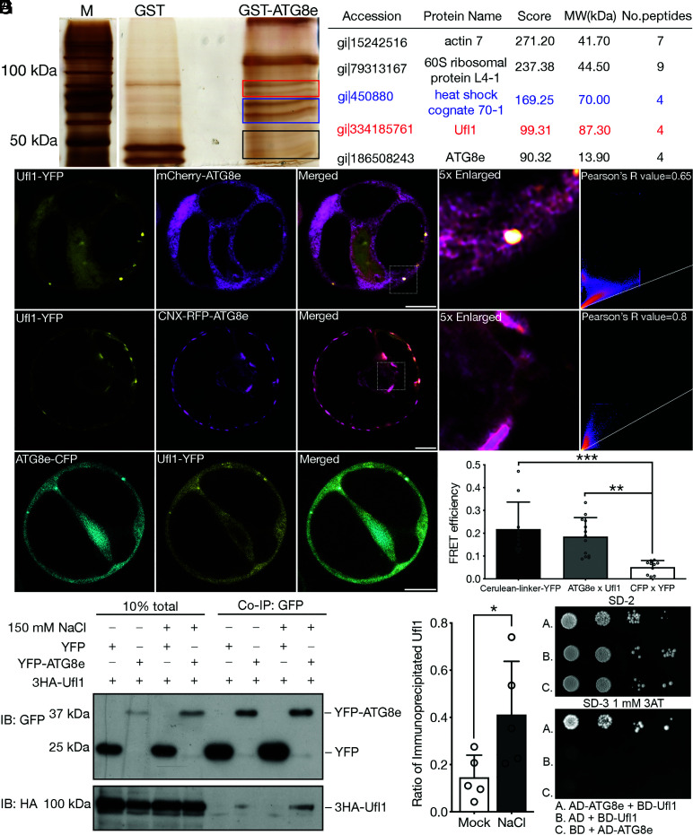
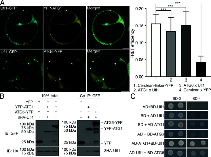
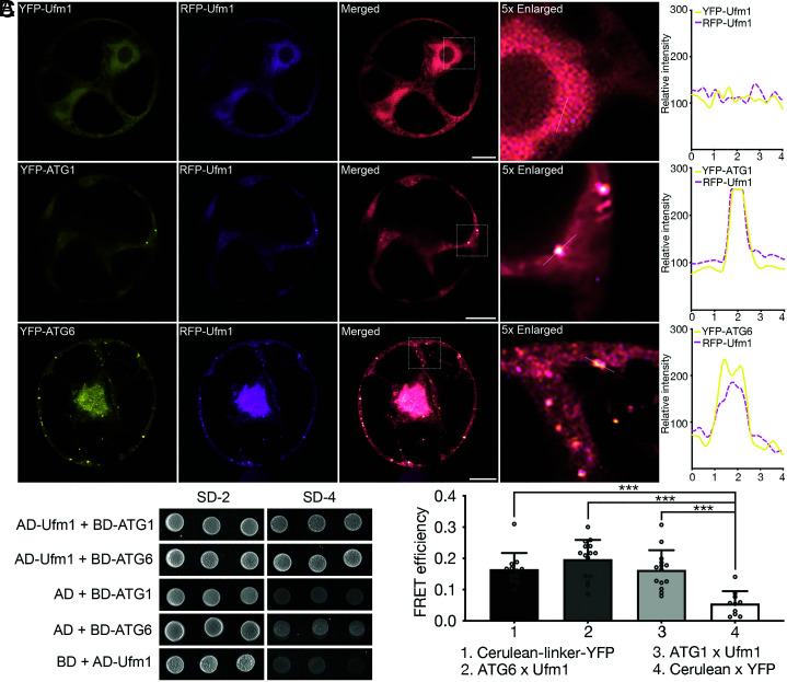
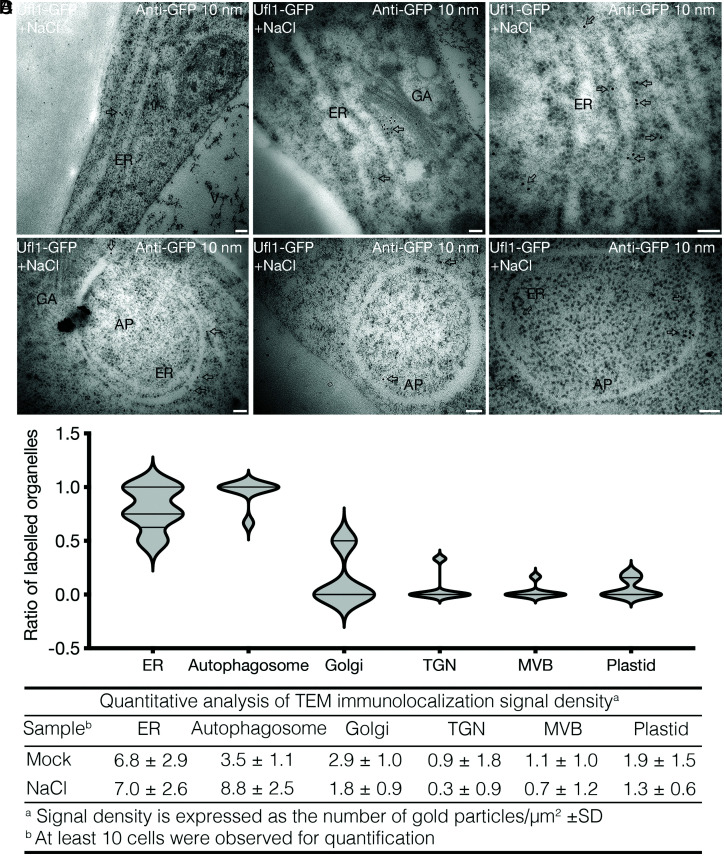
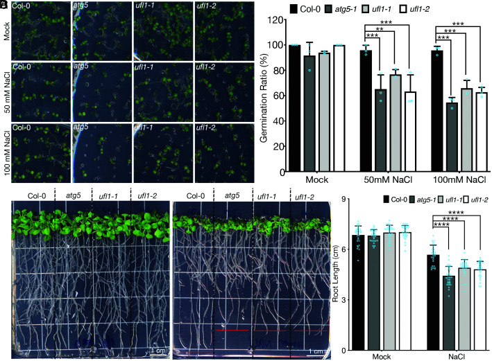
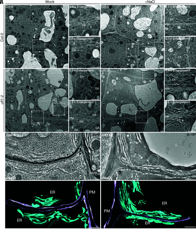
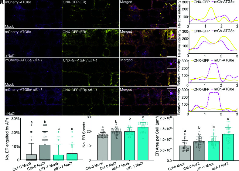
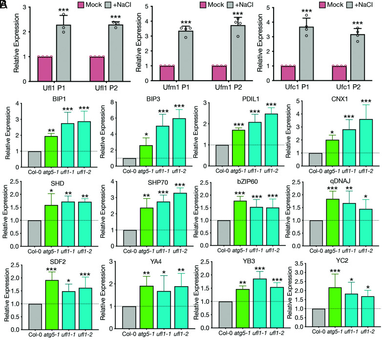
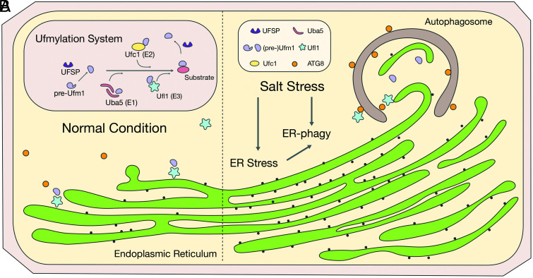
In plants, the endomembrane system is tightly regulated in response to environmental stresses for maintaining cellular homeostasis. Autophagosomes, the double membrane organelles forming upon nutrient deprivation or stress induction, degrade bulky cytosolic materials for nutrient turnover. Though abiotic stresses have been reported to induce plant autophagy, few receptors or regulators for selective autophagy have been characterized for specific stresses. Here, we have applied immunoprecipitation followed by tandem mass spectrometry using the autophagosome marker protein ATG8 as bait and have identified the E3 ligase of the ufmylation system Ufl1 as a bona fide ATG8 interactor under salt stress. Notably, core components in the ufmylation cascade, Ufl1 and Ufm1, interact with the autophagy kinase complexes proteins ATG1 and ATG6. Cellular and genetic analysis showed that Ufl1 is important for endoplasmic reticulum (ER)-phagy under persisting salt stress. Loss-of-function mutants of Ufl1 display a salt stress hypersensitive phenotype and abnormal ER morphology. Prolonged ER stress responses are detected in ufl1 mutants that phenocopy the autophagy dysfunction atg5 mutants. Consistently, expression of ufmylation cascade components is up-regulated by salt stress. Taken together, our study demonstrates the role of ufmylation in regulating ER homeostasis under salt stress through ER-phagy.


Conflict of interest statement

The authors declare no competing interest.

Figures

Fig. 1.
Fig. 1.
Identification of Ufl1 as an ATG8e interactor under salt stress condition. (A) Purified recombinant GST-ATG8e was used as bait for IP from Arabidopsis PSB-D cell lysate upon salt stress treatment. Proteins pulled-out by GST-ATG8e were separated by SDS-PAGE, and silver-stained protein bands were subjected to tandem MS analysis for subsequent protein identification. The Right panel lists promising hits in the GST-ATG8 pull down group. The label of boxes in red, blue, and black on the gel represents the portion of gel cut where candidates in the lists labeled by the same color were identified. (B) Ufl1-YFP colocalized with mCherry-ATG8e in punctate upon their co-expression in Arabidopsis protoplasts. (Scale bar, 10 μm.) (C) CNX-RFP-ATG8e recruited Ufl1-YFP to the ER upon their co-expression in Arabidopsis protoplasts. (Scale bar, 10 μm.) (D) FRET analysis of ATG8e-CFP with Ufl1-YFP in Arabidopsis protoplasts. (Scale bar, 10 μm.) **P < 0.01, ***P < 0.001. (E) Co-immunoprecipitation (Co-IP) analysis of 3HA-Ufl1 with YFP-ATG8e in Arabidopsis protoplasts with or without salt stress treatment. YFP-ATG8e or free YFP was transiently co-expressed with 3HA-Ufl1, followed by cell lysis and IP using GFP-trap magnetic beads for subsequent western blot analysis. (F) Quantification analysis of the ratio of co-immunoprecipitated Ufl1 by ATG8e shown in (E). Means ± SD; n = 5 individual experiments, two-tailed unpaired t test, *P < 0.05. (G) Y2H analysis between ATG8e and Ufl1 in serial dilution. Transformants were screened on synthetic drop-out medium lacking Trp and Leu (SD-2). Positive colonies were then grown on SD medium lacking His, Trp, and Leu (SD-3) with 1 mM 3AT to inhibit self-activation.
Fig. 2.
Fig. 2.
Ufl1 interacts with the early autophagy machinery. (A) FRET analysis of Ufl1-CFP with YFP-ATG1 or ATG6-YFP in Arabidopsis protoplasts. (Scale bar, 10 μm.) ***P < 0.001. (B) Co-IP analysis of 3HA-Ufl1 with YFP-ATG1 or ATG6-YFP in Arabidopsis protoplasts. YFP-tagged ATG1/ATG6 or free YFP was transiently co-expressed with 3HA-Ufl1, followed by cell lysis and IP using GFP-trap magnetic beads and subsequent western blot analysis. (C) Y2H analysis between Ufl1 and ATG1 or ATG6.
Fig. 3.
Fig. 3.
Ufm1 closely associates with the core autophagy machinery. (A) Transient co-expression of RFP-Ufm1 with YFP-UFM1, YFP-ATG1, or ATG6-YFP, respectively, in Arabidopsis protoplasts, followed by confocal imaging analysis. Puncta colocalization efficiency was evaluated by ImageJ. (Scale bars, 10 μm.) (B) Y2H analysis between Ufm1 and ATG1 or ATG6. (C) FRET analysis of CFP-Ufm1 with YFP-ATG1 or ATG6-YFP in Arabidopsis protoplasts. ***P < 0.001
Fig. 4.
Fig. 4.
Ufl1 localizes with the ER and shows increased membrane association under salt stress condition. (A) Confocal analysis of Ufl1-GFP in root or leave cells of 5-d-old transgenic Arabidopsis seedlings. (B) Confocal imaging analysis of Ufl1-GFP in root tip meristem cells of 5-d-old Arabidopsis seedlings under mock or salt stress conditions after ER-Tracker™ dye staining. (C) Quantification of the number of ER sheets and the ER surface area, respectively, as shown in (B) from ≥ three biological replicates. n ≥ 40 individual cells from ≥20 individual seedlings were counted. (D) Confocal imaging analysis of Ufl1-GFP and mCherry-ATG8i in root tip elongation zone cells of double transgenic seedlings under mock or salt stress conditions. (E) Western blot analysis of the subcellular distribution of Ufl1-GFP and ATG8 in transgenic 5-d-old Arabidopsis seedlings expressing Ufl1-GFP under mock or salt stress conditions. Cellular cytosolic and membrane fractions were separated by medium speed (MS, 10,000 × g) centrifugation and ultracentrifugation (100,000 × g). MSS, medium speed supernatant; S100, supernatant collected after 100,000 × g centrifugation; P100, pellet fraction after 100,000 × g centrifugation. (F) Quantification of the ratio of the membrane fraction (P100 over MSS) and the ratio of ATG8 lipidation, respectively, as shown in (D) with at least three replicates. *P < 0.05.
Fig. 5.
Fig. 5.
Ufl1 localizes in both the ER and autophagosome in Arabidopsis under salt stress condition. (A and B) Ultra-thin sections were prepared from root tip cells of 5-d-old transgenic seedlings expressing Ufl1-GFP under salt stress treatment by HPF and freeze substitution. Immunogold labeling with anti-GFP antibodies showed (A) ER and (B) autophagosome distribution. Arrows indicate examples of gold particles labeling on the corresponding organelles. AP, autophagosome; ER, endoplasmic reticulum; GA, Golgi apparatus. (Scale bars, 100 nm.) (C and D) Quantification of organelles with gold particle labeling from sections represented by (A) and (B), with at least three replicates in over five cells.
Fig. 6.
Fig. 6.
ufl1 mutants are hypersensitive to salt stress conditions. (A) Phenotypic analysis of Col-0, atg5, and ufl1 mutants (ufl1-1 and ufl1-2) directly germinated on salt stress plates with 50 mM or 100 mM NaCl. (B) Quantification of seedling germination ratio shown in (A) with at least three replicates. (C) Phenotypic analysis of seedlings of Col-0, atg5, and ufl1 mutants (ufl1-1 and ufl1-2) germinated on normal plates for 6 d, followed by another 10 d on salt stress plates with 100 mM NaCl before photography. (D) Quantification of seedling root length shown in (C) with at least three replicates. n ≥ 28 seedlings from biologically independent Col-0, atg5, ufl1-1, and ufl1-2 plants, respectively. **P < 0.01, ***P < 0.001, ****< 0.0001.
Fig. 7.
Fig. 7.
TEM and 3D-ET analysis of massive ER network in ufl1 mutants under salt stress condition. (A) Ultra-thin sections were prepared from HPF and freeze substitution root tip cells of 5-d-old Arabidopsis seedlings under mock or salt stress conditions. TEM analysis shows cortical cells in transition zones in Col-0 and ufl1-2 mutant root tips. GA, Golgi Apparatus; ER, endoplasmic reticulum; PM, plasma membrane; V, vacuole. (Scale bars, 500 nm.) (B) 3D-ET analysis of high-pressure-frozen 5-d-old ufl1-1 mutant root tip comparing with Col-0 seedlings under salt stress. Cortical cells in transition zones were selected for analysis. In the 3D model, ER was highlighted in turquoise and PM in lilac. MVB, multivesicular body. (Scale bars, 500 nm.)
Fig. 8.
Fig. 8.
Loss of Ufl1 impedes normal ER-phagy with ER accumulation in plants under salt stress condition. (A) Confocal imaging analysis showing root tip transition zone cortical cells of 5-d-old double transgenic lines co-expressing mCherry-ATG8e and CNX-GFP in Col-0 and ufl1-1 backgrounds treated with or without 100 mM NaCl. (Scale bars, 10 μm.) (B) Quantification of the total number of ER in close association with or engulfed by autophagosomes, the number of ER sheets, and the ER surface area, respectively, as shown in (A) from ≥ three biological replicates. n ≥ 30 individual cells from ≥15 individual seedlings were counted.
Fig. 9.
Fig. 9.
Ufmylation components are regulated by salt stress and contribute to salt stress-triggered UPRs. (A) qRT-PCR analysis comparing Ufl1, Ufc1 or Ufm1 expression levels in 5-d-old Col-0 seedlings under mock or salt stress conditions. Two sets of primers (P1, P2) targeting different sites were designed for each gene. n ≥ three biological replicates. (B) qRT-PCR analysis comparing selective ER stress marker genes in 5-d-old Col-0 seedlings with atg5, ufl1-1 and ufl1-2 mutants under salt stress condition. n ≥ three biological replicates. *P < 0.05, **P < 0.01, ***P < 0.001.
Fig. 10.
Fig. 10.
Working model of ufmylation functions in mediating ER turnover via ER-phagy under salt stress condition in plants. (A) Under normal condition, ER functions normally as the secretory protein factory. (B) Under salt stress condition, UPR and ER stress responses are triggered, where ER-localized Ufl1 complex interacts with ATG8s for the active degradation and turnover of the expanded or dysfunctional ER under stress condition.

Similar articles

Cited by

References

    1. Millar A. H., et al. , The scope, functions, and dynamics of posttranslational protein modifications. Annu. Rev. Plant Biol. 70, 119–151 (2019). - PubMed
    1. Komander D., Rape M., The ubiquitin code. Annu. Rev. Biochem. 81, 203–229 (2012). - PubMed
    1. Pickart C. M., Eddins M. J., Ubiquitin: Structures, functions, mechanisms. Biochim. Biophys. Acta 1695, 55–72 (2004). - PubMed
    1. Popovic D., Vucic D., Dikic I., Ubiquitination in disease pathogenesis and treatment. Nat. Med. 20, 1242–1253 (2014). - PubMed
    1. Komatsu M., et al. , A novel protein-conjugating system for Ufm1, a ubiquitin-fold modifier. EMBO J. 23, 1977–1986 (2004). - PMC - PubMed

Publication types

MeSH terms

LinkOut - more resources