. 2023 Jan 25;19(1):e1011023.
doi: 10.1371/journal.ppat.1011023. eCollection 2023 Jan.

Genome-wide screen in human plasma identifies multifaceted complement evasion of Pseudomonas aeruginosa

Affiliations

Genome-wide screen in human plasma identifies multifaceted complement evasion of Pseudomonas aeruginosa

Manon Janet-Maitre et al. PLoS Pathog. .

Abstract

Pseudomonas aeruginosa, an opportunistic Gram-negative pathogen, is a leading cause of bacteremia with a high mortality rate. We recently reported that P. aeruginosa forms a persister-like sub-population of evaders in human plasma. Here, using a gain-of-function transposon sequencing (Tn-seq) screen in plasma, we identified and validated previously unknown factors affecting bacterial persistence in plasma. Among them, we identified a small periplasmic protein, named SrgA, whose expression leads to up to a 100-fold increase in resistance to killing. Additionally, mutants in pur and bio genes displayed higher tolerance and persistence, respectively. Analysis of several steps of the complement cascade and exposure to an outer-membrane-impermeable drug, nisin, suggested that the mutants impede membrane attack complex (MAC) activity per se. Electron microscopy combined with energy-dispersive X-ray spectroscopy (EDX) revealed the formation of polyphosphate (polyP) granules upon incubation in plasma of different size in purD and wild-type strains, implying the bacterial response to a stress signal. Indeed, inactivation of ppk genes encoding polyP-generating enzymes lead to significant elimination of persisting bacteria from plasma. Through this study, we shed light on a complex P. aeruginosa response to the plasma conditions and discovered the multifactorial origin of bacterial resilience to MAC-induced killing.


Conflict of interest statement

The authors have declared that no competing interests exist.

Figures

Fig 1
Fig 1. Gain-of-function Tn-seq reveals common and novel pathways contributing to P. aeruginosa survival in plasma.
A. Schematic representation of the screening method used for this study. A transposon-insertion mutant library was generated in the plasma-sensitive clinical isolate P. aeruginosa IHMA87 and exposed either to human plasma or to HIP (input) for 3 h. Aliquots were plated on LB and individual transposon mutants were stored for further analysis. B. Bioinformatics analyses presented as Volcano plots. Insertions in coding regions (left) and intergenic regions (right) were analyzed separately. Significant hits in genes involved in the same pathway are shown in the same color. C. Survival of isolated transposon mutants following incubation for 3 h in plasma. Survival rates were calculated based on CFU measurements. The dataset was log-transformed, and rates for all mutants were significantly different from rates for the wild-type strain (p-value < 0.005). Log2(Fold-Change (FC)) obtained from the Tn-seq analysis for each gene or intergenic region is indicated above the histogram.
Fig 2
Fig 2. Overexpression of srgA increases survival 100-fold.
A. Schematic representation of the predicted srgABC operon and position of the transposon in the isolated Tn::Psrg mutant. Corresponding genes from PAO1 are indicated. Note that the transposon is inserted 13 bp upstream of the predicted transcriptional start site. B. Zoom in on the Tn-seq profiles in the region surrounding the srg operon, showing the number of normalized reads in input (HIP) and output (plasma). C. Role of individual srg genes in the plasma resilience phenotype of the Tn::Psrg strain. Deletion of srgA restores sensitivity to plasma. D. Differential gene expression between Tn::Psrg and the parental IHMA87 strain represented in a volcano plot showing overexpression of srgABC and alginate-related genes. RNA was extracted from cultures grown on LB, and whole transcriptomes were determined by an RNA-seq pipeline E-F. Survival in plasma and alginate production for P. aeruginosa strains. Alginate synthesis was visualized based on colony morphology and quantified by carbazole assay, as described in Materials and Methods. Alginate overproduction appears as a darker bacterial spot (F). The data shown are from one representative experiment, performed in biological triplicates.
Fig 3
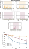
Fig 3. Inactivation of biotin and purine biosynthetic pathways increases survival rates of IHMA87 in plasma.
A. Zoom in on Tn-seq profiles of bio and pur genes and operons, showing normalized numbers of reads in input (HIP) and output (plasma) samples. B. IHMA87 wild-type strain, ΔbioB, and ΔpurD mutants’ survival kinetics in plasma over 6 h incubation, as measured by CFU counting (n = 5). Note the biphasic killing curves for the parental strain and ΔbioB mutant, indicating increased persistence of ΔbioB. The ΔpurD mutant displayed increased tolerance compared to the parental strain.
Fig 4
Fig 4. ATP and biotin influence bacterial sensitivity to plasma.
A. Schematic view of the purine pathway (adapted from KEGG database [42]). Significantly enriched insertions in corresponding genes of the Tn-seq screen are indicated. B. Measurement of intra-bacterial ATP levels in LB and after 2h-incubation in HIP normalized to the CFU counts. C. Trans-complementation of ΔbioB and ΔpurD phenotype by exogenous biotin or ATP, respectively. Biotin (1 μM) and ATP (1.5 mM) were added at the beginning of the incubation of bacteria in plasma. The survival was estimated by CFU counting and the median of all independent experiments is represented by the histogram. Statistical analysis was performed and p-value <0.05 or 0.01 are indicated with ‘*’ and ‘**’, respectively. D. The biotin biosynthetic pathway. Significantly enriched hits in Tn-seq are highlighted in green.
Fig 5
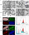
Fig 5. Polyphosphate granules are formed upon P. aeruginosa incubation in plasma.
A. Transmission electron microscopy images of indicated strains after growth in LB (left) or 1h-incubation in human plasma (right). Red and blue arrows show big- and small-sized granules respectively. Scale bar = 500 nm. B. EDX elemental composition maps of oxygen (green), phosphorus (red) and nitrogen (blue) of IHMA87 wild-type and ΔpurD in plasma (left). EDX spectra (right) of highlighted locations (red = granules, cyan = cytoplasm), major peaks are assigned. Data show elevated levels of phosphorus and oxygen in the granule, indicating that the identified objects are polyP.
Fig 6
Fig 6. Polyphosphates are critical for evaders’ survival in plasma.
A. Zoom in on Tn-seq profiles of ppk1 gene with 2000 bp upstream and downstream, showing normalized numbers of reads in input (HIP) and output (plasma) samples. B. Transmission electron microscopy images of IHMA87, Δppk1, Δppk2 or Δppk1Δppk2 after 1h-incubation in human plasma. Scale bar = 500 nm. C. Survival of IHMA87 wild-type strain, individual Δppk1, Δppk2 or double Δppk1Δppk2 deletion mutants following incubation for 3 h in plasma. Survival rates were calculated based on CFU measurements and the median of all independent experiments is represented by the histogram. Statistical analysis was performed and p-value <0.05 is indicated with ‘*’.
Fig 7
Fig 7. Mutants’ response to MAC-dependent killing.
Residual complement activity of the classical (A.) and alternative (B.) complement pathways was measured after bacterial challenge in plasma, using lysis kinetics of erythrocytes, as described in Materials and Methods. Area under the curve (AUC) values were determined for each series and expressed relative to the 90% plasma pool control. n = 3. C. C3b deposition on bacterial surface after incubation for 15 min in 0% (Control) or 90% plasma. Deposition was determined by FACS using the C3b specific PE-labeled antibody. D. Mean fluorescence intensities of the plasma condition presented in C. n = 3. **p-value < 0.001. E. C9-AF647 insertion into bacterial membrane after 30 min in 0% (Control) or 3% pooled human serum and F. mean fluorescence intensities (*p-value < 0.05). G. C5b-9 deposition on bacterial surface after 15 min incubation in 0% (Control) and 90% plasma. Deposition was determined by FACS; mean fluorescence intensities are plotted (H.). n = 3. I. Bacterial survival in pooled human serum or in the presence of nisin with or without the complement inhibitor OmCI.
Fig 8
Fig 8. Complement evasion by P. aeruginosa is a multifactorial phenomenon.
In addition to described complement-mediated killing evasion mechanisms including exopolysaccharides (alginates, Psl and Pel) and long O-specific antigen, which limit bacterial opsonization or MAC insertion, we herein describe three novel evasion determinants. Decreased biotin production and SrgA overexpression, increase P. aeruginosa persistence and tolerance, respectively. Finally, bacterial incubation in plasma leads to the production of polyP granules, which are critical for the formation of evaders. The mutants in ppk genes, unable to produce polyP, have a significant decrease in the number of evaders formed upon plasma challenge. The three mechanisms described here impact bacterial survival in plasma by impeding MAC lytic activity.

References

    1. Bartell J.A., Cameron D.R., Mojsoska B., Haagensen J.A.J., Pressler T., Sommer L.M. et al.. (2020). Bacterial persisters in long-term infection: Emergence and fitness in a complex host environment. PLoS Pathog. 16, e1009112. doi: 10.1371/journal.ppat.1009112 - DOI - PMC - PubMed
    1. Fauvart M., De Groote V.N., and Michiels J. (2011). Role of persister cells in chronic infections: clinical relevance and perspectives on anti-persister therapies. J. Med. Microbiol. 60, 699–709. doi: 10.1099/jmm.0.030932-0 - DOI - PubMed
    1. Balaban N.Q., Helaine S., Lewis K., Ackermann M., Aldridge B., Andersson D.I. et al.. (2019). Definitions and guidelines for research on antibiotic persistence (vol 17, pg 441, 2019). Nat. Rev. Microbiol. 17, 460–460. doi: 10.1038/s41579-019-0207-4 - DOI - PMC - PubMed
    1. Manuse S., Shan Y., Canas-Duarte S.J., Bakshi S., Sun W.-S., Mori H. et al.. (2021). Bacterial persisters are a stochastically formed subpopulation of low-energy cells. PLOS Biol. 19, e3001194. doi: 10.1371/journal.pbio.3001194 - DOI - PMC - PubMed
    1. Shan Y., Brown Gandt A., Rowe S.E., Deisinger J.P., Conlon B.P., and Lewis K. (2017). ATP-Dependent Persister Formation in Escherichia coli. mBio 8. doi: 10.1128/mBio.02267-16 - DOI - PMC - PubMed

Publication types

MeSH terms

Substances