. 2023 Jan 25;9(4):eadf4904.
doi: 10.1126/sciadv.adf4904. Epub 2023 Jan 25.

Stacking the odds: Multiple sites for HSV-1 latency

Affiliations

Stacking the odds: Multiple sites for HSV-1 latency

Shaohui Wang et al. Sci Adv. .

Abstract

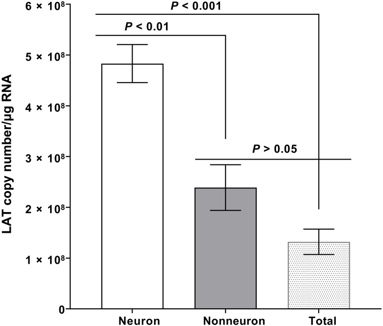
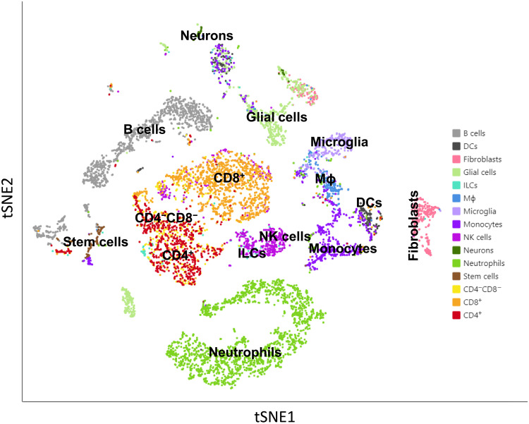
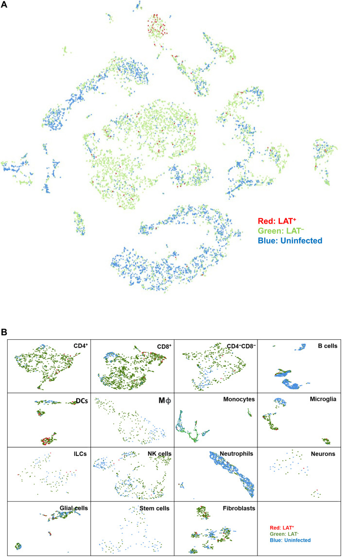
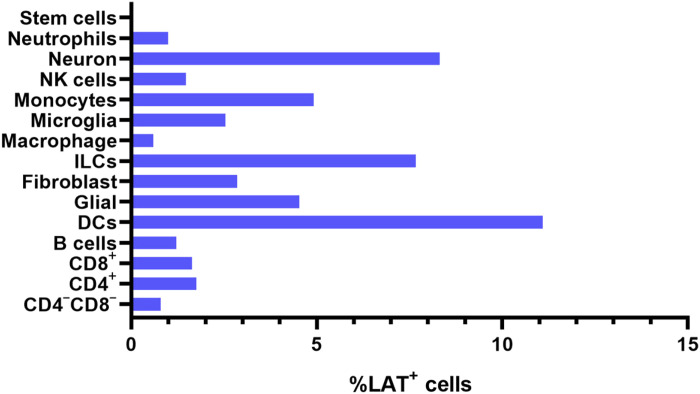
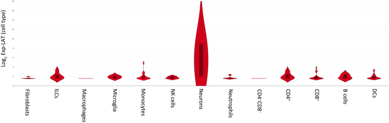
A hallmark of herpes simplex virus (HSV) infection is the establishment of latent virus in peripheral sensory ganglia of the latently infected host. We and others originally reported that the latency-associated transcript (LAT) is the only abundantly expressed viral gene in neurons within trigeminal ganglia (TG) of a latently infected host. Here, we investigated the possible contribution of various cells [i.e., B cells, dendritic cells (DCs), fibroblasts, glial cells, innate lymphoid cells (ILCs), macrophages, microglia, monocytes, natural killer cells, neurons, neutrophils, and T cells] isolated from TG of latently infected mice. Our results demonstrated that all of these cell types contain LAT, with DCs, neurons, and ILCs having the most LAT+ cells. These results suggest that HSV-1 can establish a quiescent/latent infection in a subset of nonneuronal cells, which enhances the chances that the virus will survive in its host.


Figures

Fig. 1.
Fig. 1.. Detection of LAT in the nonneuronal fraction of latently infected TG.
Wild-type (WT) mice were ocularly infected with 2 × 105 PFU per eye of HSV-1 strain McKrae without corneal scarification. TG were harvested on day 35 PI. Four latently infected TG from two mice were pooled together and dissociated into a single-cell suspension by digesting with collagenase D. Neuronal and nonneuronal fractions were isolated using the Miltenyi Biotec neuron isolation kit, as described in the company protocol. Briefly, dissociated cells were stained with the antibody cocktail, mixed with magnetic beads, and neuronal and nonneuronal cells were collected. Total RNA was extracted from each fraction, and unfractionated RNA from individual TG was used as a control. LAT copy number was measured by qRT-PCR using a standard curve generated from pGEM5317, as we described previously (73). Glyceraldehyde-3-phosphate dehydrogenase (GAPDH) expression was used to normalize relative levels of LAT RNA expression. Each bar represents the mean LAT copy number/μg RNA ± SEM from 12 mice for nonneuronal RNA and neuronal RNA and from 12 mice for total TG RNA.
Fig. 2.
Fig. 2.. tSNE map of individual cell types from mouse TG.
Mice were infected with HSV-1 McKrae. TG were harvested 35 days PI, pooled together (30 TG), and dissociated to single cells. Dead cells were removed using a dead cell removal kit, and CD45+ immune cells were isolated as described in Materials and Methods. Purified cells were mixed back with the original single-cell suspension for single-cell sequencing. ImmGen reference was used to determine immune cell types, while multiple mouse single-cell sequence references were used to determine neurons.
Fig. 3.
Fig. 3.. Detection of LAT+ cells in TG of latently infected mice.
(A) Aggregated tSNE map of single-cell sequencing from uninfected (blue), infected (green), or LAT+ cells (red). As described in Fig. 2 legend, the aggregated cell separation map was created by Loupe to visualize LAT+ cells. Cells expressing LAT (expression level greater than 1) were marked as red. (B) Individual UMAP of each cell type containing LAT. Each cell type was divided into three groups according to infection status or LAT expression status (greater than 1). Red, infected and LAT+; green, infected and LAT; and blue, uninfected and LAT.
Fig. 4.
Fig. 4.. Percentage of LAT+ cells in different cell populations.
The percentage of LAT+ cells in different infected cell populations are shown. In addition, number of LAT+/total cells for CD4CD8 T cells, 3 of 372; CD4+ T cells, 13 of 737; CD8+ T cells, 17 of 1029; B cells, 5 of 408; DCs, 24 of 216; glial cells, 26 of 572; fibroblasts, 9 of 214; ILCs, 6 of 78; macrophages, 1 of 164; microglia, 5 of 196; monocytes, 17 of 345; NK cells, 5 of 337; neurons, 2 of 24; and neutrophils, 5 of 498.
Fig. 5.
Fig. 5.. LAT expression levels in different cell types.
Violin plots using Loupe software for single-cell sequencing analysis were used to measure LAT expression levels in LAT+ cells from each cell type.
Fig. 6.
Fig. 6.. Detection of gB and gD DNA in neuronal and nonneuronal fractions of latently infected TG.
WT mice were infected ocularly with HSV-1 McKrae as described in Fig. 1 legend. TG were isolated on day 35 PI, and eight TG from four mice were pooled and dissociated to single cells. Immune cells enriched by Percoll density gradient were stained with CD45, CD3, CD4, CD8, F4/80, and CD11c antibodies. CD4, CD8, F4/80, and CD11c populations were sorted, and genomic DNA from each population was purified. gB (A) and gD (B) copy numbers were measured by qPCR, and GAPDH was used as internal control. Experiments were repeated three times.

References

    1. M. L. Cook, V. B. Bastone, J. G. Stevens, Evidence that neurons harbor latent herpes simplex virus. Infect. Immun. 9, 946–951 (1974). - PMC - PubMed
    1. M. L. Cook, J. G. Stevens, Latent herpetic infections following experimental viraemia. J. Gen. Virol. 31, 75–80 (1976). - PubMed
    1. J. M. Hill, F. Sedarati, R. T. Javier, E. K. Wagner, J. G. Stevens, Herpes simplex virus latent phase transcription facilitates in vivo reactivation. Virology 174, 117–125 (1990). - PubMed
    1. D. L. Rock, A. B. Nesburn, H. Ghiasi, J. Ong, T. L. Lewis, J. R. Lokensgard, S. L. Wechsler, Detection of latency-related viral RNAs in trigeminal ganglia of rabbits latently infected with herpes simplex virus type 1. J. Virol. 61, 3820–3826 (1987). - PMC - PubMed
    1. J. G. Stevens, Human herpesviruses: A consideration of the latent state. Microbiol. Rev. 53, 318–332 (1989). - PMC - PubMed