. 2023 Feb 9;66(3):1790-1808.
doi: 10.1021/acs.jmedchem.2c01617. Epub 2023 Jan 25.

N-Substituted l-Iminosugars for the Treatment of Sanfilippo Type B Syndrome

Affiliations

N-Substituted l-Iminosugars for the Treatment of Sanfilippo Type B Syndrome

Valeria De Pasquale et al. J Med Chem. .

Abstract

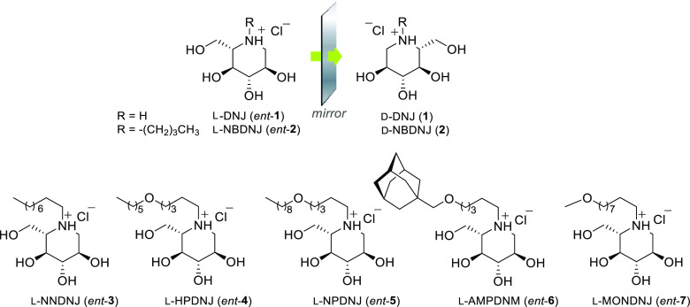
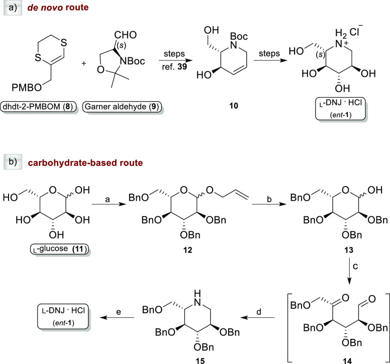
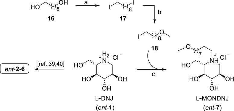
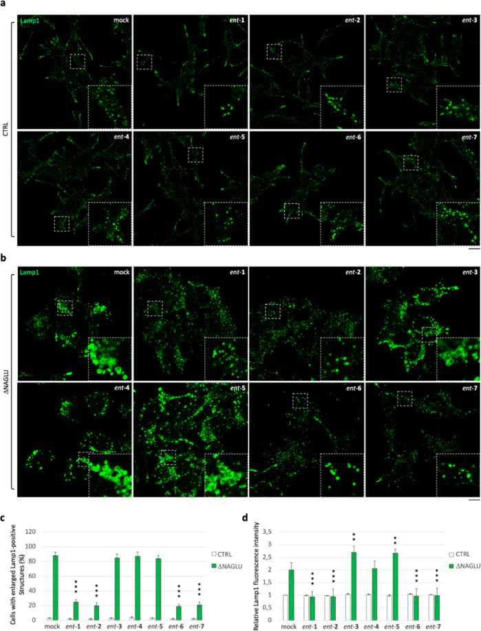
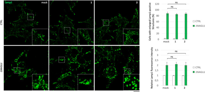
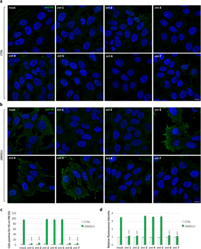
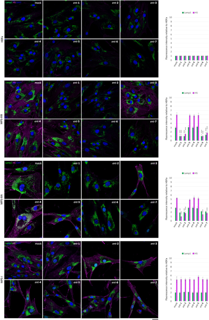
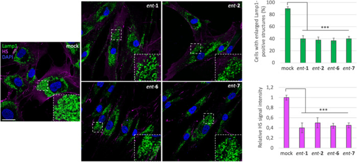
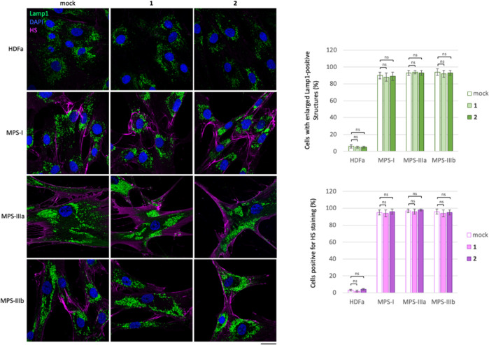
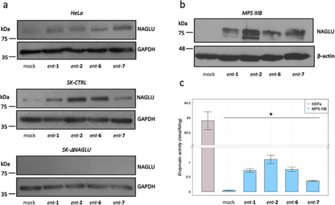
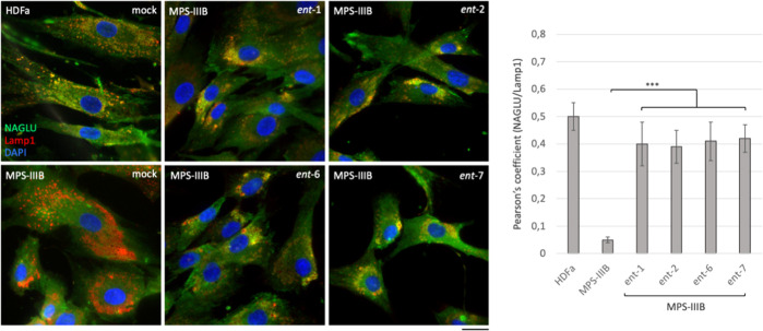
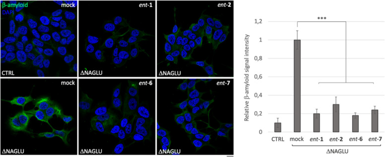
Sanfilippo syndrome comprises a group of four genetic diseases due to the lack or decreased activity of enzymes involved in heparan sulfate (HS) catabolism. HS accumulation in lysosomes and other cellular compartments results in tissue and organ dysfunctions, leading to a wide range of clinical symptoms including severe neurodegeneration. To date, no approved treatments for Sanfilippo disease exist. Here, we report the ability of N-substituted l-iminosugars to significantly reduce substrate storage and lysosomal dysfunctions in Sanfilippo fibroblasts and in a neuronal cellular model of Sanfilippo B subtype. Particularly, we found that they increase the levels of defective α-N-acetylglucosaminidase and correct its proper sorting toward the lysosomal compartment. Furthermore, l-iminosugars reduce HS accumulation by downregulating protein levels of exostosin glycosyltransferases. These results highlight an interesting pharmacological potential of these glycomimetics in Sanfilippo syndrome, paving the way for the development of novel therapeutic approaches for the treatment of such incurable disease.


Conflict of interest statement

The authors declare the following competing financial interest(s): Luigi Michele Pavone, Annalisa Guaragna, Valeria De Pasquale, Anna Esposito, Massimo DAgostino have a patent pending application comprising therapeutic compositions with the described L-iminosugars for the treatment of mucopolysaccharidoses, cancers and other diseases with abnormal accumulation of heparan sulfate (patent application n 102022000007808). The authors declare no additional competing financial interests.

Figures

Figure 1
Figure 1
l-Iminosugars evaluated in Sanfilippo disease cellular models.
Scheme 1
Scheme 1. Synthesis of ent-1 by (a) De Novo Route and (b) Carbohydrate-Based Route
Reagents and conditions: (a) (i): AllOH, BF3·OEt; Δ, on; (ii): BnBr, NaH, DMF, 0 °C, then rt, on, 91% overall yield; (b) PdCl2, MeOH, rt, on, 95% (c) (i): LiAlH4, THF, 0 °C, 20 h at rt; (ii): (COCl)2, DMSO, TEA, DCM; (d) NaBH3CN, NH4OAc, Na2SO4, MeOH, 75% over three steps; and (e) 1 M, BCl3, DCM, 0 °C, 12 h, 92%.
Scheme 2
Scheme 2. Synthesis of ent-(2–7)
Reagents and conditions: (a) PS-TPP, I2, DCM, rt, 1 h, 95%; (b) MeOH, NaH, THF, 0 °C for 1 h then rt, 48 h, 75%; (c) (i): l-DNJ (ent-1), K2CO3, DMF, 80 °C, on, 75%; (ii): MeOH, HCl 1 M, rt, quantitative.
Figure 2
Figure 2
Activity of ent-(17) in rescuing the lysosomal phenotype in the Sanfilippo B cellular model. (a) Lysosomal staining of CTRL clone treated with ent-(17): control stable clone seeded on coverslips was grown for 48 h in the presence of 20 μM of each l-iminosugar and then processed for indirect immunofluorescence by using a specific antibody against Lamp1 (lysosomal marker). (b) Lysosomal staining of the NAGLU-silenced stable clone (ΔNAGLU): ΔNAGLU clone seeded on coverslips was grown for 48 h in the presence of 20 μM of each l-iminosugar and then processed as for the CTRL clone. Magnifications are relative to the dashed white squares. (c,d) Quantification of immunofluorescence staining: the histograms represent, respectively, the quantification of cells with enlarged Lamp1 positive structures (bottom left) and the mean fluorescence intensity of Lamp1 (bottom right) among the different experimental conditions. 50 randomly chosen cells from three independent experiments were used for quantification. Single focal sections are shown. Scale bar: 20 μm. Asterisks indicate the statistically significant differences: (**) p-value < 0.001, (***) p-value < 0.0001.
Figure 3
Figure 3
Effect of d-iminosugars in rescuing the lysosomal phenotype in the Sanfilippo B cellular model. Lysosomal staining of control clone and NAGLU silenced stable clone (ΔNAGLU) treated with iminosugars 1 and 2: stable clones seeded on coverslips were grown for 48 h in the presence of 20 μM of each d-iminosugar and then processed for indirect immunofluorescence by using a specific antibody against Lamp1 (lysosomal markers). Quantification of immunofluorescence staining: histograms represent, respectively, the quantification of cells with an enlarged Lamp1 positive structure (bottom left) and the mean fluorescence intensity of Lamp1 (bottom right) among the different experimental conditions. 50 randomly chosen cells from three independent experiments were used for quantifications. Single focal sections are shown. Scale bar: 20 μm. (ns) p-value not statistically significant.
Figure 4
Figure 4
Activity of ent-(17) in rescuing the HS accumulation in the ΔNAGLU clone. (a) HS staining of CTRL clone treated with ent-(17): control stable clone seeded on coverslips was grown for 48 h in the presence of 20 μM of each l-iminosugar and then processed for indirect immunofluorescence by using the anti-HS antibody 10E4. (b) HS staining of ΔNAGLU clone treated with ent-(17): ΔNAGLU clone seeded on coverslips was grown for 48 h in the presence of 20 μM of each l-iminosugar and then processed as for the CTRL clone. (c,d) Quantification of immunofluorescence staining: histograms represent, respectively, (c) the quantification of cells positive for HS on plasma membrane (%) and (d) the mean fluorescence intensity of HS among the different experimental conditions. 50 randomly chosen cells from three independent experiments were used for quantification. Single focal sections are shown. Scale bar: 10 μm. Asterisks indicate the statistically significant differences: (***) p-value < 0.0001.
Figure 5
Figure 5
Activity of iminosugars ent-(17) in rescuing lysosomal phenotype and HS accumulation in Sanfilippo B and A fibroblasts. HS and Lamp1 staining of the HDFa and Sanfilippo B (MPS IIIB), Sanfilippo A (MPS IIIA), and MPS I fibroblasts treated with ent-(17): fibroblasts seeded on coverslips were grown for 48 h in the presence of 20 μM of each l-iminosugar and then processed for indirect immunofluorescence by using specific antibodies against HS and Lamp1 and decorated with DAPI (nuclear marker). Quantifications of immunofluorescence staining: the histograms represent the quantification of the mean fluorescence intensity of each MPS sample relative to the untreated cells. 50 randomly chosen cells from three independent experiments were used for quantifications. Single focal sections are shown. Scale bar: 50 μm. Asterisks indicate the statistically significant differences: (***) p-value < 0.0001.
Figure 6
Figure 6
Activity of ent-1, ent-2, ent-6, and ent-7 in rescuing the lysosomal phenotype and HS accumulation in fibroblasts from MPS I patients. HS and Lamp1 staining of the l-iminosugar-treated and -untreated MPS I fibroblasts. Fibroblasts seeded on coverslips were grown for 48 h in the presence of 40 μM of each l-iminosugar, then processed for indirect immunofluorescence by using specific antibodies against HS and Lamp1, and decorated with DAPI (nuclear marker). Quantification of immunofluorescence staining: histograms represent the quantification of the mean fluorescence intensity of each MPS sample relative to untreated cells. 50 randomly chosen cells from three independent experiments were used for the quantification. Single focal sections are shown. Scale bar: 50 μm. Asterisks indicate the statistically significant differences: (***) p-value < 0.0001.
Figure 7
Figure 7
No effect of d-iminosugars in rescuing the lysosomal phenotype in fibroblasts from MPS patients. HS and Lamp1 staining of d-iminosugars-treated and -untreated HDFa, MPS I, MPS IIIA, and MPS IIIB fibroblasts. Fibroblasts seeded on coverslips were grown for 48 h in the presence of 20 μM of each d-iminosugar, then processed for indirect immunofluorescence by using specific antibodies against HS and Lamp1, and decorated with DAPI (nuclear marker). Quantification of immunofluorescence staining: histograms represent the quantification based on the percentage of cells positive for enlarged Lamp1 structures and HS staining. 50 randomly chosen cells from three independent experiments were used for quantification. Single focal sections are shown. Scale bar: 50 μm. Asterisks indicate the statistically significant differences: (ns) p-value not statistically significant.
Figure 8
Figure 8
Effect of l-iminosugars on NAGLU protein levels and enzymatic activity. (a) Western blotting analysis of NAGLU protein levels in untreated mock (lane 1) and treated with ent-1 (lane 2), ent-2 (lane 3), ent-6 (lane 4), and ent-7 (lane 5) HeLa cells, SK-NBE clones (CTRL), and NAGLU silenced clone. To monitor equal loading of the proteins in the gel lanes, the blots were re-probed using an anti-GAPDH antibody. (b) Western blotting analysis of NAGLU protein levels in untreated and treated fibroblasts from MPS IIIB patients. To monitor equal loading of the proteins in the gel lanes, the blots were re-probed using an anti-β-actin antibody. (c) Enzymatic activity of mutated NAGLU extracted from Sanfilippo B fibroblasts: extracts from HDFa (control) and Sanfilippo B fibroblasts were incubated or not with 20 μM of l-iminosugars together with 4-methylumbelliferyl-N-acetyl-α-d-glucosaminide as the fluorogenic substrate.
Figure 9
Figure 9
Effect of the active l-iminosugars on NAGLU sorting to the lysosomal compartment. HDFa and MPS IIIB seeded on coverslips were grown for 48 h in the presence or not (mock) of 20 μM of ent-1, ent-2, ent-6, and ent-7, then processed for indirect immunofluorescence by using specific antibodies against NAGLU and Lamp1, and decorated with DAPI (nuclear marker). Quantification of immunofluorescence staining: histograms represent the quantification of the mean fluorescence intensity of each MPS IIIB sample relative to the untreated cells. 50 randomly chosen cells from three independent experiments were used for quantifications. Single focal sections are shown. Scale bar: 50 μm. Asterisks indicate the statistically significant differences: (***) p-value < 0.0001.
Figure 10
Figure 10
Activity of ent-1, ent-2, ent-6, and ent-7 in rescuing the accumulation of Aβ1-42 peptide in the Sanfilippo B cellular model. Aβ1-42 peptide immunostaining of control (CTRL) and ΔNAGLU clone untreated and treated with ent-1, ent-2, ent-6, and ent-7: ΔNAGLU clone seeded on the coverslips were grown for 48 h in the presence of 20 μM of each l-iminosugar and then processed for indirect immunofluorescence by using a specific Aβ1-42 peptide antibody. Quantification of immunofluorescence staining: the histograms represent, respectively, the quantification of cells with Aβ1-42 peptide positive structures among the different experimental conditions. 50 randomly chosen cells from three independent experiments were used for quantification. Single focal sections are shown. Scale bar: 10 μm. Asterisks indicate the statistically significant differences: (***) p-value < 0.0001.

Similar articles

Cited by

References

    1. Neufeld E.; Muenzer J.. The Mucopolysaccharidoses. The Online Metabolic and Molecular Bases of Inherited Disease, OMMBID; McGraw-Hill Medical, 2001.
    1. Heon-Roberts R.; Nguyen A. L. A.; Pshezhetsky A. V. Molecular Bases of Neurodegeneration and Cognitive Decline, the Major Burden of Sanfilippo Disease. J. Clin. Med. 2020, 9, 344.10.3390/jcm9020344. - DOI - PMC - PubMed
    1. Muenzer J. Overview of the Mucopolysaccharidoses. Rheumatology 2011, 50, v4–12. 10.1093/rheumatology/ker394. - DOI - PubMed
    1. Wagner V. F.; Northrup H.. Mucopolysaccharidosis Type III; GeneReviews, 1993.
    1. Pearse Y.; Iacovino M. A Cure for Sanfilippo Syndrome? A Summary of Current Therapeutic Approaches and Their Promise. Med. Res. Arch. 2020, 8 (2), 1.10.18103/mra.v8i2.2045. - DOI - PMC - PubMed

Publication types

MeSH terms

Substances