. 2023 Jul;56(7):e13402.
doi: 10.1111/cpr.13402. Epub 2023 Jan 25.

The sodium new houttuyfonate suppresses NSCLC via activating pyroptosis through TCONS-14036/miR-1228-5p/PRKCDBP pathway

Affiliations

The sodium new houttuyfonate suppresses NSCLC via activating pyroptosis through TCONS-14036/miR-1228-5p/PRKCDBP pathway

Rilei Jiang et al. Cell Prolif. 2023 Jul.

Abstract

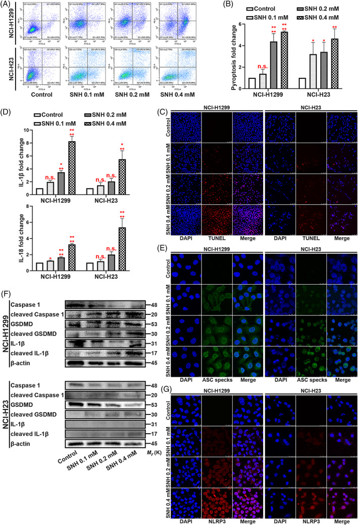
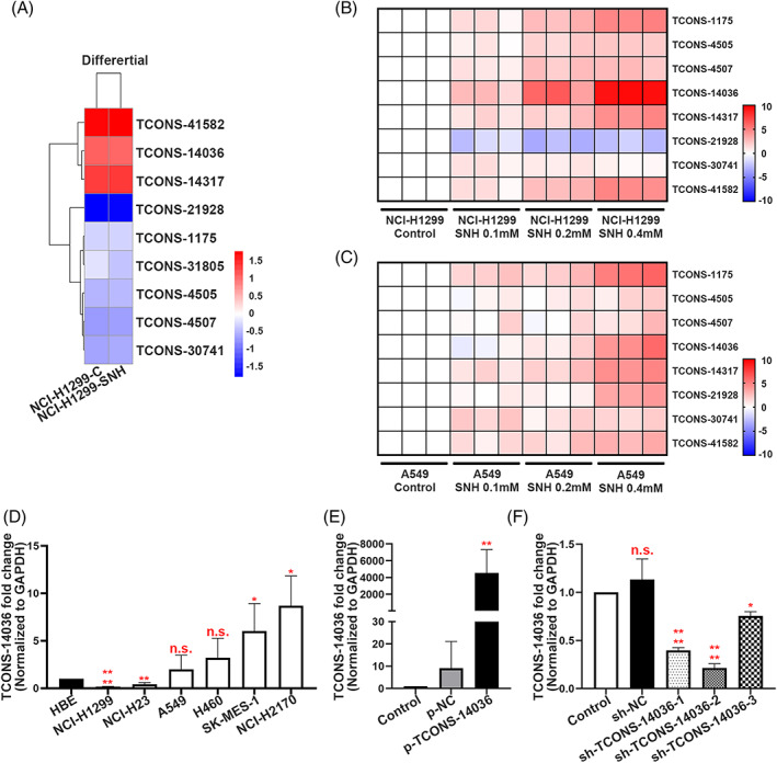
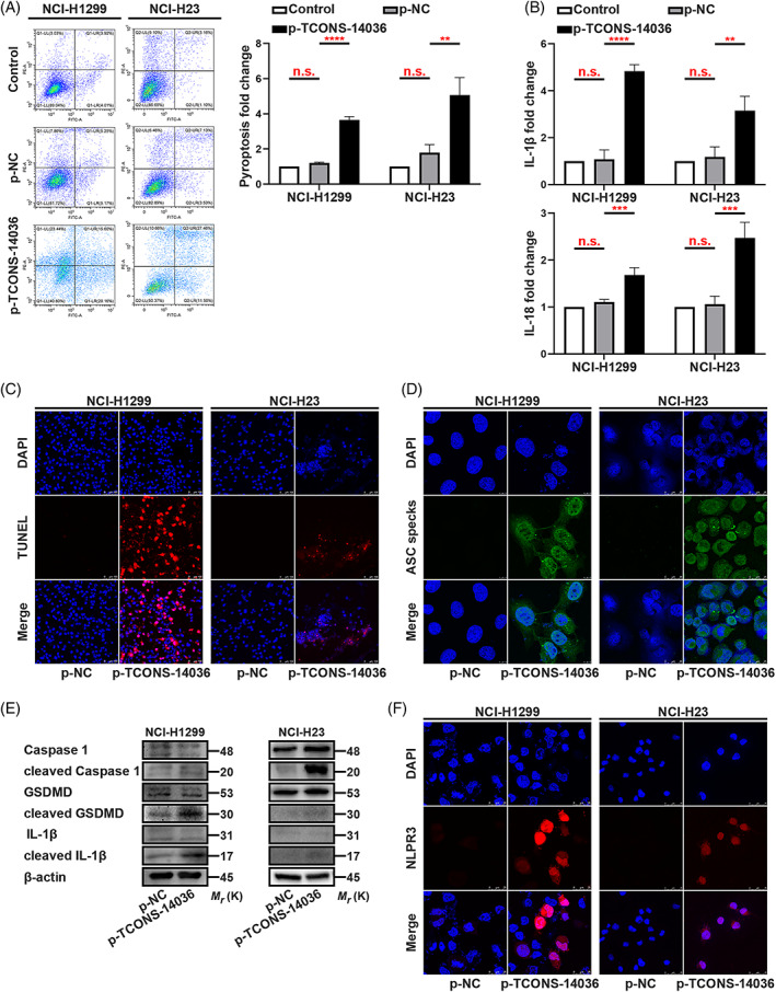
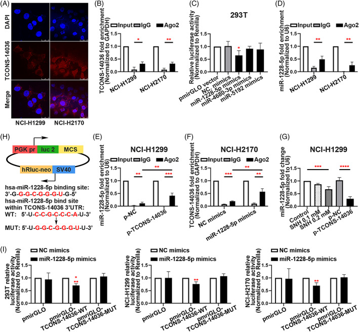
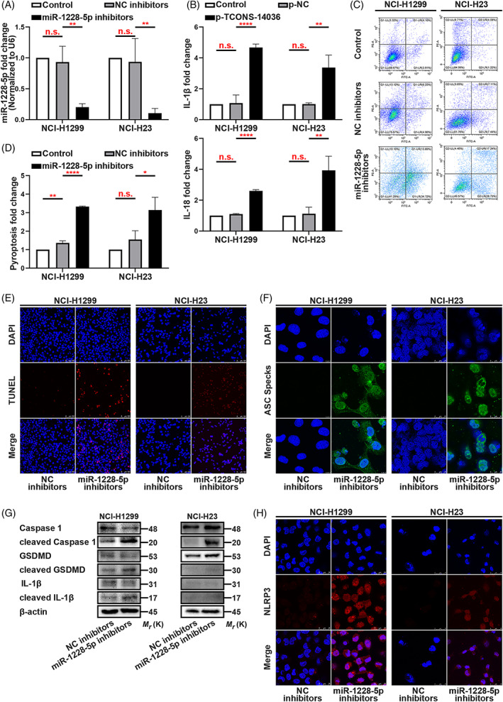
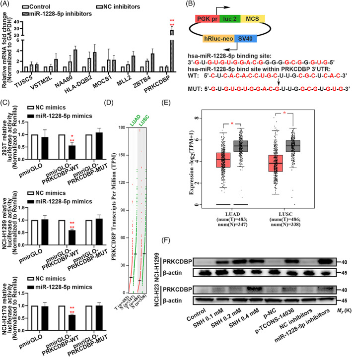
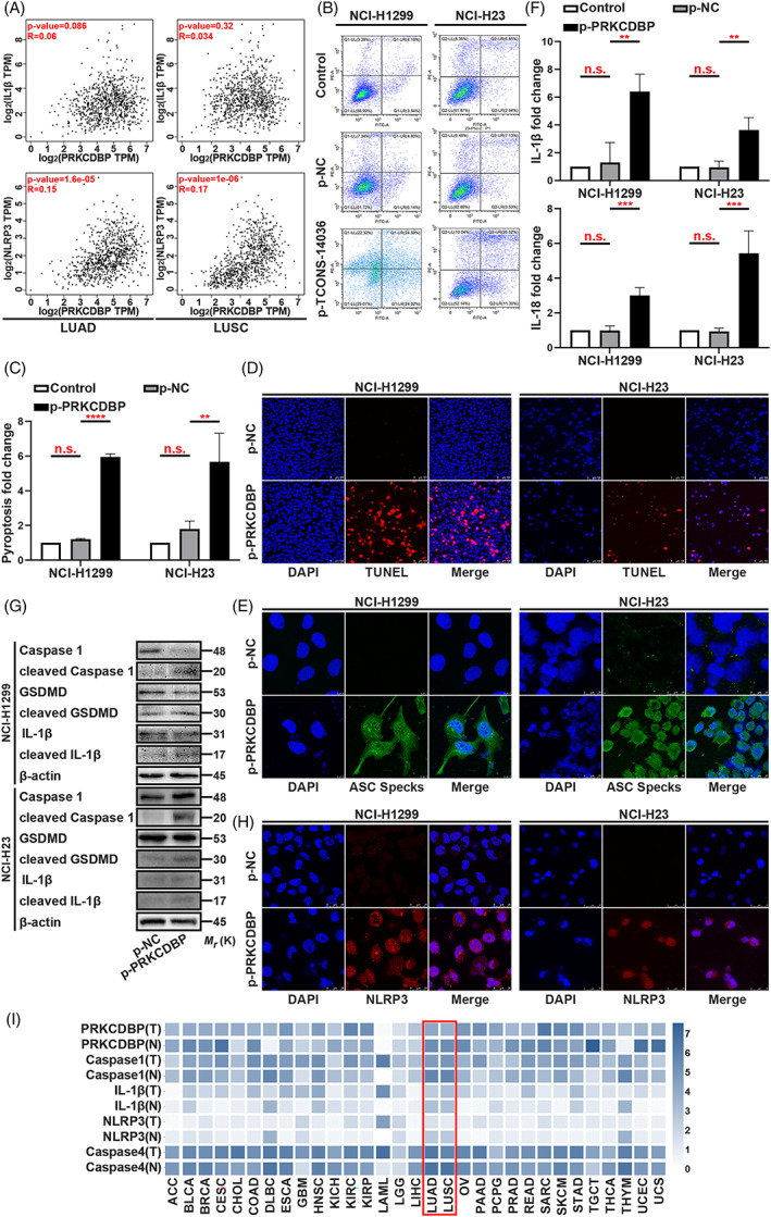
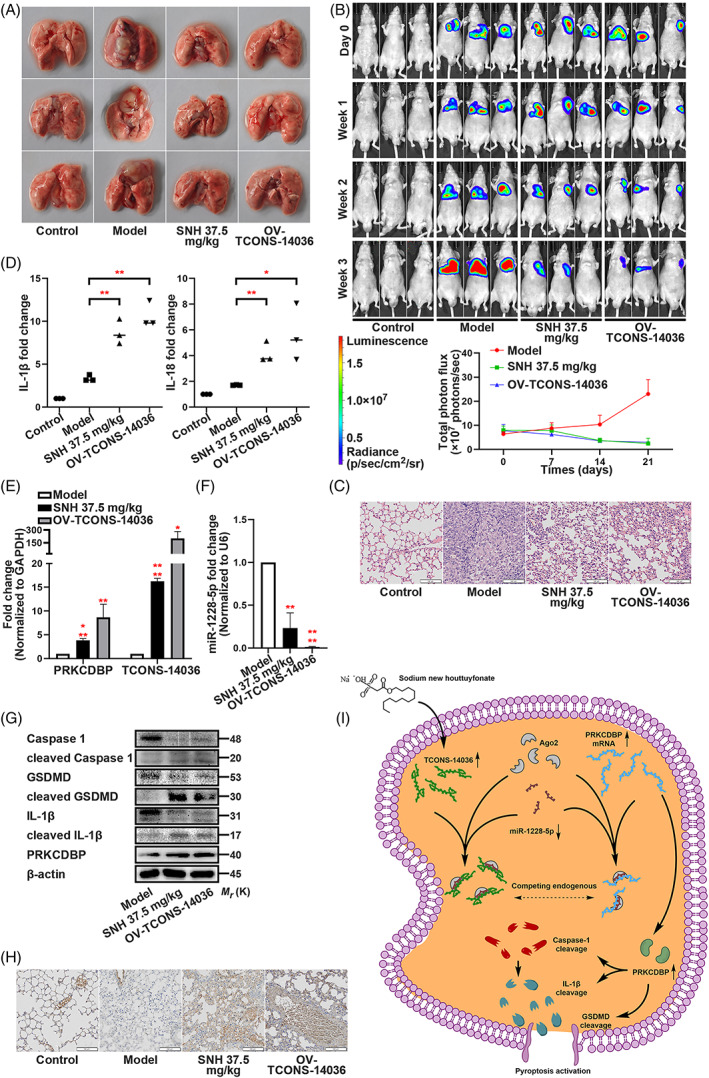
Several studies have suggested the potential value of Houttuynia cordata as a therapeutic agent in lung cancer, but direct evidence is still lacking. The study aimed to determine the regulatory impact of a major H. cordata constituent derivative (sodium new houttuyfonate [SNH]) on lncRNA networks in non-small cell lung cancer (NSCLC) to identify new potential therapeutic targets. After exposing NSCLC cells to SNH, we analysed the following: cell death (via flow cytometry, TUNEL and ASC speck formation assays), immune factors (via ELISA), gene transcription (via RT-qPCR), subcellular localisation (via FISH), gene-gene and gene-protein interactions (via dual-luciferase reporter and RNA immunoprecipitation assays, respectively) and protein expression and distribution (via western blotting and immunocytochemistry or immunohistochemistry). In addition, statistical analysis (via one-way ANOVA or unpaired t-tests) was performed. Exposure to SNH promoted NSCLC cell pyroptosis, concomitant with significant up-regulation of TCONS-14036, a novel lncRNA. Mechanistic research demonstrated that TCONS-14036 functions as a competing endogenous (ce)RNA by sequestering microRNA (miR)-1228-5p, thereby up-regulating PRKCDBP-encoding transcript levels. Indeed, PRKCDBP promoted pyroptosis by activating the NLRP3 inflammasome, resulting in CASP1, IL-1β and GSDMD cleavage. Our findings elucidate the potential molecular mechanisms underlying the ability of SNH to suppress NSCLC growth through activation of pyroptosis via the TCONS-14036/miR-1228-5p/PRKCDBP pathway. Thus, we identify a new potential therapeutic targets for NSCLC.


Conflict of interest statement

The authors declare that they have no competing interests.

Figures

FIGURE 1
FIGURE 1
SNH promotes NSCLC cell pyroptosis. (A,B) Annexin V and PI staining flow cytometric analysis of NCI‐H1299 and NCI‐H23 cells death with 0, 0.1, 0.2 and 0.4 mM SNH treatment. (C) TUNEL stain assay in NCI‐H1299 and NCI‐H23 with the treatment of SNH. (D) ELISA assay tested the IL‐1β and IL‐18, which released in NCI‐H1299 and NCI‐H23 cells culture mediums by the treatment of SNH. (E) Immunofluorescence staining of ASC specks expression in NCI‐H1299 with the treatment of SNH. (F) Expression of pyroptosis associated proteins in NCI‐H1299 and NCI‐H23 cells with the treatment of SNH as determined by western blot analysis. (G) Immunofluorescence staining of NLRP3 expression in the treatment of SNH in NCI‐H1299 and NCI‐H23 cells. The bars and error bars indicate the mean ± SD. n.s.p > 0.05, *p < 0.05, **p < 0.01, ***p < 0.005 and ****p < 0.001.
FIGURE 2
FIGURE 2
TCONS‐14036 is a potential target of SNH. (A) Heat map of the differential expression of screened novel lncRNA sequences in NCI‐H1299 cells treated or not treated with SNH for 24 H. (B,C) qRT‐PCR retrospective experiment in NCI‐H1299 and A549 cells screened out TCONS‐14036 was the most significant regulated novel lncRNA. (D) TCONS‐14036 expression in 6 NSCLC cell lines and 1 human bronchial epithelial cell line (HBE) as determined by qRT‐PCR. (E) Validation of TCONS‐14036 overexpression by p‐TCONS‐14036 in NCI‐H1299 cells as determined by qRT‐PCR. (F) Validation of sh‐RNA knockdown efficiency in NCI‐H2170 cells as determined by qRT‐PCR. The bars and error bars indicate the mean ± SD. n.s.p > 0.05, *p < 0.05, **p < 0.01, ***p < 0.005 and ****p < 0.001.
FIGURE 3
FIGURE 3
TCONS‐14036 activates pyroptosis in NSCLC. (A) Annexin V and PI staining flow cytometric analysis of NCI‐H1299 and NCI‐H23 cells death with p‐TCONS‐14036 transfection. (B) ELISA assay tested the IL‐1β and IL‐18, which released in NCI‐H1299 and NCI‐H23 cells culture mediums by p‐TCONS‐14036 transfection. (C) TUNEL stain assay in NCI‐H1299 and NCI‐H23 with the transfection of p‐TCONS‐14036. (D) Immunofluorescence staining of ASC specks expression in NCI‐H1299 and NCI‐H23 with the transfection of p‐TCONS‐14036. (E) Expression of pyroptosis associated proteins in NCI‐H1299 and NCI‐H23 cells with the transfection of p‐TCONS‐14036 as determined by western blot analysis. (F) Immunofluorescence staining of NLRP3 expression in the transfection of p‐TCONS‐14036 in NCI‐H1299 and NCI‐H23 cells. The bars and error bars indicate the mean ± SD. n.s.p > 0.05, *p < 0.05, **p < 0.01, ***p < 0.005 and ****p < 0.001.
FIGURE 4
FIGURE 4
TCONS‐14036 functions as a ceRNA and sponges miR‐1228‐5p in NSCLC cells. (A) The RNA florescent in situ hybridization of TCONS‐14036 in NCI‐H1299 and NCI‐H2170 cell lines. (B) RNA immunoprecipitation (RIP) assay of TCONS‐14036 binding to Ago2 in NCI‐H1299 and NCI‐H2170 cell extracts. (C) Dual‐luciferase reporter assays were used to determine the interaction between miRNAs and TCONS‐14036. (D) RIP assay of miR‐1228‐5p binding to Ago2 in NCI‐H1299 and NCI‐H2170 cell extracts. (E) RIP assay of miR‐1228‐5p binding to Ago2 in p‐NC/p‐TCONS‐14036 transfected NCI‐H1299 cell extracts. (F) RIP assay of TCONS‐14036 binding to Ago2 in NC/miR‐1228‐5p mimics transfected NCI‐H2170 cell extracts. (G) qRT‐PCR analysis of miR‐1228‐5p in NCI‐H1299 cells after the treatment of SNH or p‐NC/p‐TCONS‐14036 transfection. (H) Predicted binding sites for miR‐1228‐5p on TCONS‐14036 and a diagram depicting the construction of the wild type (WT) and mutant type (MUT) pmirGLO‐TCONS‐14036 plasmids. (I) 293T, NCI‐H1299 and NCI‐H2170 cells were co‐transfected with miR‐1228‐5p mimics or NC mimics and pmirGLO or pmirGLO‐TCONS‐14036‐WT or pmirGLO‐TCONS‐14036‐MUT. Luciferase activity was detected 24 H after transfection using a dual‐luciferase assay. The bars and error bars indicate the mean ± SD. n.s.p > 0.05, *p < 0.05, **p < 0.01, ***p < 0.005 and ****p < 0.001.
FIGURE 5
FIGURE 5
Knockdown of miR‐1228‐5p induces pyroptosis in NSCLC. (A) MiR‐1228‐5p expression in miR‐1228‐5p inhibitors transfected NCI‐H1299 and NCI‐H23 cells as determined by qRT‐PCR. (B) ELISA assay tested the IL‐1β and IL‐18, which released in NCI‐H1299 and NCI‐H23 cells culture mediums by miR‐1228‐5p inhibitors transfection. (C,D) Annexin V and PI staining flow cytometric analysis of NCI‐H1299 and NCI‐H23 cell death with miR‐1228‐5p inhibitors transfection. (E) TUNEL stain assay in NCI‐H1299 and NCI‐H23 with the transfection of miR‐1228‐5p inhibitors. (F) Immunofluorescence staining of ASC specks expression in NCI‐H1299 and NCI‐H23 with the transfection of miR‐1228‐5p inhibitors. (G) Expression of pyroptosis associated proteins in NCI‐H1299 cells with the transfection of miR‐1228‐5p inhibitors as determined by western blot analysis. (H) Immunofluorescence staining of NLRP3 expression in the transfection of miR‐1228‐5p inhibitors in NCI‐H1299 and NCI‐H23 cells. The bars and error bars indicate the mean ± SD. n.s.p > 0.05, *p < 0.05, **p < 0.01, ***p < 0.005 and ****p < 0.001.
FIGURE 6
FIGURE 6
MiR‐1228‐5p directly targets PRKCDBP. (A) Verification of miR‐1228‐5p downstream targets by qRT‐PCR in NCI‐H1299 and NCI‐H23. (B) Predicted binding sites for miR‐1228‐5p on PRKCDBP and a diagram depicting the construction of the wild type (WT) and mutant type (MUT) pmirGLO‐PRKCDBP plasmids. (C) 293T, NCI‐H1299 and NCI‐H2170 cells were co‐transfected with miR‐1228‐5p mimics or NC mimics and pmirGLO or pmirGLO‐PRKCDBP‐WT or pmirGLO‐PRKCDBP‐MUT. Luciferase activity was detected 24 H after transfection using a dual‐luciferase assay. (D,E) TCGA analysis of PRKCDBP Transcripts Per Million (TPM) of lung adenocarcinoma (LUAD) and lung squamous carcinoma (LUSC). (F) Expression of PRKCDBP in NCI‐H1299 and NCI‐H23 cells with the treatment of SNH and transfection of p‐TCONS‐14036 and miR‐1228‐5p inhibitors as determined by western blot analysis. The bars and error bars indicate the mean ± SD. n.s.p > 0.05, *p < 0.05, **p < 0.01, ***p < 0.005 and ****p < 0.001.
FIGURE 7
FIGURE 7
PRKCDBP overexpression confers pyroptosis. (A) Scatter plot of Transcripts Per Million (TPM) of clinic between PRKCDBP and IL‐1β or NLRP3. (B,C) Annexin V and PI staining flow cytometric analysis of NCI‐H1299 and NCI‐H23 cells death with p‐PRKCDBP transfection. (D) TUNEL stain assay in NCI‐H1299 and NCI‐H23 with the transfection of p‐PRKCDBP. (E) Immunofluorescence staining of ASC specks expression in NCI‐H1299 and NCI‐H23 with the transfection of p‐PRKCDBP. (F) ELISA assay tested the IL‐1β and IL‐18, which released in NCI‐H1299 and NCI‐H23 cells culture mediums by p‐PRKCDBP transfection. (G) Expression of pyroptosis associated proteins in NCI‐H1299 and NCI‐H23 cells with the transfection of p‐PRKCDBP as determined by western blot analysis. (H) Immunofluorescence staining of NLRP3 expression in the transfection of p‐PRKCDBP in NCI‐H1299 and NCI‐H23 cells. (I) TCGA analysis of PRKCDBP, Caspase‐1, IL‐1β, NLRP3 and Caspase‐4 expression in clinic. The bars and error bars indicate the mean ± SD. n.s.p > 0.05, *p < 0.05, **p < 0.01, ***p < 0.005 and ****p < 0.001.
FIGURE 8
FIGURE 8
SNH controls the transcriptional pathway TCONS‐14036/miR‐1228‐5p/PRKCDBP in vivo. (A) Representative images of the lungs of nude mice which were injected with NCI‐H1299‐luc cells and treated with SNH or OV‐TCONS‐14036 lentivirus. (B) Bioluminescent imaging and quantification of photon flux of SNH or OV‐TCONS‐14036 lentivirus treatment NSCLC mice model. (C) Representative images showing haematoxylin and eosin staining of lung samples from the different groups. (D) ELISA assay tested the IL‐1β and IL‐18 which released in irrigating solutions of lungs. (E,F) PRKCDBP mRNA, TCONS‐14036 and miR‐1228‐5p levels of mice lung tissues as determined by qRT‐PCR. (G) Expression of pyroptosis associated proteins and PRKCDBP of mice lung tissues as determined by western blot analysis. (H) IHC staining showing PRKCDBP expression in the different groups. (I) Schematic diagram of mechanism on this research. The bars and error bars indicate the mean ± SD. n.s.p > 0.05, *p < 0.05, **p < 0.01, ***p < 0.005 and ****p < 0.001.

Similar articles

Cited by

References

    1. Hirsch FR, Scagliotti GV, Mulshine JL, et al. Lung cancer: current therapies and new targeted treatments. Lancet. 2017;389:299‐311. - PubMed
    1. Siegel RL, Miller KD, Jemal A. Cancer statistics, 2020. CA Cancer J Clin. 2020;70:7‐30. - PubMed
    1. Fang Y, Tian S, Pan Y, et al. Pyroptosis: a new frontier in cancer. Biomed Pharmacother. 2020;121:109595. - PubMed
    1. Cookson BT, Brennan MA. Pro‐inflammatory programmed cell death. Trends Microbiol. 2001;9:113‐114. - PubMed
    1. Man SM, Karki R, Kanneganti TD. Molecular mechanisms and functions of pyroptosis, inflammatory caspases and inflammasomes in infectious diseases. Immunol Rev. 2017;277:61‐75. - PMC - PubMed

MeSH terms