. 2023 Mar 1;43(9):1627-1642.
doi: 10.1523/JNEUROSCI.1366-22.2023. Epub 2023 Jan 25.

Peroxynitrite Contributes to Behavioral Responses, Increased Trigeminal Excitability, and Changes in Mitochondrial Function in a Preclinical Model of Migraine

Affiliations

Peroxynitrite Contributes to Behavioral Responses, Increased Trigeminal Excitability, and Changes in Mitochondrial Function in a Preclinical Model of Migraine

Jacob Lackovic et al. J Neurosci. .

Abstract

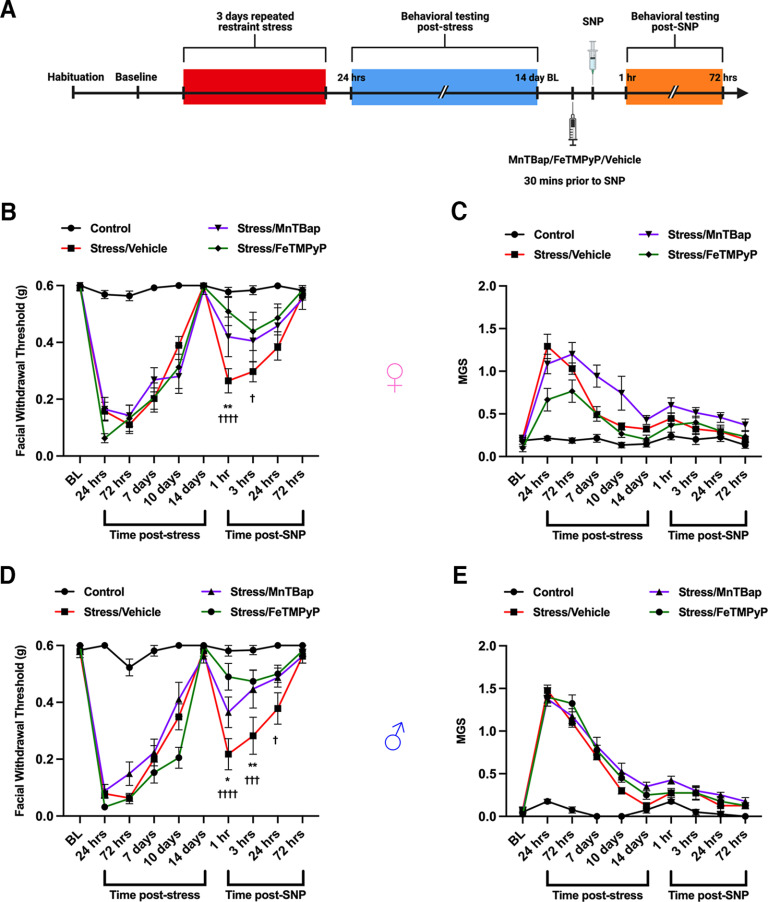
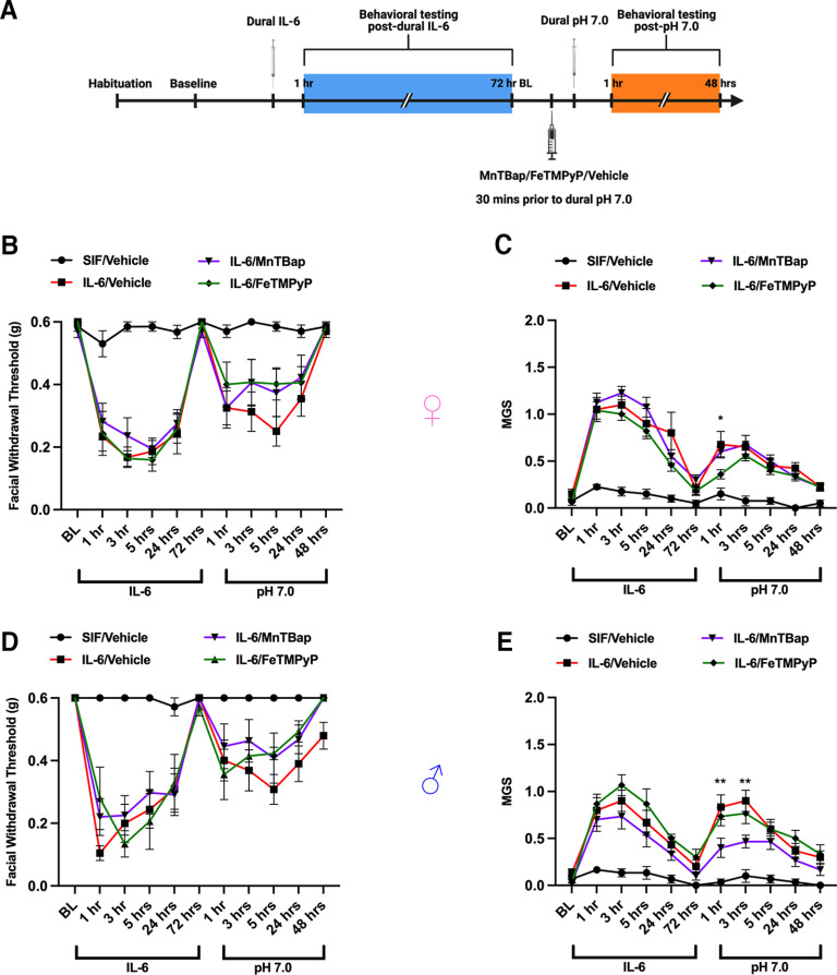
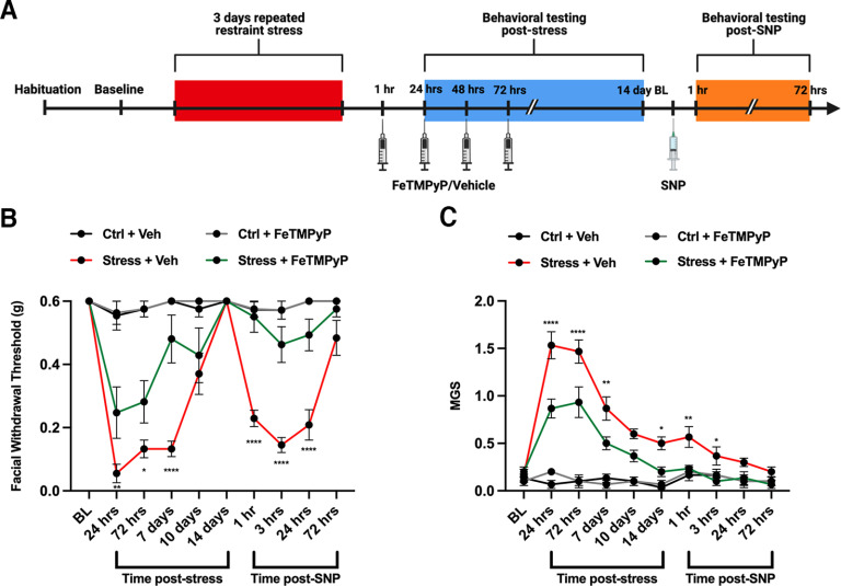
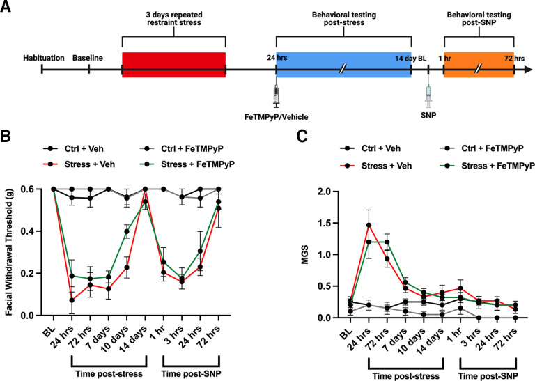
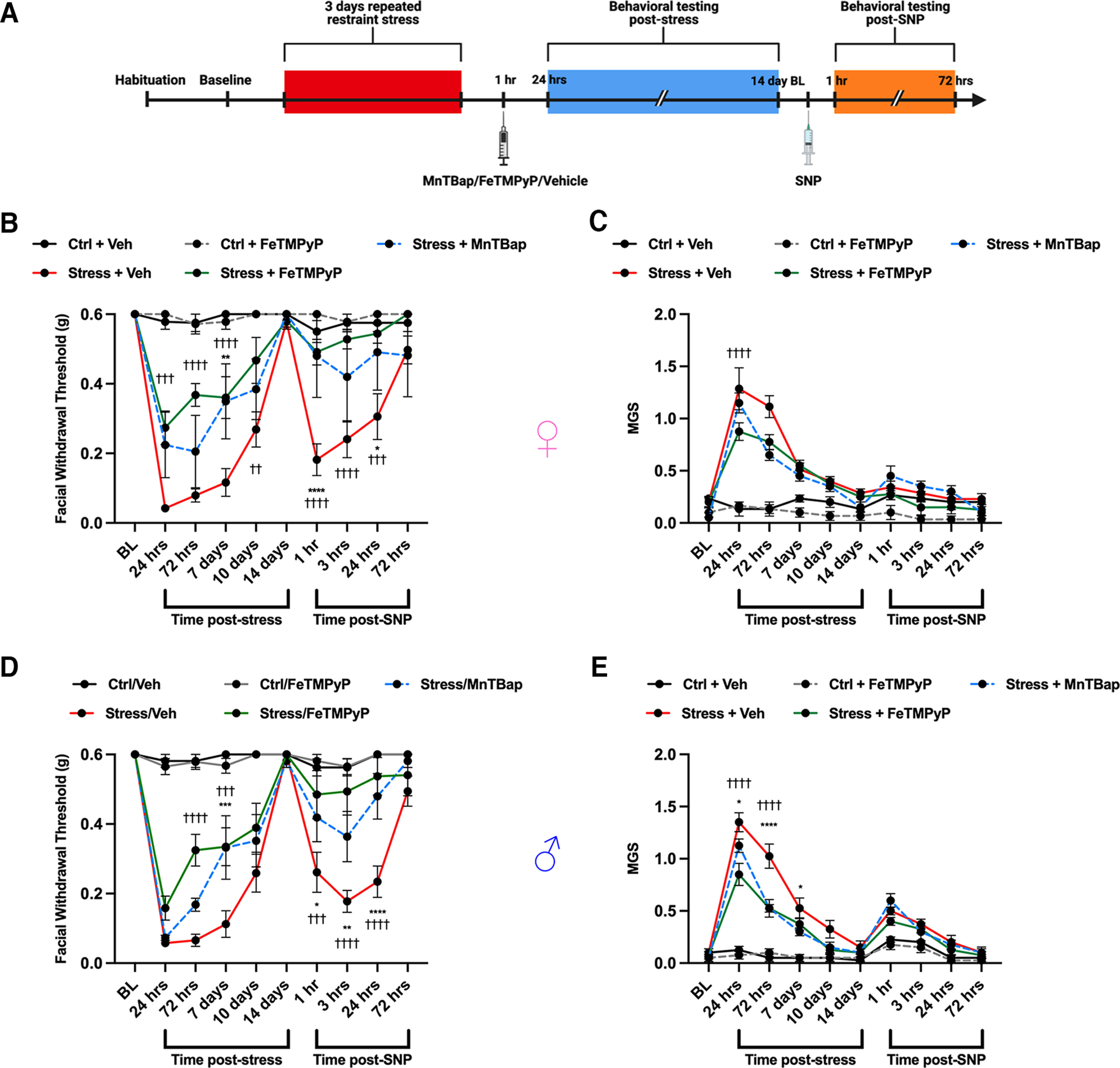
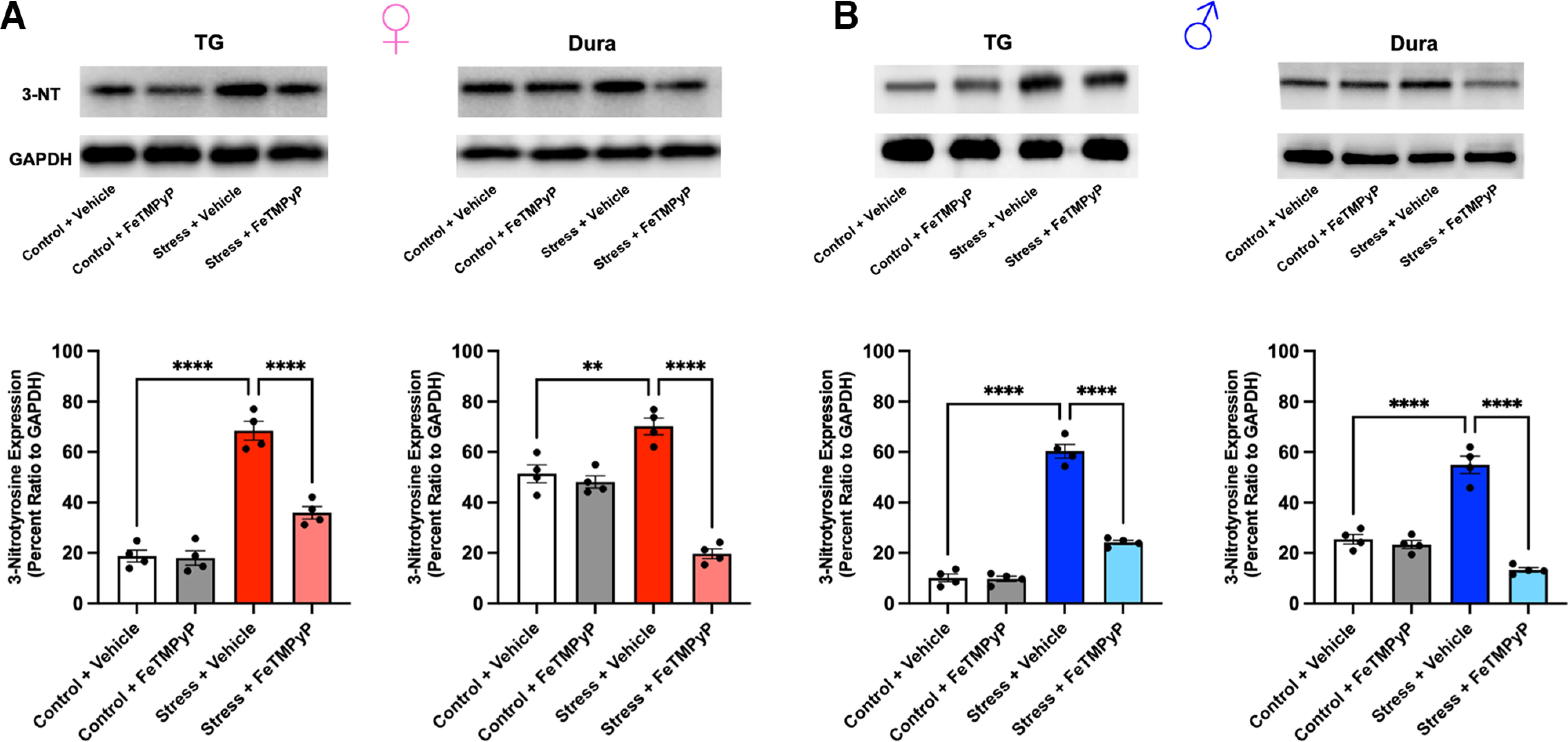
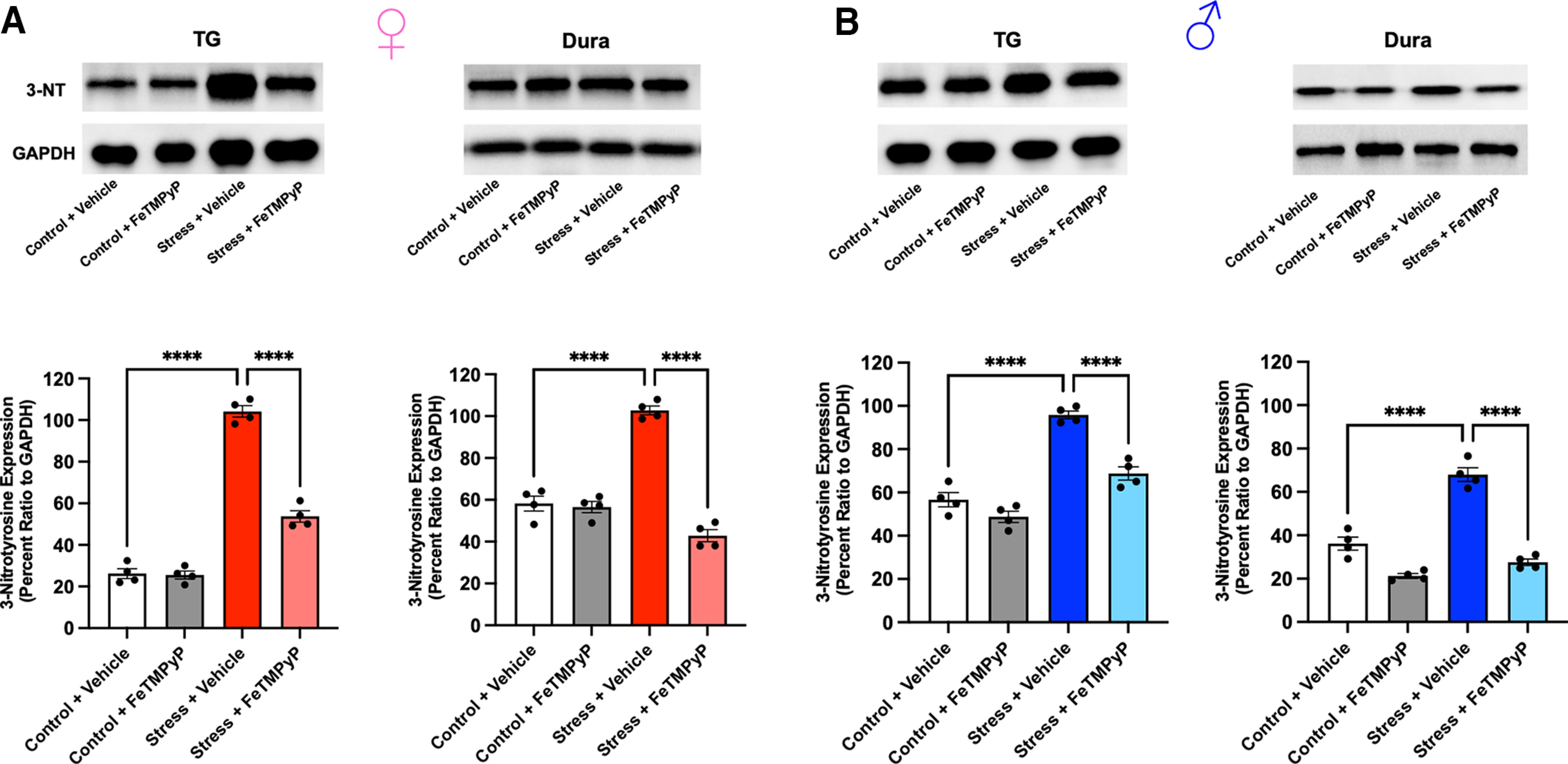
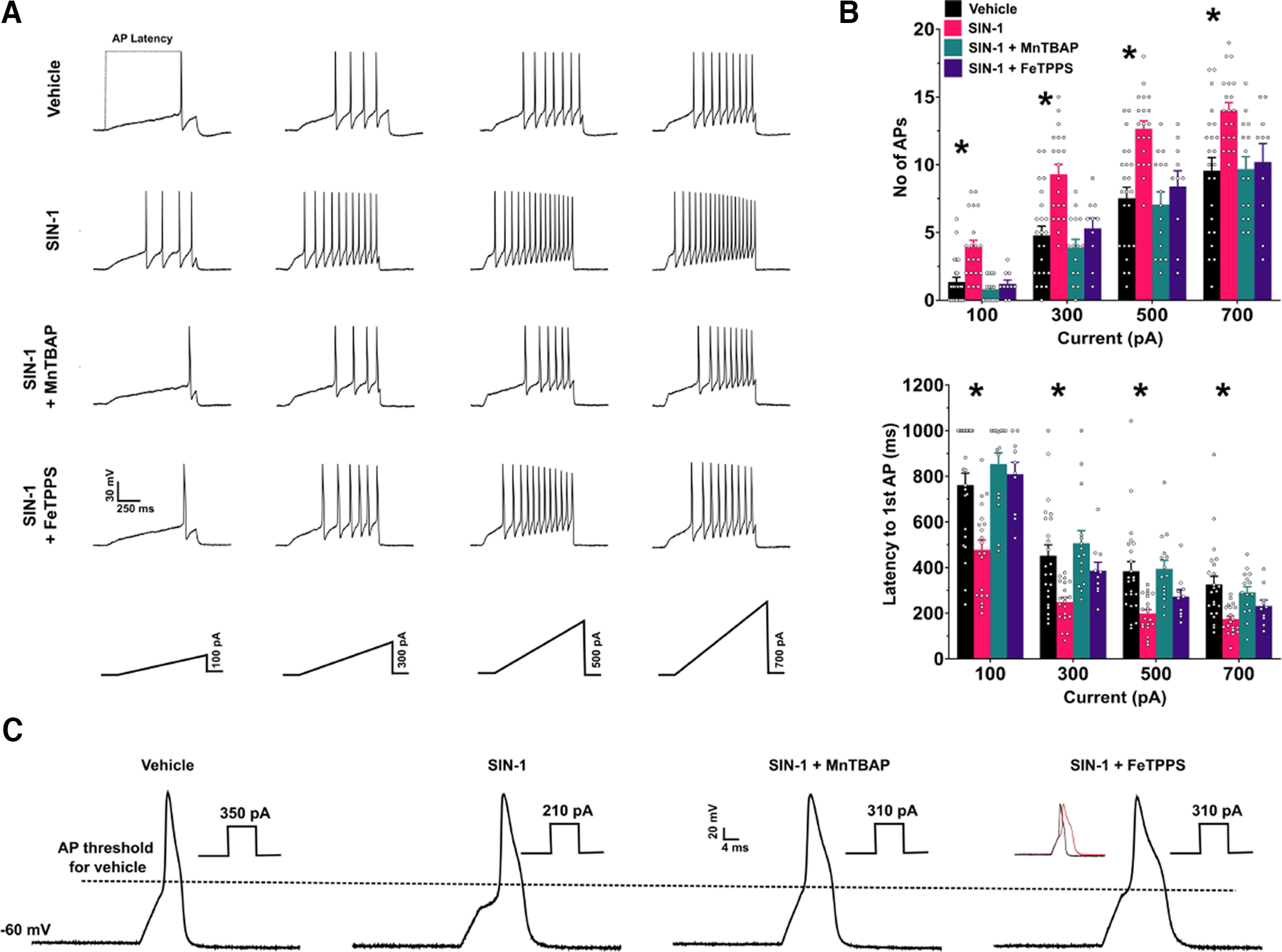
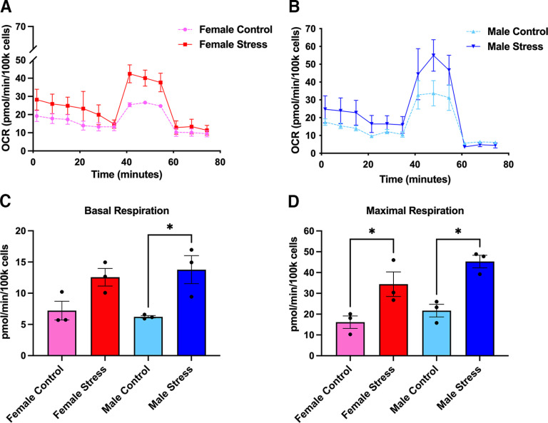
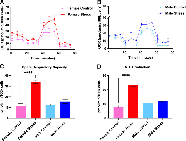
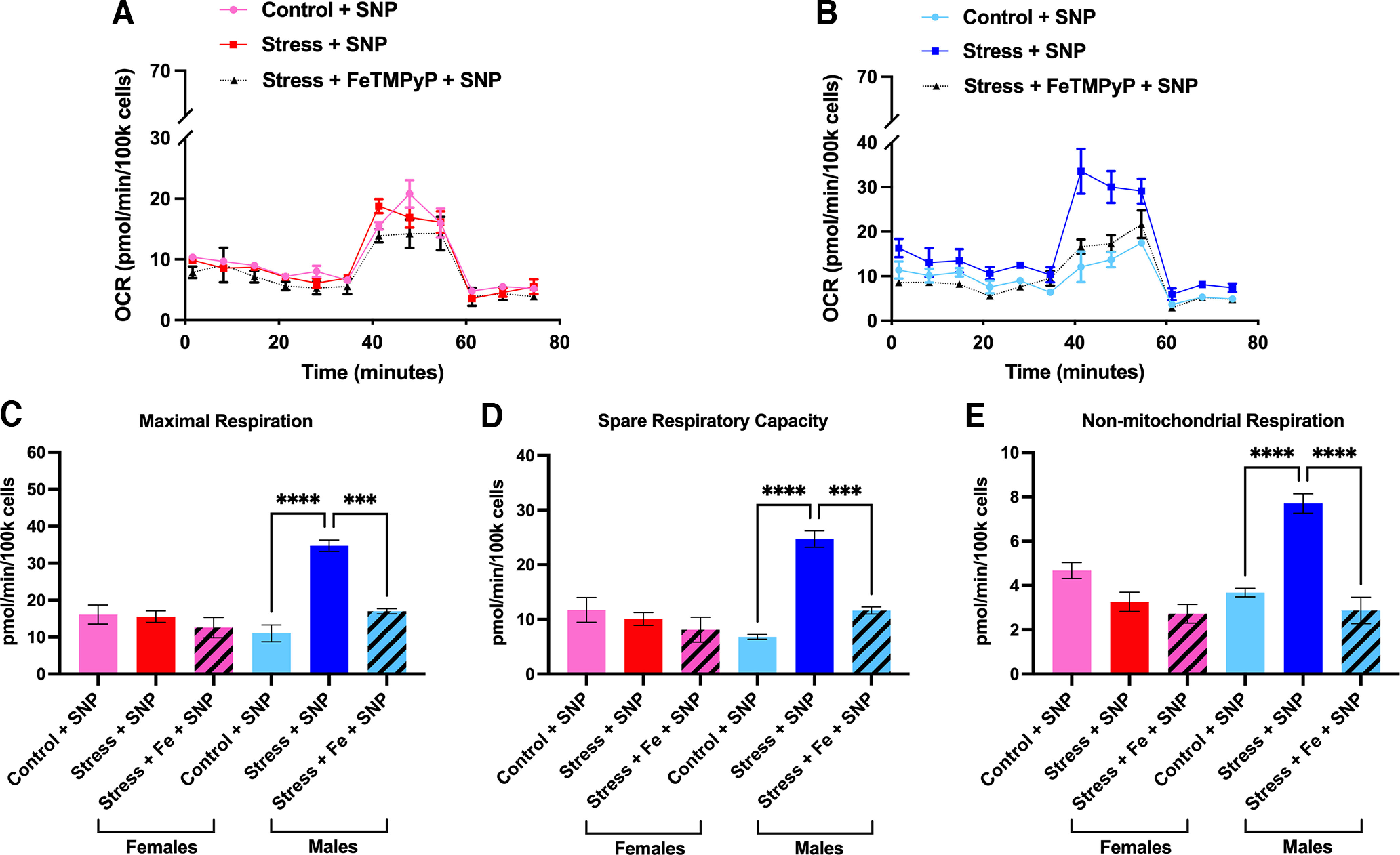
Administration of a nitric oxide (NO) donor triggers migraine attacks, but the mechanisms by which this occurs are unknown. Reactive nitroxidative species, including NO and peroxynitrite (PN), have been implicated in nociceptive sensitization, and neutralizing PN is antinociceptive. We determined whether PN contributes to nociceptive responses in two distinct models of migraine headache. Female and male mice were subjected to 3 consecutive days of restraint stress or to dural stimulation with the proinflammatory cytokine interleukin-6. Following resolution of the initial poststimulus behavioral responses, animals were tested for hyperalgesic priming using a normally non-noxious dose of the NO donor sodium nitroprusside (SNP) or dural pH 7.0, respectively. We measured periorbital von Frey and grimace responses in both models and measured stress-induced changes in 3-nitrotyrosine (3-NT) expression (a marker for PN activity) and trigeminal ganglia (TGs) mitochondrial function. Additionally, we recorded the neuronal activity of TGs in response to the PN generator SIN-1 [5-amino-3-(4-morpholinyl)-1,2,3-oxadiazolium chloride]. We then tested the effects of the PN decomposition catalysts Fe(III)5,10,15,20-tetrakis(N-methylpyridinium-4-yl) porphyrin (FeTMPyP) and FeTPPS [Fe(III)5,10,15,20-tetrakis(4-sulfonatophenyl)porphyrinato chloride], or the PN scavenger MnTBAP [Mn(III)tetrakis(4-benzoic acid)porphyrin] against these behavioral, molecular, and neuronal changes. Neutralizing PN attenuated stress-induced periorbital hypersensitivity and priming to SNP, with no effect on priming to dural pH 7.0. These compounds also prevented stress-induced increases in 3-NT expression in both the TGs and dura mater, and attenuated TG neuronal hyperexcitability caused by SIN-1. Surprisingly, FeTMPyP attenuated changes in TG mitochondrial function caused by SNP in stressed males only. Together, these data strongly implicate PN in migraine mechanisms and highlight the therapeutic potential of targeting PN.SIGNIFICANCE STATEMENT Among the most reliable experimental triggers of migraine are nitric oxide donors. The mechanisms by which nitric oxide triggers attacks are unclear but may be because of reactive nitroxidative species such as peroxynitrite. Using mouse models of migraine headache, we show that peroxynitrite-modulating compounds attenuate behavioral, neuronal, and molecular changes caused by repeated stress and nitric oxide donors (two of the most common triggers of migraine in humans). Additionally, our results show a sex-specific regulation of mitochondrial function by peroxynitrite following stress, providing novel insight into the ways in which peroxynitrite may contribute to migraine-related mechanisms. Critically, our data underscore the potential in targeting peroxynitrite formation as a novel therapeutic for the treatment of migraine headache.

Keywords: dura mater; headache; migraine; nitric oxide; peroxynitrite; trigeminal ganglia.


Conflict of interest statement

The authors declare no competing financial interests.

Figures

Figure 1.
Figure 1.
Peroxynitrite mediates NO donor-induced mechanical hypersensitivity in stress-primed mice. A, A schematic of the stress paradigm used is shown. Mice were subjected to repeated restraint stress or control conditions and tested for facial allodynia via von Frey assessment and mean grimace scores. Upon returning to baseline thresholds 14 d after stress, mice received a 30 mg/kg intraperitoneal injection of a PN scavenger (MnTBAP), a PN decomposition catalyst (FeTMPyP), or vehicle (PBS) 30 min before injection of the NO donor SNP (0.1 mg/kg, i.p.) and were again tested for facial allodynia. B, D, MnTBAP and FeTMPyP both significantly attenuated facial hypersensitivity caused by SNP in stress-primed female (B) and male (D) mice. C, E, No differences in grimace scoring were found in either sex (C, E). All control groups received vehicle before SNP. Two-way ANOVA followed by Bonferroni’s post hoc analysis revealed significant differences in the priming phase between stressed mice that received vehicle before SNP and stressed mice that received MnTBAP (denoted by *) or FeTMPyP (denoted by ) before SNP. n ≥ 6 for all groups in B and C; n = 8 for all groups in D and E. Data are represented as the mean ± SEM. Table 1, see for F-values. *p < 0.05, **p < 0.01, ***†††p < 0.001, ****††††p < 0.0001.
Figure 2.
Figure 2.
Modulating PN does not attenuate facial priming to dural pH 7.0. A, Dural injections and behavioral testing timelines are presented. B–E, Female (B, C) and male (D, E) mice received a 5 µl dural injection of vehicle (SIF) or IL-6 (0.1 ng) to induce acute periorbital hypersensitivity and grimacing that lasted out to 72 h. After the pain resolved, mice were given a 30 mg/kg intraperitoneal injection of MnTBAP, FeTMPyP, or vehicle (SIF) 30 min before a second 5 µl dural injection of a SIF pH 7.0 solution to check for the presence of hyperalgesic priming. Mice that received a PN-modulating compound did not exhibit significant differences in nociceptive thresholds from those that received vehicle after IL-6; however, a two-way ANOVA with Bonferroni’s post hoc analysis of the priming phase revealed significantly lower grimace scores between the group that received MnTBAP (denoted by *) and the IL-6/vehicle group within the first 3 h following dural pH 7.0. All control mice received pH 7.0 solution in the priming phase. n ≥ 8 for all groups in A and B; n = 6 for all groups in D and E. Data are represented as the mean ± SEM. Table 1, see for F-values. *p < 0.05, **p < 0.01.
Figure 3.
Figure 3.
Multiple dosing with a PNMC attenuates stress-induced hypersensitivity and priming to a NO donor. A, Stress paradigm and dosing regimen are shown. B, C, Following 3 d of repeated stress, female ICR mice were administered FeTMPyP (30 mg/kg, i.p.) or vehicle at 1, 24, 48, and 72 h poststress and tested for acute facial hypersensitivity (B) and grimacing (C). Upon returning to baseline thresholds, mice were checked for priming to low-dose SNP (0.1 mg/kg, i.p.). Stress-induced acute mechanical hypersensitivity and grimace responses in mice that received multiple injections of vehicle; however, these effects were attenuated by multiple injections of FeTMPyP, determined by a two-way ANOVA with Bonferroni’s post hoc analysis. *Significance between stressed mice that received FeTMPyP and those that received vehicle. All control groups received vehicle and were administered SNP before the priming phase (n = 6 for all groups). Data are represented as the mean ± SEM. Table 1, see for F-values. *p < 0.05, **p < 0.01, ****p < 0.0001.
Figure 4.
Figure 4.
Administration of a PNMC 24 h following repeated stress does not block facial allodynia. A, Stress paradigm and dosing regimen are shown. B, C, Following stress, female ICR mice exhibited robust facial hypersensitivity (B) and grimacing (C), and were primed to low-dose SNP (0.1 mg/kg, i.p.). A two-way ANOVA with Bonferroni’s post hoc analysis revealed no significant differences in acute hypersensitivity or priming in stressed mice that received a PNMC at 24 h following stress compared with stressed mice that received vehicle (n = 3–5 for all groups). Data are represented as the mean ± SEM. Table 1, see for F-values.
Figure 5.
Figure 5.
Administration of a PNMC at 1 h following stress results in attenuation of acute facial hypersensitivity and prevents priming to a NO donor. A, Following repeated stress, mice were administered FeTMPyP or MnTBAP (30 mg/kg, i.p.) 1 h poststress and tested for facial hypersensitivity, grimacing, and priming to low-dose SNP (0.1 mg/kg, i.p.). B–E, Compared with stressed mice that received vehicle, stressed mice that received a PNMC were found to have significant attenuation of acute allodynia and grimace scores, and did not prime to SNP in both females (B, C) and males (D, E). *Significance between stressed mice that received MnTBAP and those that received vehicle. Significance between Stress/FeTMPyP and Stress/Vehicle groups. All control groups received vehicle and were administered SNP before the priming phase (n ≥ 4 in B and C; n = 8 in D and E). Data are represented as the mean ± SEM. Table 1, see for F-values. *p < 0.05, **††p < 0.01, ***†††p < 0.001, ****††††p < 0.0001. Ctrl, Control.
Figure 6.
Figure 6.
PN mediates stress-induced increases in 3-NT expression in the TGs and dura. A, B, Approximately 24 h following the final day of repeated stress, an increase in 3-NT expression, a marker for PN, was observed in the TGs and dura of female (A) and male (B) mice compared with controls, suggesting an increase in PN activity. These changes were prevented by the administration of FeTMPyP (30 mg/kg) at 1 h poststress, as indicated by a one-way ANOVA with post hoc Bonferroni’s correction (n = 4 independent replicates/group). Representative Western blots are shown. Data are represented as the mean ± SEM. **p < 0.01, ****p < 0.0001.
Figure 7.
Figure 7.
A, B, Administration of low-dose SNP (0.1 mg/kg, i.p.) ∼14 d after repeated stress (when mice typically return to baseline withdrawal thresholds) induces a robust increase in 3-NT expression in the TGs and dura of stressed female (A) and male (B) mice compared with controls. Notably, pretreatment with FeTMPyP (30 mg/kg, i.p.) 30 min before injection of SNP prevented this increase in 3-NT expression, as indicated by one-way ANOVA with Bonferroni’s post hoc correction (n = 4 independent replicates/group). Representative Western blots shown. Data are represented as the mean ± SEM. ****p < 0.0001.
Figure 8.
Figure 8.
SIN-1 (1 mm) increases the excitability of TG neurons, and this hyperexcitability is prevented by the coapplication with SIN-1 of the peroxynitrite scavenger MnTBAP (100 μm) or the peroxynitrite decomposition catalyst FeTPPS (100 μm). A, Example traces of action potentials in the different treatment groups in response to the slow ramp injection of varying intensities. B, Top, Bar graphs representing the number of APs evoked by ramps. Main effect of treatment, F(3,64) = 11.01, p < 0.0001; *p < 0.05 for vehicle versus SIN-1 groups, post hoc Dunnett’s test. Bottom, Bar graphs representing the latency to the first AP at each ramp injection. Main effect of treatment, F(3,64) = 9.38, p < 0.0001, *p < 0.05 for vehicle versus SIN-1 groups, post hoc Dunnett’s test. Bottom, Bar graphs representing the latency to the first AP at each ramp injection. Main effect of treatment, F(3,64) = 9.38, p < 0.0001, *p < 0.05 for vehicle versus SIN-1 groups, post hoc Dunnett’s test. C, Raw traces of single APs evoked by a depolarizing step protocol. The dotted line is used to represent the lowering of threshold to fire an AP by SIN-1 and the prevention of this change in relation to vehicle treatment. The corresponding current rheobase used to evoke the APs is shown near each AP trace. Inset, Comparison of the AP half-width between vehicle group (black) and the SIN-1 + FeTPPS 100 μm group (red). Vehicle, n = 23 cells from 18 mice; SIN-1, n = 20 cells from 11 mice; SIN-1 + MnTBAP, n = 15 cells from 6 mice; SIN-1 + FeTPPS, n = 10 cells from 3 mice.
Figure 9.
Figure 9.
Mitochondrial respiration is increased in the TGs of male and female mice at 24 h following repeated restraint stress. A, B, Mitochondrial OCRs were measured for females (A) and males (B) from which several metabolic parameters were calculated. C, D, Overall, male mice exhibited an increase in basal respiration (C) levels while both sexes were found to have increased levels of maximal respiration (D), indicating that stress increases respiration rates in TG mitochondria (n = 3 replicate runs). Data are represented as the mean ± SEM. *p < 0.05.
Figure 10.
Figure 10.
At day 14 following repeated stress (when mice typically return to baseline withdrawal thresholds), spare respiratory capacity and ATP production are increased in female mice. A, B, Mitochondrial OCRs were measured for females (A) and males (B) from which several metabolic parameters were calculated. C, D, Female mice exhibited increased mitochondrial spare respiratory capacity (C) and ATP production (D) in their TGs, an effect that was not observed in male mice, suggesting a potential sex difference in the long-term effects of stress on mitochondrial function (n = 3 replicate runs). Data are represented as the mean ± SEM. ****p < 0.0001.
Figure 11.
Figure 11.
Administration of low-dose SNP induces robust changes in mitochondrial function in the TGs of male, but not female mice, an effect that is attenuated by pretreatment with FeTMPyP. Fourteen days following repeated stress, mice were administered either FeTMPyP (30 mg/kg, i.p.) or vehicle and were primed with SNP (0.1 mg/kg, i.p.). A, B, Mitochondrial OCRs were measured for females (A) and males (B) from which several metabolic parameters were calculated. C–E, Interestingly, stress-primed male mice exhibited robust increases in maximal respiration (C), spare respiratory capacity (D), and nonmitochondrial respiration (E) in response to SNP. Notably, these changes were attenuated by pretreatment with FeTMPyP. No changes were observed in female mice (n = 3 replicate runs). Data are represented as the mean ± SEM. **p < 0.01, ***p < 0.001, ***p < 0.0001.

Similar articles

Cited by

References

    1. Afridi SK, Kaube H, Goadsby PJ (2004) Glyceryl trinitrate triggers premonitory symptoms in migraineurs. Pain 110:675–680. 10.1016/j.pain.2004.05.007 - DOI - PubMed
    1. Agalave NM, Mody PH, Szabo-Pardi TA, Jeong HS, Burton MD (2021) Neuroimmune consequences of eIF4E phosphorylation on chemotherapy-induced peripheral neuropathy. Front Immunol 12:642420. 10.3389/fimmu.2021.642420 - DOI - PMC - PubMed
    1. Akerman S, Williamson DJ, Kaube H, Goadsby PJ (2002) Nitric oxide synthase inhibitors can antagonize neurogenic and calcitonin gene-related peptide induced dilation of dural meningeal vessels. Br J Pharmacol 137:62–68. 10.1038/sj.bjp.0704842 - DOI - PMC - PubMed
    1. Akerman S, Salvemini D, Romero-Reyes M (2021) Targeting reactive nitroxidative species in preclinical models of migraine. Cephalalgia 41:1187–1200. 10.1177/03331024211017884 - DOI - PubMed
    1. Avona A, Burgos-Vega C, Burton MD, Akopian AN, Price TJ, Dussor G (2019) Dural calcitonin gene-related peptide produces female-specific responses in rodent migraine models. J Neurosci 39:4323–4331. 10.1523/JNEUROSCI.0364-19.2019 - DOI - PMC - PubMed

Publication types