Dynamic Behavior of Polymer Microbubbles During Long Ultrasound Tone-Burst Excitation and Its Application for Sonoreperfusion Therapy
- PMID: 36697268
- PMCID: PMC9974862
- DOI: 10.1016/j.ultrasmedbio.2022.12.013
Dynamic Behavior of Polymer Microbubbles During Long Ultrasound Tone-Burst Excitation and Its Application for Sonoreperfusion Therapy
Abstract
Objective: Ultrasound (US)-targeted microbubble (MB) cavitation (UTMC)-mediated therapies have been found to restore perfusion and enhance drug/gene delivery. Because of the potentially longer circulation time and relative ease of storage and reconstitution of polymer-shelled MBs compared with lipid MBs, we investigated the dynamic behavior of polymer microbubbles and their therapeutic potential for sonoreperfusion (SRP) therapy.
Methods: The fate of polymer MBs during a single long tone-burst exposure (1 MHz, 5 ms) at various acoustic pressures and MB concentrations was recorded via high-speed microscopy and passive cavitation detection (PCD). SRP efficacy of the polymer MBs was investigated in an in vitro flow system and compared with that of lipid MBs.
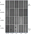
Discussion: Microscopy videos indicated that polymer MBs formed gas-filled clusters that continued to oscillate, fragment and form new gas-filled clusters during the single US burst. PCD confirmed continued acoustic activity throughout the 5-ms US excitation. SRP efficacy with polymer MBs increased with pulse duration and acoustic pressure similarly to that with lipid MBs but no significant differences were found between polymer and lipid MBs.
Conclusion: These data suggest that persistent cavitation activity from polymer MBs during long tone-burst US excitation confers excellent reperfusion efficacy.
Keywords: High-speed microscopy; Microbubble dynamics; Sonoreperfusion; Ultrasound contrast agents; Ultrasound therapy; Ultrasound-targeted microbubble cavitation.
Copyright © 2022 World Federation for Ultrasound in Medicine & Biology. Published by Elsevier Inc. All rights reserved.
Conflict of interest statement
Conflict of interest The authors declare no competing interests.
Figures


Similar articles
-
Dynamic Behavior of Microbubbles during Long Ultrasound Tone-Burst Excitation: Mechanistic Insights into Ultrasound-Microbubble Mediated Therapeutics Using High-Speed Imaging and Cavitation Detection.Ultrasound Med Biol. 2016 Feb;42(2):528-538. doi: 10.1016/j.ultrasmedbio.2015.09.017. Epub 2015 Nov 18. Ultrasound Med Biol. 2016. PMID: 26603628 Free PMC article.
-
Fibrin-targeted phase shift microbubbles for the treatment of microvascular obstruction.Nanotheranostics. 2024 Jan 1;8(1):33-47. doi: 10.7150/ntno.85092. eCollection 2024. Nanotheranostics. 2024. PMID: 38164499 Free PMC article.
-
Effect of Thrombus Composition and Viscosity on Sonoreperfusion Efficacy in a Model of Micro-Vascular Obstruction.Ultrasound Med Biol. 2016 Sep;42(9):2220-31. doi: 10.1016/j.ultrasmedbio.2016.04.004. Epub 2016 May 17. Ultrasound Med Biol. 2016. PMID: 27207018 Free PMC article.
-
Methods of preparation of multifunctional microbubbles and their in vitro / in vivo assessment of stability, functional and structural properties.Curr Pharm Des. 2012;18(15):2135-51. doi: 10.2174/138161212800099874. Curr Pharm Des. 2012. PMID: 22352769 Review.
-
Italian Society of Cardiovascular Echography (SIEC) Consensus Conference on the state of the art of contrast echocardiography.Ital Heart J. 2004 Apr;5(4):309-34. Ital Heart J. 2004. PMID: 15185894 Review.
Cited by
-
Quantifying the Effect of Acoustic Parameters on Temporal and Spatial Cavitation Activity: Gauging Cavitation Dose.Ultrasound Med Biol. 2023 Nov;49(11):2388-2397. doi: 10.1016/j.ultrasmedbio.2023.08.002. Epub 2023 Aug 28. Ultrasound Med Biol. 2023. PMID: 37648590 Free PMC article.
-
Long-pulsed ultrasound-mediated microbubble thrombolysis in a rat model of microvascular obstruction.Open Med (Wars). 2024 Mar 27;19(1):20240935. doi: 10.1515/med-2024-0935. eCollection 2024. Open Med (Wars). 2024. PMID: 38584836 Free PMC article.
-
Mediation of long-pulsed ultrasound enhanced microbubble recombinant tissue plasminogen activator thrombolysis in a rat model of platelet-rich thrombus.Cardiovasc Diagn Ther. 2024 Feb 15;14(1):51-58. doi: 10.21037/cdt-23-356. Epub 2024 Feb 1. Cardiovasc Diagn Ther. 2024. PMID: 38434566 Free PMC article.
-
Synergistic sonothrombolysis based on coaxial confocal dual-frequency focused ultrasound and vortex beams.Ultrason Sonochem. 2025 May;116:107314. doi: 10.1016/j.ultsonch.2025.107314. Epub 2025 Mar 17. Ultrason Sonochem. 2025. PMID: 40121707 Free PMC article.
References
-
- Rezkalla SH, Kloner RA. Coronary no-reflow phenomenon: from the experimental laboratory to the cardiac catheterization laboratory. Catheter Cardiovasc Interv. 2008;72(7):950–7. - PubMed
-
- Lee CH, Tse HF. Microvascular obstruction after percutaneous coronary intervention. Catheter Cardiovasc Interv. 2010;75(3):369–77. - PubMed
-
- Ito H, Tomooka T, Sakai N, Yu H, Higashino Y, Fujii K, et al. Lack of myocardial perfusion immediately after successful thrombolysis. A predictor of poor recovery of left ventricular function in anterior myocardial infarction. Circulation. 1992;85(5):1699–705. - PubMed
Publication types
MeSH terms
Substances
Grants and funding
LinkOut - more resources
Full Text Sources
Miscellaneous

