. 2023 Jan 25;9(1):31.
doi: 10.1038/s41420-023-01298-0.

Radiation-induced gastrointestinal (GI) syndrome as a function of age

Affiliations

Radiation-induced gastrointestinal (GI) syndrome as a function of age

Hongyan Li et al. Cell Death Discov. .

Erratum in

Abstract

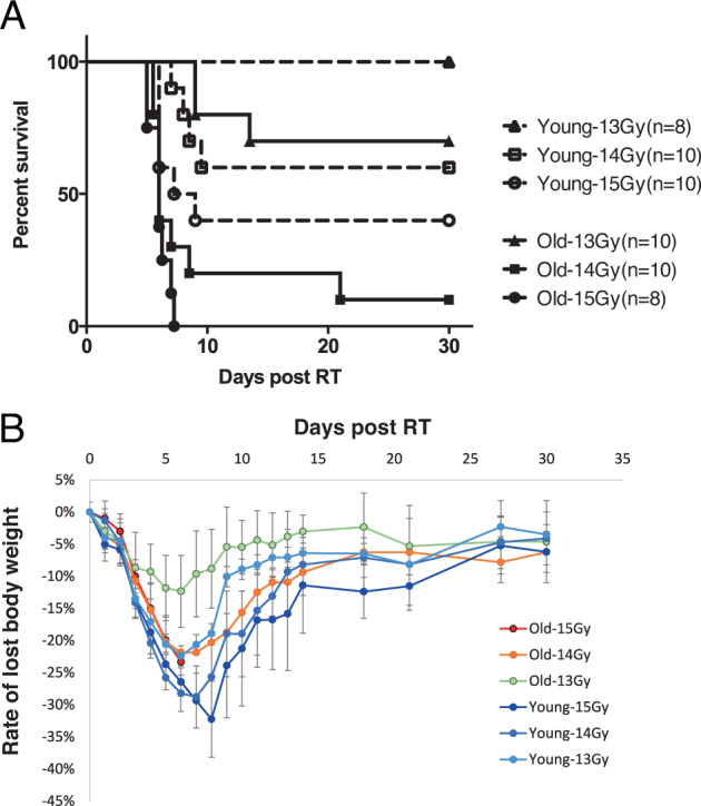
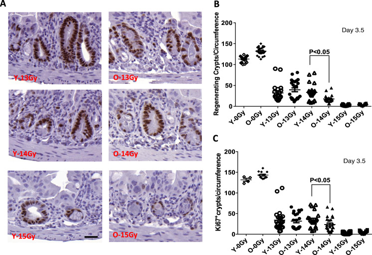
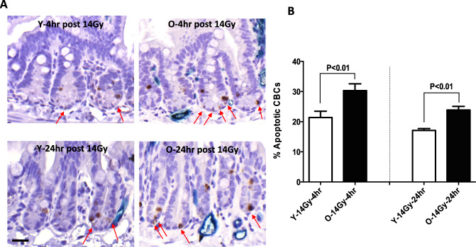
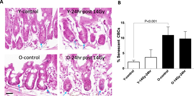
Previous studies show increased sensitivity of older mice (28-29 months) compared with young adult mice (3 months, possessing a mature immune system) to radiation-induced GI lethality. Age-dependent lethality was associated with higher levels of apoptotic stem cells in small intestinal crypts that correlated with sphingomyelinase activity, a source of pro-apoptotic ceramide. The objective of this study is to determine whether the cycling crypt base columnar cells (CBCs) in aging animals are specifically more sensitive to radiation effects than the CBCs in young adult mice, and to identify factors that contribute to increased radiosensitivity. Mortality induced by subtotal body radiation was assessed at different doses (13 Gy, 14 Gy, and 15 Gy) in young adult mice versus older mice. Each dose was evaluated for the occurrence of lethal GI syndrome. A higher death rate due to radiation-induced GI syndrome was observed in older mice as compared with young adult mice: 30 vs. 0% at 13 Gy, 90 vs. 40% at 14 Gy, and 100 vs. 60% at 15 Gy. Radiation-induced damage to crypts was determined by measuring crypt regeneration (H&E staining, Ki67 expression), CBC biomarkers (lgr5 and ascl2), premature senescence (SA-β-gal activity), and apoptosis of CBCs. At all three doses, crypt microcolony survival assays showed that the older mice had fewer regenerating crypts at 3.5 days post-radiation treatment. Furthermore, in the older animals, baseline CBCs numbers per circumference were significantly decreased, correlating with an elevated apoptotic index. Analysis of tissue damage showed an increased number of senescent CBCs per crypt circumference in older mice relative to younger mice, where the latter was not significantly affected by radiation treatment. It is concluded that enhanced sensitivity to radiation-induced GI syndrome and higher mortality in older mice can be attributed to a decreased capacity to regenerate crypts, presumably due to increased apoptosis and senescence of CBCs.


Conflict of interest statement

The authors declare no competing interests.

Figures

Fig. 1
Fig. 1. Survival curves of 3 months young mice (Y) and 29 months old mice (O) post subtotal irradiation of 13 Gy, 14 Gy, and 15 Gy.
A Percent of the survival curve. Numbers in parentheses indicate animal number/group. B Rate of lost body weight curve.
Fig. 2
Fig. 2. Regenerating crypts per jejunum circumference at 3.5 days post subtotal RT.
The sections of proximal jejunum were obtained from animals sacrificed at 3.5 days post-radiation. A Representative images of regenerating crypt 3.5 days post subtotal RT. B Quantitation of regenerating crypt numbers. A regenerating crypt was defined as a crypt containing at least 1 Paneth cell, over 10 non-Paneth cells and 1 lumen; appearing intensely stained body in HE-section. C Quantitation of regenerating crypt number was scored by counting Ki67-positive crypts. Data are presented as means ± SD. N = 3 mice/group, 10 circumferences per mouse. *P < 0.05 Y-14Gy vs O-14 Gy, Student’s t-test. Scale bar 20 μm.
Fig. 3
Fig. 3. Quantification of CBC per jejunum circumference at 3.5 days post subtotal RT.
Red arrows indicate the positive staining of Lgr5 in-situ hybridization staining fluorescence (Upper left & center, 40X magnification) andAscl2 in-situ hybridization bright field (Lower left & center, 40X magnification). A Representative images of data of Lgr5 positive CBC per circumference young (upper left panel) and older mice (upper center panel). B Quantification of Lgr5 positive per jejuna circumference. Lgr5 positive CBC are presented as means ± SD. N = 3 mice/group, 10 circumferences per mouse. C Representative images of Ascl2 positive CBC per jejuna circumference young (bottom left panel) and older positive Ascl2 per circumference (bottom center panel). D Quantification of Ascl2 positive CBC per jejuna circumference. *P < 0.05 Y-13Gy vs O-13 Gy and Y-14 Gy vs O-14 Gy, Student’s t-test.Scale bar 20 μm.
Fig. 4
Fig. 4. Apoptotic CBCs in response in young vs old mice at 4 h and 24 h post-RT.
A The sections of proximal jejunum were obtained from young adult and old mice sacrificed at 4h (upper left and center panels) and 24 h post 14 Gy irradiation(bottom left and center panels). The apoptosis was detected with cleaved caspase-3 by IHC staining. Apoptosis Index = positive apoptotic CBC/total CBC per jejuna circumference x 100%. Red arrows indicate apoptotic CBCs. B Quantification of percent apoptotic CBC per jejuna circumference. Data are presented as means ± SD. N = 3 mice/group, 10 circumferences per mouse. **P < 0.01 Y-14 Gy-4 h vs O-14 Gy-4 h and Y-14 Gy-24 h vs O-14 Gy-24 h, Student’s t-test. Scale bar 20 μm.
Fig. 5
Fig. 5. The sections of proximal jejunum were obtained from young adult and old mice sacrificed at 24 h post 14 Gy irradiation.
A The senescence-associated β-gal (SA-β-Gal) staining was detected with a commercial kit. Senescent index = positive β-gal CBC/total CBC per jejuna circumference × 100%. Blue arrows indicate senescent CBCs. B Quantification of percent senescent CBC per jejuna circumference. Data were presented as means ± SD. N = 3 mice/group, 10 circumferences per mouse. ***P < 0.001 Y-control vs O-control, Student’s t-test. Scale bar 20 µm.
Fig. 6
Fig. 6. The sections of proximal jejunum were obtained from young adult and old mice sacrificed at 12, 24, and 48 h post 14 Gy irradiation.
Paneth cell proliferation was assessed by IHF double staining of nuclear protein Ki67 (red) and Paneth cell marker lysozyme (green). Green arrows indicate lysozyme staining. Representative images containing 4 to 6 Paneth cells were shown. Scale bar 20 µm.

References

    1. Hendry JH, Potten CS, Roberts NP. The gastrointestinal syndrome and mucosal clonogenic cells: relationships between target cell sensitivities, LD50 and cell survival, and their modification by antibiotics. Radiat Res. 1983;96:100–12. doi: 10.2307/3576169. - DOI - PubMed
    1. Potten CS. A comprehensive study of the radiobiological response of the murine (BDF1) small intestine. Int J Radiat Biol. 1990;58:95–73. doi: 10.1080/09553009014552281. - DOI - PubMed
    1. Potten CS, Booth C, Pritchard DM. The intestinal epithelial stem cell: the mucosal governor. Int J Exp Pathol. 1997;78:219–43. doi: 10.1046/j.1365-2613.1997.280362.x. - DOI - PMC - PubMed
    1. Booth C, Potten CS. Gut instincts: thoughts on intestinal epithelial stem cells. J Clin Invest. 2000;105:1493–9. doi: 10.1172/JCI10229. - DOI - PMC - PubMed
    1. Paris F, Fuks Z, Capodicci P, Juan G, Ehleiter D, Schwartz J, et al. Endothelial apoptosis as the primary lesion initiating intestinal radiation damage in mice. Science. 2001;293:293–7. doi: 10.1126/science.1060191. - DOI - PubMed