. 2023 Jan 25;14(1):59.
doi: 10.1038/s41419-023-05598-2.

Characterization of circSCL38A1 as a novel oncogene in bladder cancer via targeting ILF3/TGF-β2 signaling axis

Affiliations

Characterization of circSCL38A1 as a novel oncogene in bladder cancer via targeting ILF3/TGF-β2 signaling axis

Peilong Li et al. Cell Death Dis. .

Abstract

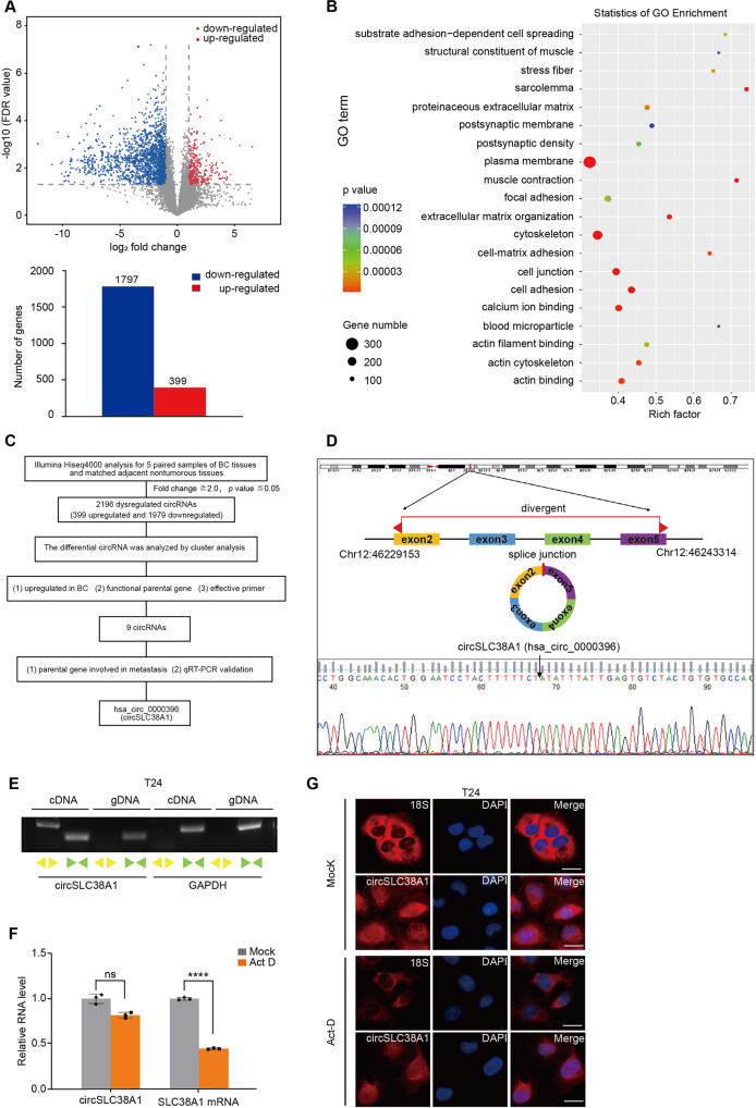
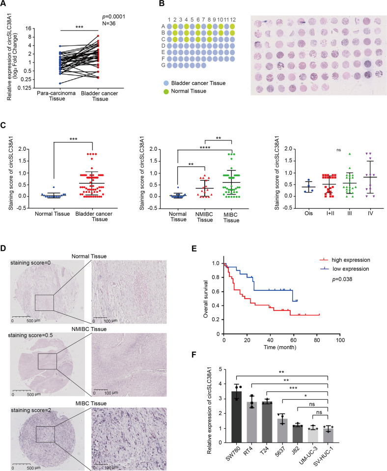
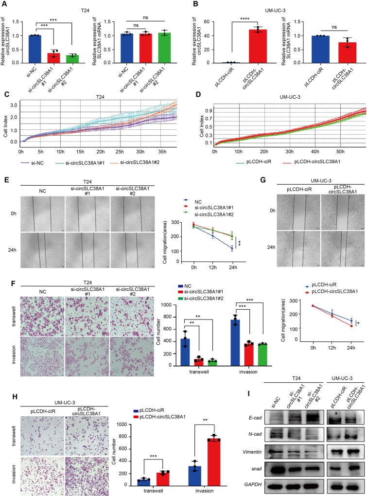
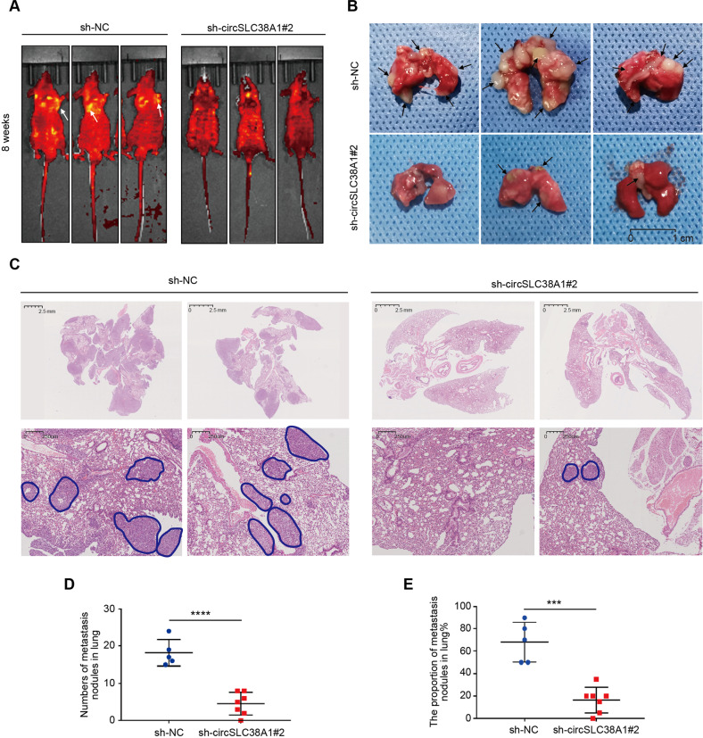
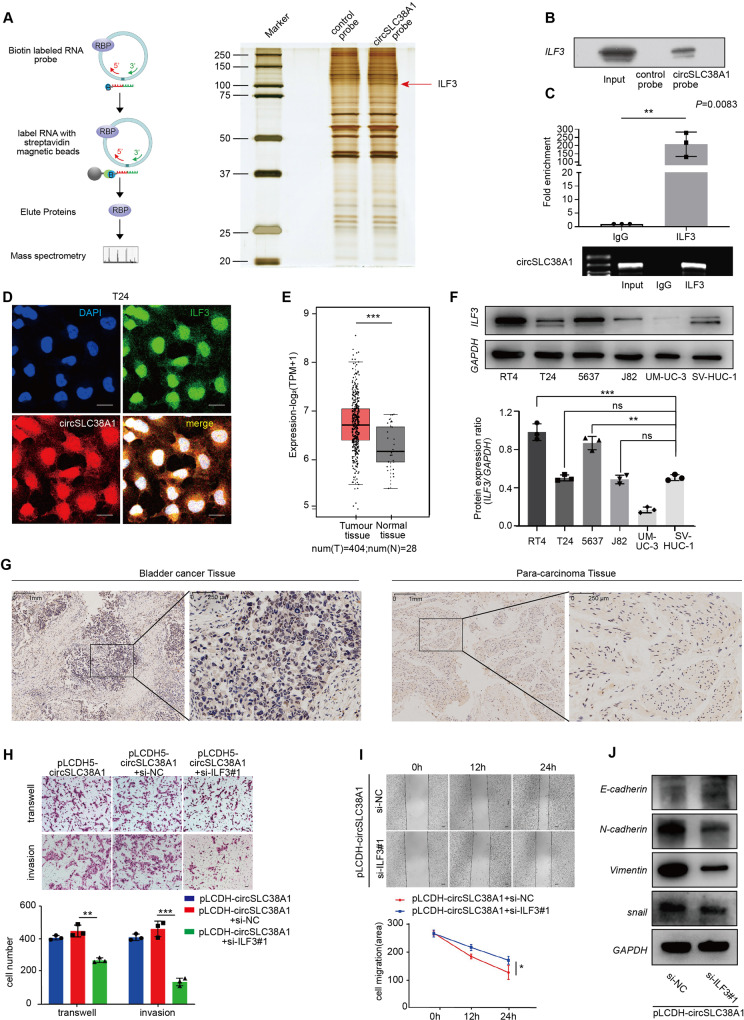
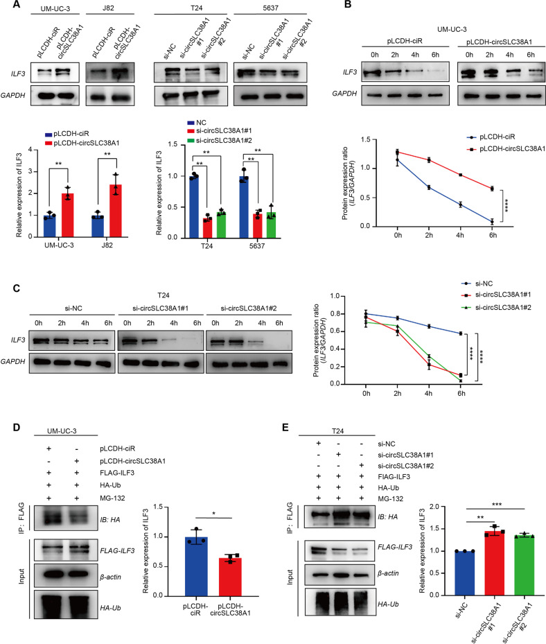
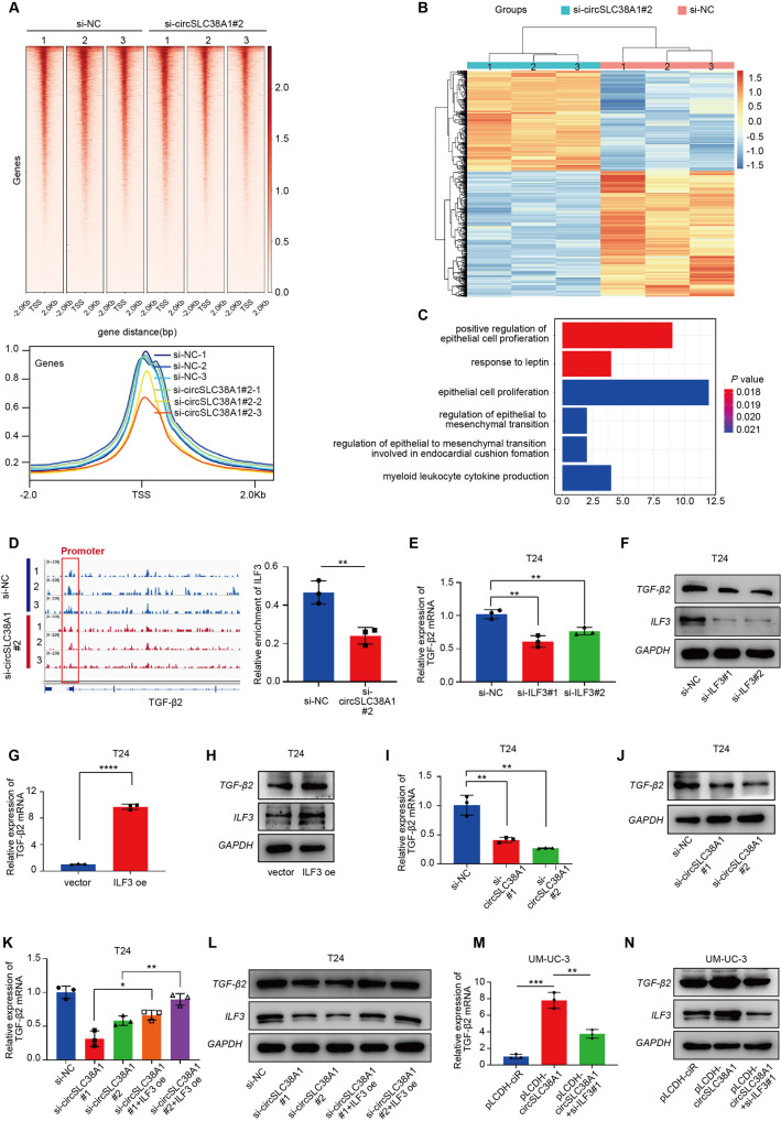
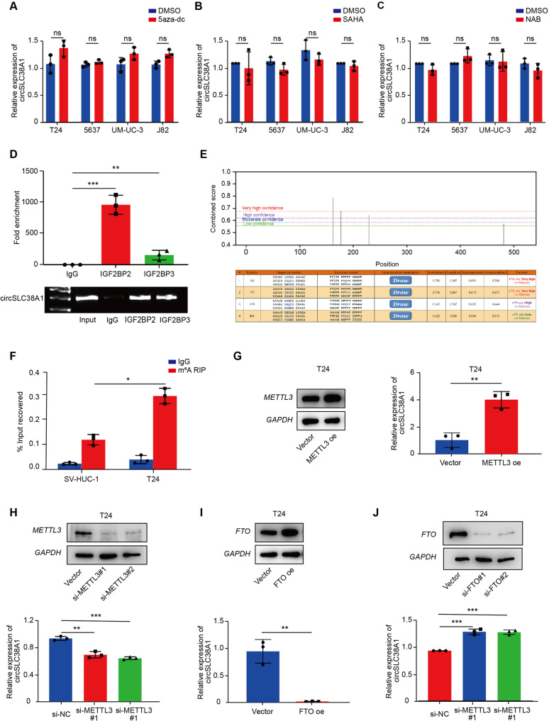
The regulatory role of circRNAs in cancer metastasis has become a focused issue in recent years. To date, however, the discovery of novel functional circRNAs and their regulatory mechanisms via binding with RBPs in bladder cancer (BC) are still lacking. Here, we screened out circSLC38A1 based on our sequencing data and followed validation with clinical tissue samples and cell lines. Functional assays showed that circSLC38A1 promoted BC cell invasion in vitro and lung metastasis of mice in vivo. By conducting RNA pull-down, mass spectrum, and RIP assays, circSLC38A1 was found to interact with Interleukin enhancer-binding factor 3 (ILF3), and stabilize ILF3 protein via modulating the ubiquitination process. By integrating our CUT&Tag-seq and RNA-seq data, TGF-β2 was identified as the functional target of the circSLC38A1-ILF3 complex. In addition, m6A methylation was enriched in circSLC38A1 and contributed to its upregulation. Clinically, circSLC38A1 was identified in serum exosomes of BC patients and could distinguish BC patients from healthy individuals with a diagnostic accuracy of 0.878. Thus, our study revealed an essential role and clinical significance of circSLC38A1 in BC via activating the transcription of TGF-β2 in an ILF3-dependent manner, extending the understanding of the importance of circRNA-mediated transcriptional regulation in BC metastasis.


Conflict of interest statement

The authors declare no competing interests.

Figures

Fig. 1
Fig. 1. circRNA expression profiles in BC.
A Top: volcano plot of the differentially expressed circRNAs in five pairs of BC tissues and corresponding adjacent nontumorous tissues by RNA-seq analysis. X-axis: log2 ratio of circRNA expression levels between normal and tumor tissues. Y-axis: the false discovery rate value (−log10 transformed) of circRNAs. Bottom: a total of 2196 dysregulated circRNAs were identified in BC tissues, of which 399 circRNAs were upregulated and 1797 circRNAs were downregulated (P < 0.05 and fold change >2.0). B GO analysis for the 2196 dysregulated circRNAs. C The flowchart delineates the steps for identifying circRNAs in BC. D Illustration of the annotated genomic region of SLC38A1, the putative different mRNA splicing forms (linear splicing and ‘head-to-tail’ splicing), circSLC38A1 is back-spliced by exon 2–5 of SLC38A1. Sanger sequencing following PCR conducted using the indicated divergent flanking primers showed the ‘head-to-tail’ splicing of circSLC38A1 in T24 cells. E CircSLC38A1 and GAPDH were amplified from cDNA or gDNA from T24 cells with divergent and convergent primers, respectively. F Stability of circSLC38A1 and linear SLC38A1 in T24 cells with or without 2 μg/mL Act D treatment for 24 h compared by qRT-PCR. ****p < 0.0001, error bars represent three independent experiments. G FISH with junction-specific probe of circSLC38A1 and 18 S probe was used to detect the localization of circSLC38A1; scale bar: 25 μm.
Fig. 2
Fig. 2. circSLC38A1 is upregulated in BC.
A Expression levels of circSLC38A1 in 36 pairs of BC and adjacent normal tissues were detected by the divergent primers (P = 0.0001). B The expression level of circSLC38A1 was measured by RNA ISH staining tissue microarrays (n = 79). C The difference in staining score between normal tissues and BC tissues (left), the difference in staining score between NMIBC tissues and MIBC tissues (middle), and the correlation of circSCL38A1 expression with tumor node metastasis classification (TNM) of bladder cancer (left). The staining score = staining intensity score * staining positive rate. (staining intensity score 0 is defined as negative, staining intensity score 1+ is defined as weak expression, staining intensity score 2+ is defined as moderate expression, and staining intensity score 3+ is defined as a strong expression). D The representative staining images were shown. E Kaplan–Meier analysis of the correlation between circSLC38A1 expression and overall survival. Patients with high levels of circSLC38A1 had significantly shorter overall survival (P = 0.038). The P value was determined by a Log-rank test. F Relative expression of circSLC38A1 in BC cell lines and human normal urothelial cell line measured by qRT-PCR. Data are presented as means ± standard deviation from three independent experiments. *p < 0.05, **p < 0.01, ***p < 0.001, ****p < 0.0001.
Fig. 3
Fig. 3. circSLC38A1 promotes migration and invasion capacities of BC cells in vitro.
A Relative expression levels of circSLC38A1 and SLC38A1 mRNA in T24 cells treated with circSLC38A1 siRNA or corresponding negative control (si-NC). B Relative expression levels of circSLC38A1 and SLC38A1 mRNA in UM-UC-3 cells after transduction with circSLC38A1 overexpression plasmid or vector plasmid. C The cell viability of T24 cell lines transfected with si-circSLC38A1 or si-NC were recorded by RTCA system. D The cell viability of UM-UC-3 cell lines transfected with circSLC38A1 overexpression plasmid or vector plasmid were recorded by RTCA system. E, F Wound healing assay (E), transwell migration, and matrigel invasion assay (F) showing decreased cell invasion in circSLC38A1 knockdown T24 cells. Scale bar, 100 µm (E), 25 µm (F). G, H Wound healing assay (G), transwell migration and matrigel invasion assay (H) showing that increased cell invasion in circSLC38A1 overexpression UM-UC-3 cells. Scale bar, 100 µm (G), 25 µm (H). I The expression levels of E-cadherin, N-cadherin, Vimentin, and snail in BC cells with knockdown or overexpression of circSLC38A1 were detected by western blot. Data are presented as means ± standard deviation from three independent experiments. *P < 0.05, **P < 0.01, ***P < 0.001.
Fig. 4
Fig. 4. circSLC38A1 promotes metastasis of BC in vivo.
A Representative fluorescent images for each experimental group at 8 weeks. B Representative images about tumor metastasis formed in the lungs of mice through vein tail injection of circSLC38A1-NC or circSLC38A1- knockdown BC cells. Metastatic nodules formed in the lungs of mice (Black Arrow). C Left, representative H&E staining of lung metastatic lesion through vein tail injection of circSLC38A1-NC BC cells. Multiple tumor metastases can be seen in the lung tissue. Right, representative H&E staining of lung metastatic lesion through vein tail injection of circSLC38A1-knockdown BC cells. The area circled with blue lines showed the tumor nests, among which the tumor cells exhibited solid growth mode. D The number of metastatic nodules formed in the lungs of mice for each group. E The proportion of metastatic nodules in the lungs of mice for each group. Data represent mean ± SD, and the P values were determined by a two-tailed unpaired Student’s t test, ***p < 0.001, ****p < 0.0001.
Fig. 5
Fig. 5. CircSLC38A1 interacted with ILF3 protein.
A Left, schematic diagram showing the process of circSLC38A1-pull down. The biotin-labeled probes were used to pull down interacting proteins in T24 cells, the pull-down proteins were identified by MS. Right, identification of the circSLC38A1-protein complex pulled down by circSLC38A1 probe with protein extracts from T24 cells. The arrows indicate the additional band presented in circSLC38A1-protein complex. B Immunoblot analysis of ILF3 after pulldown assay showing its specific association with circSLC38A1. C RIP assays showing the association of ILF3 with circSLC38A1 in T24 cells. Top, fold enrichment representing RNA levels associated with ILF3 relative to IgG, IgG antibody served as a control. Bottom, agarose gel electrophoresis for products of RIP assay. D Co-localization of circSLC38A1 and ILF3 visualized by coupling circRNA FISH assay with ILF3 Immunofluorescence staining; scale bar: 25 μm. E ILF3 is up-regulated in BC based on data available from TCGA database. F Relative expression of ILF3 in BC cell lines and human normal urothelial cell line detected by western blot. G Expression levels of ILF3 in BC and adjacent normal tissues were detected by immunohistochemical staining. H, I Interference with ILF3 can effectively reverse the migratory and invasive potential of bladder cancer cells induced by circSLC38A1. J The expression levels of E-cadherin, Vimentin, ILF3, and snail between pLCDH-circSLC38A1+si-NC and pLCDH-circSLC38A1 + siILF3#1 group were detected by western blot. Data represent mean ± SD from three independent experiments; *P < 0.05, **P < 0.01, ***P < 0.001.
Fig. 6
Fig. 6. circSLC38A1 increased ILF3 expression via inhibition of protein degradation.
A Expression levels of ILF3 in BC cells with circSLC38A1 deficient or circSLC38A1 overexpression. B Protein level of ILF3 in UM-UC-3 cells over-expressed circSLC38A1, followed by 100 µg/ml CHX treatment for the indicated time points. C Protein level of ILF3 in circSLC38A1 knockdown T24 cells, followed by 100 µg/ml CHX treatment for the indicated time points. D, E Immunoblot analysis of ILF3 in polyubiquitination assays of 293 T cells transfected with the indicated constructs and treated with 20 µM MG132 for 6 h. Data represent mean ± SD from three independent experiments; *P < 0.05, **P < 0.01, ***P < 0.001, ****P < 0.0001.
Fig. 7
Fig. 7. CircSLC38A1 recruits ILF3 to initiate TGF-β2 expression.
A Top: Heatmap of CUT&Tag-seq peaks associated with the ILF3 in T24 cell lines transfected with si-circSLC38A1#2 or si-NC, signals are displayed from -2.0 kb to +2.0 kb surrounding the TSS. Bottom: the average intensity curves for ILF3 signals at TSSs in a region comprising ±2 Kb in T24 cell lines transfected with si-circSLC38A1 or si-NC. B Heatmap of the differentially expressed RNAs in si-circSLC38A1 and si-NC cells by RNA-seq analysis. Red, upregulated RNAs; blue, downregulated RNAs. C Functional enrichment analysis of overlapped downregulated genes that were uniquely aligned to the genome using the online DAVID software. The results show that these genes are significantly associated with functional modules, such as regulation of epithelial cell proliferation and regulation of epithelial to mesenchymal transition. D Right: the IGV shows the CUT&Tag signals of ILF3 at the TGF-β2 gene loci. Left: quantitative diagram of the degree of enrichment of ILF3 signal on the TGF-β2 promoter. E, F Relative expression levels of TGF-β2 in T24 cells treated with ILF3 siRNA or corresponding negative control were measured by qRT-PCR (E) or western blot (F). G, H Relative expression levels of TGF-β2 in T24 cells transfected with vector control or ILF3 overexpression plasmid were measured by qRT-PCR (G) or western blot (H). I, J Relative expression levels of TGF-β2 in T24 cells treated with circSLC38A1 siRNA or corresponding negative control were measured by qRT-PCR (I) or western blot (J). K, L The expression of TGF-β2 in T24 cells with circSLC38A1 deficiency could be effectively reversed by overexpression of ILF3. M, N The expression of TGF-β2 that was upregulated with circSLC38A1 overexpression could be reduced by knocking down the expression of ILF3. Data represent mean ± SD from three independent experiments; *P < 0.05, **P < 0.01, ***P < 0.001, ****P < 0.0001.
Fig. 8
Fig. 8. m6A modification is involved in the upregulation of circSLC38A1 in BC cells.
A 4 BC cells were treated with or without 5 µM 5-aza-dC for 7 days, and circSLC38A1 expression was measured by qRT-PCR. B 4 BC cells were treated with or without 2 µM SAHA for 24 h, and circSLC38A1 expression was measured by qRT-PCR. C 4 BC cells were treated with or without 2 µM NAB for 24 h, and circSLC38A1 expression was measured. D RIP assays shows the association between IGF2BP2 or IGF2BP3 with circSLC38A1 in T24 cells. Top, fold enrichment representing RNA levels associated with IGF2BP2 or IGF2BP3 relative to IgG, IgG antibody served as a control. Bottom, agarose gel electrophoresis for products of RIP assay. E Three m6A modification sites on circSLC38A1 with high or very high confidence were identified by using online software tool SRAMP. F m6A RIP qRT-PCR analysis of circSLC38A1 in T24 and SV-HUC-1 cells. G The expression levels of circSLC38A1 in T24 cells transfected with vector control or METTL3 overexpression plasmid were measured by qRT-PCR. H The expression levels of circSLC38A1 in T24 cells transfected with si-NC or METTL3 siRNAs were measured by qRT-PCR. I The expression levels of circSLC38A1 in T24 cells transfected with vector control or FTO overexpression plasmid were measured by qRT-PCR. J The expression levels of circSLC38A1 in T24 cells transfected with si-NC or FTO siRNAs were measured by qRT-PCR. Data represent mean ± SD from three independent experiments; *P < 0.05, **P < 0.01, ***P < 0.001.

Similar articles

Cited by

References

    1. Antoni S, Ferlay J, Soerjomataram I, Znaor A, Jemal A, Bray F. Bladder cancer incidence and mortality: a global overview and recent trends. Eur Urol. 2017;71:96–108. doi: 10.1016/j.eururo.2016.06.010. - DOI - PubMed
    1. Sung H, Ferlay J, Siegel RL. Global cancer statistics 2020: GLOBOCAN estimates of incidence and mortality worldwide for 36 cancers in 185 countries. CA: a cancer J Clinicians. 2021;71:209–49. - PubMed
    1. Robertson AG, Kim J, Al-Ahmadie H, Bellmunt J, Guo G, Cherniack AD, et al. Comprehensive molecular characterization of muscle-invasive bladder cancer. Cell. 2017;171:540–556.e525. doi: 10.1016/j.cell.2017.09.007. - DOI - PMC - PubMed
    1. Kamat AM, Hahn NM, Efstathiou JA, Lerner SP, Malmstrom PU, Choi W, et al. Bladder cancer. Lancet. 2016;388:2796–810. doi: 10.1016/S0140-6736(16)30512-8. - DOI - PubMed
    1. Alfred Witjes J, Lebret T, Comperat EM, Cowan NC, De Santis M, Bruins HM, et al. Updated 2016 EAU guidelines on muscle-invasive and metastatic bladder cancer. Eur Urol. 2017;71:462–75. doi: 10.1016/j.eururo.2016.06.020. - DOI - PubMed

Publication types

Substances