

Encapsulation within a coordination cage modulates the reactivity of redox-active dyes
- PMID: 36697669
- PMCID: PMC9814915
- DOI: 10.1038/s42004-022-00658-8
Encapsulation within a coordination cage modulates the reactivity of redox-active dyes
Abstract
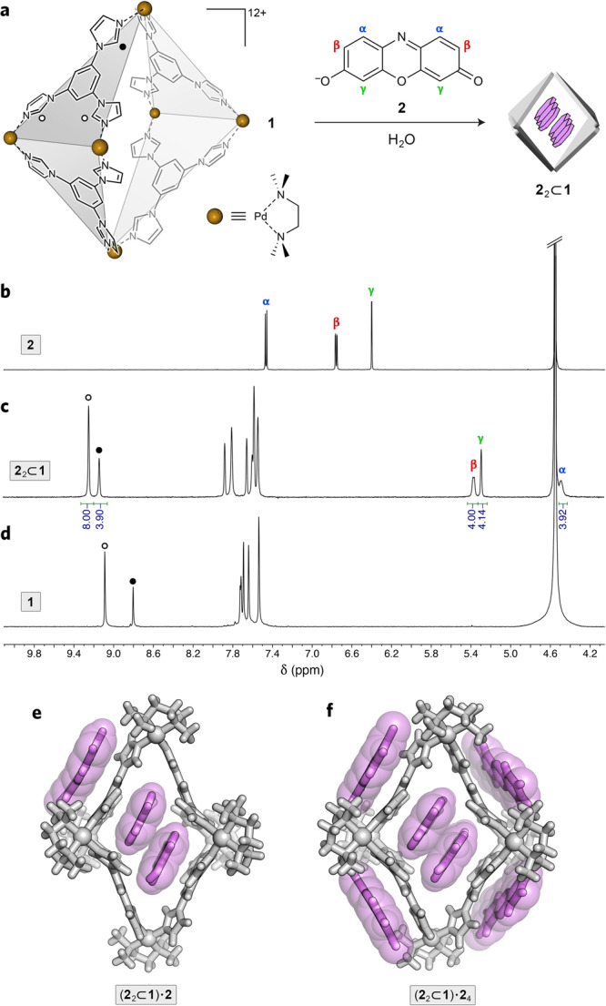
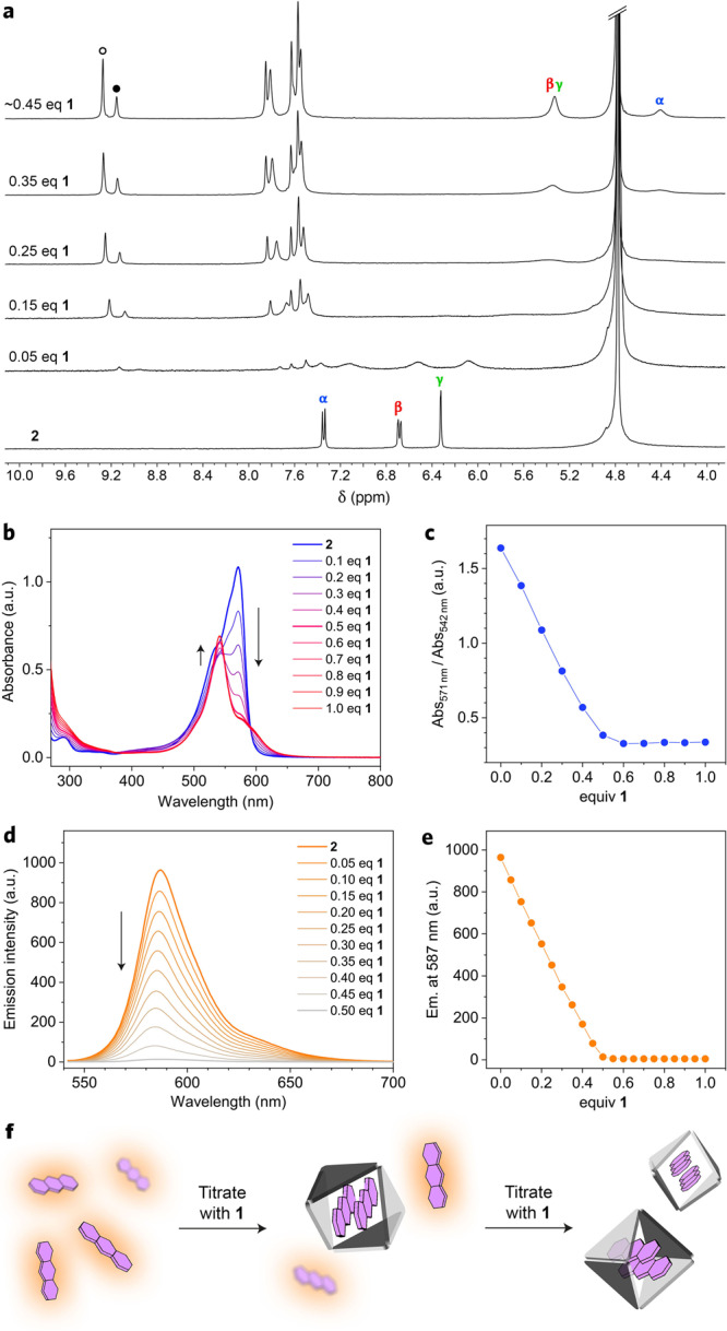
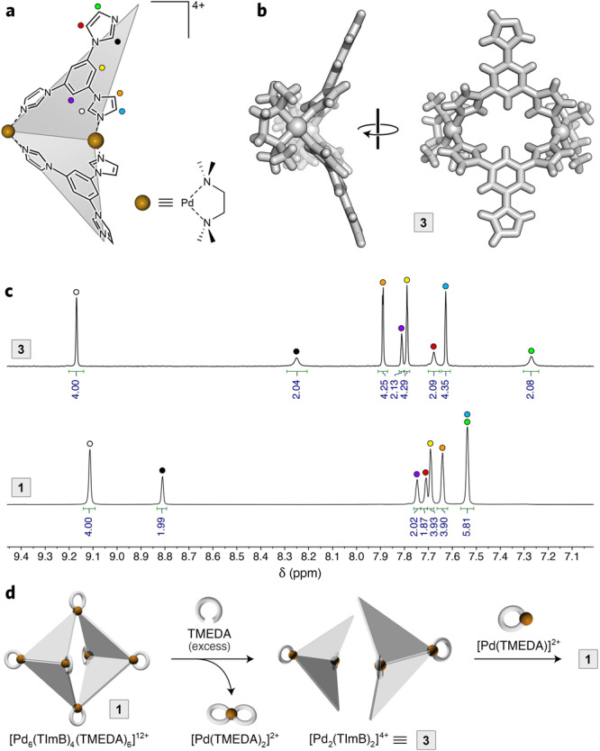
Confining molecules within well-defined nanosized spaces can profoundly alter their physicochemical characteristics. For example, the controlled aggregation of chromophores into discrete oligomers has been shown to tune their optical properties whereas encapsulation of reactive species within molecular hosts can increase their stability. The resazurin/resorufin pair has been widely used for detecting redox processes in biological settings; yet, how tight confinement affects the properties of these two dyes remains to be explored. Here, we show that a flexible PdII6L4 coordination cage can efficiently encapsulate both resorufin and resazurin in the form of dimers, dramatically modulating their optical properties. Furthermore, binding within the cage significantly decreases the reduction rate of resazurin to resorufin, and the rate of the subsequent reduction of resorufin to dihydroresorufin. During our studies, we also found that upon dilution, the PdII6L4 cage disassembles to afford PdII2L2 species, which lacks the ability to form inclusion complexes - a process that can be reversed upon the addition of the strongly binding resorufin/resazurin guests. We expect that the herein disclosed ability of a water-soluble cage to reversibly modulate the optical and chemical properties of a molecular redox probe will expand the versatility of synthetic fluorescent probes in biologically relevant environments.
© 2022. The Author(s).
Conflict of interest statement
The authors declare no competing interests.
Figures


Similar articles
-
Modulating the Optical Properties of BODIPY Dyes by Noncovalent Dimerization within a Flexible Coordination Cage.J Am Chem Soc. 2020 Oct 14;142(41):17721-17729. doi: 10.1021/jacs.0c08589. Epub 2020 Oct 2. J Am Chem Soc. 2020. PMID: 33006898 Free PMC article.
-
Complex Formation of Resorufin and Resazurin with Β-Cyclodextrins: Can Cyclodextrins Interfere with a Resazurin Cell Viability Assay?Molecules. 2018 Feb 10;23(2):382. doi: 10.3390/molecules23020382. Molecules. 2018. PMID: 29439432 Free PMC article.
-
Reversible chromism of spiropyran in the cavity of a flexible coordination cage.Nat Commun. 2018 Feb 13;9(1):641. doi: 10.1038/s41467-017-02715-6. Nat Commun. 2018. PMID: 29440687 Free PMC article.
-
Resorufin-based responsive probes for fluorescence and colorimetric analysis.J Mater Chem B. 2021 Jan 7;9(1):53-79. doi: 10.1039/d0tb01628d. Epub 2020 Nov 23. J Mater Chem B. 2021. PMID: 33226060 Review.
-
Selenium- and tellurium-containing fluorescent molecular probes for the detection of biologically important analytes.Acc Chem Res. 2014 Oct 21;47(10):2985-98. doi: 10.1021/ar500187v. Epub 2014 Sep 23. Acc Chem Res. 2014. PMID: 25248146 Review.
Cited by
-
Guest Encapsulation Alters the Thermodynamic Landscape of a Coordination Host.J Am Chem Soc. 2023 Nov 2;145(45):24755-64. doi: 10.1021/jacs.3c08666. Online ahead of print. J Am Chem Soc. 2023. PMID: 37917939 Free PMC article.
-
Ternary host-guest complexes with rapid exchange kinetics and photoswitchable fluorescence.Chem. 2022 Sep 8;8(9):2362-2379. doi: 10.1016/j.chempr.2022.05.008. Chem. 2022. PMID: 36133801 Free PMC article.
-
Programmable synthesis of well-defined, glycosylated iron(ii) supramolecular assemblies with multivalent protein-binding capabilities.Chem Sci. 2022 Dec 20;14(4):1018-1026. doi: 10.1039/d2sc05689e. eCollection 2023 Jan 25. Chem Sci. 2022. PMID: 36755719 Free PMC article.
-
Encapsulation of reactive species within metal-organic cages.Chem Sci. 2025 Jul 22. doi: 10.1039/d5sc02081f. Online ahead of print. Chem Sci. 2025. PMID: 40747319 Free PMC article. Review.
-
Reconstructing reactivity in dynamic host-guest systems at atomistic resolution: amide hydrolysis under confinement in the cavity of a coordination cage.Chem Sci. 2022 Aug 29;13(37):11232-11245. doi: 10.1039/d2sc02000a. eCollection 2022 Sep 28. Chem Sci. 2022. PMID: 36320487 Free PMC article.
References
-
- Varçin M, Bilgenur Şener B, Bayraç C. Adsorption of resazurin by poly(acrylic acid) hydrogels and evaluation of its use in reduction assay for quantification of cell viability. Dyes Pigments. 2021;186:109038. doi: 10.1016/j.dyepig.2020.109038. - DOI
-
- Twigg RS. Oxidation-reduction aspects of resazurin. Nature. 1945;155:401–402. doi: 10.1038/155401a0. - DOI
-
- Dallan E, Regier P, Marion A, González-Pinzón R. Does the mass balance of the reactive tracers resazurin and resorufin close at the microbial scale? J. Geophys. Res. Biogeosci. 2020;125:e2019JG005435. doi: 10.1029/2019JG005435. - DOI
LinkOut - more resources
Full Text Sources
