

Directly imaging emergence of phase separation in peroxidized lipid membranes
- PMID: 36697756
- PMCID: PMC9845225
- DOI: 10.1038/s42004-022-00809-x
Directly imaging emergence of phase separation in peroxidized lipid membranes
Abstract
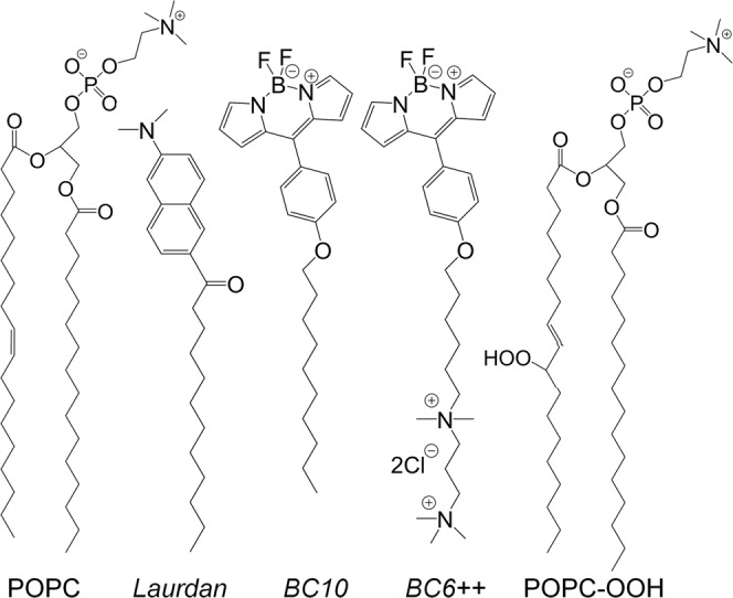
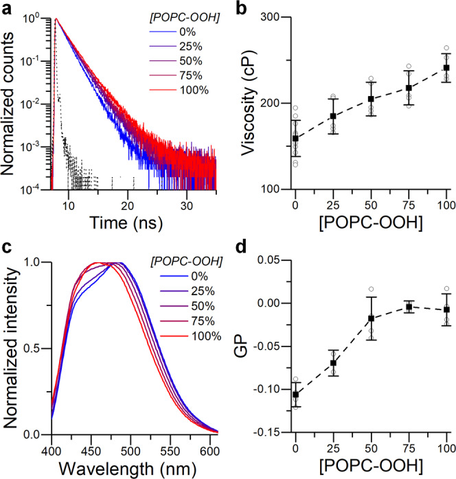
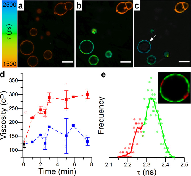
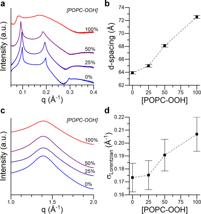
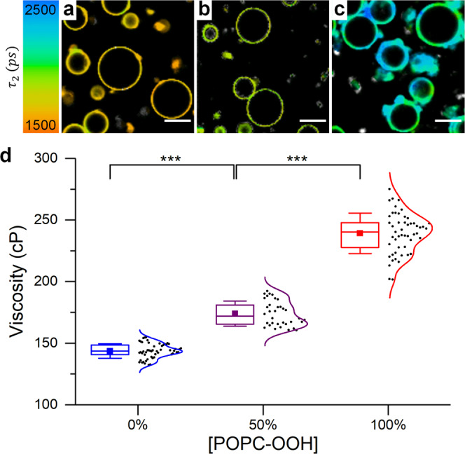
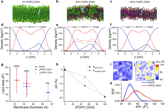
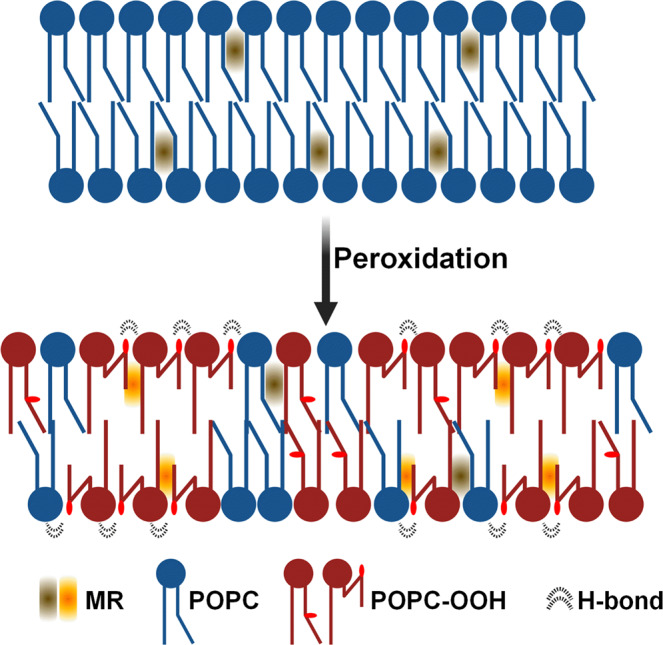
Lipid peroxidation is a process which is key in cell signaling and disease, it is exploited in cancer therapy in the form of photodynamic therapy. The appearance of hydrophilic moieties within the bilayer's hydrocarbon core will dramatically alter the structure and mechanical behavior of membranes. Here, we combine viscosity sensitive fluorophores, advanced microscopy, and X-ray diffraction and molecular simulations to directly and quantitatively measure the bilayer's structural and viscoelastic properties, and correlate these with atomistic molecular modelling. Our results indicate an increase in microviscosity and a decrease in the bending rigidity upon peroxidation of the membranes, contrary to the trend observed with non-oxidized lipids. Fluorescence lifetime imaging microscopy and MD simulations give evidence for the presence of membrane regions of different local order in the oxidized membranes. We hypothesize that oxidation promotes stronger lipid-lipid interactions, which lead to an increase in the lateral heterogeneity within the bilayer and the creation of lipid clusters of higher order.
© 2023. The Author(s).
Conflict of interest statement
The authors declare no competing interests.
Figures
Similar articles
-
A Molecular Rotor that Measures Dynamic Changes of Lipid Bilayer Viscosity Caused by Oxidative Stress.Chemistry. 2016 Sep 5;22(37):13210-7. doi: 10.1002/chem.201601925. Epub 2016 Aug 3. Chemistry. 2016. PMID: 27487026 Free PMC article.
-
Imaging non-classical mechanical responses of lipid membranes using molecular rotors.Chem Sci. 2020 Dec 22;12(7):2604-2613. doi: 10.1039/d0sc05874b. Chem Sci. 2020. PMID: 34164028 Free PMC article.
-
Lipid Hydroperoxide Compromises the Membrane Structure Organization and Softens Bending Rigidity.Langmuir. 2021 Aug 24;37(33):9952-9963. doi: 10.1021/acs.langmuir.1c00830. Epub 2021 Aug 10. Langmuir. 2021. PMID: 34374545
-
Do sterols reduce proton and sodium leaks through lipid bilayers?Prog Lipid Res. 2001 Jul;40(4):299-324. doi: 10.1016/s0163-7827(01)00009-1. Prog Lipid Res. 2001. PMID: 11412894 Review.
-
Biophysical consequences of lipid peroxidation in membranes.Chem Phys Lipids. 1987 Jul-Sep;44(2-4):175-89. doi: 10.1016/0009-3084(87)90049-1. Chem Phys Lipids. 1987. PMID: 3311416 Review.
Cited by
-
Activation energy for pore opening in lipid membranes under an electric field.Proc Natl Acad Sci U S A. 2023 Mar 14;120(11):e2213112120. doi: 10.1073/pnas.2213112120. Epub 2023 Mar 7. Proc Natl Acad Sci U S A. 2023. PMID: 36881617 Free PMC article.
-
Molecular Rotors: Fluorescent Sensors for Microviscosity and Conformation of Biomolecules.Angew Chem Int Ed Engl. 2024 Feb 5;63(6):e202311233. doi: 10.1002/anie.202311233. Epub 2023 Nov 14. Angew Chem Int Ed Engl. 2024. PMID: 37856157 Free PMC article. Review.
-
Viscosity-Sensitive Membrane Dyes as Tools To Estimate the Crystalline Structure of Lipid Bilayers.Anal Chem. 2023 Aug 15;95(32):12006-12014. doi: 10.1021/acs.analchem.3c01747. Epub 2023 Aug 1. Anal Chem. 2023. PMID: 37526607 Free PMC article.
-
Cancer Cell Identification via Lysosomal Membrane Microviscosities Using a Green-Emitting BODIPY Molecular Rotor.JACS Au. 2025 Apr 14;5(4):2004-2014. doi: 10.1021/jacsau.5c00253. eCollection 2025 Apr 28. JACS Au. 2025. PMID: 40313834 Free PMC article.
-
Tensing Flipper: Photosensitized Manipulation of Membrane Tension, Lipid Phase Separation, and Raft Protein Sorting in Biological Membranes.J Am Chem Soc. 2024 Aug 28;146(34):24114-24124. doi: 10.1021/jacs.4c08580. Epub 2024 Aug 20. J Am Chem Soc. 2024. PMID: 39162019 Free PMC article.
References
Grants and funding
LinkOut - more resources
Full Text Sources
