Review
. 2023 Apr;24(4):195-212.
doi: 10.1038/s41583-022-00674-6. Epub 2023 Jan 25.

Spectrins: molecular organizers and targets of neurological disorders

Affiliations
Review

Spectrins: molecular organizers and targets of neurological disorders

Damaris N Lorenzo et al. Nat Rev Neurosci. 2023 Apr.

Abstract

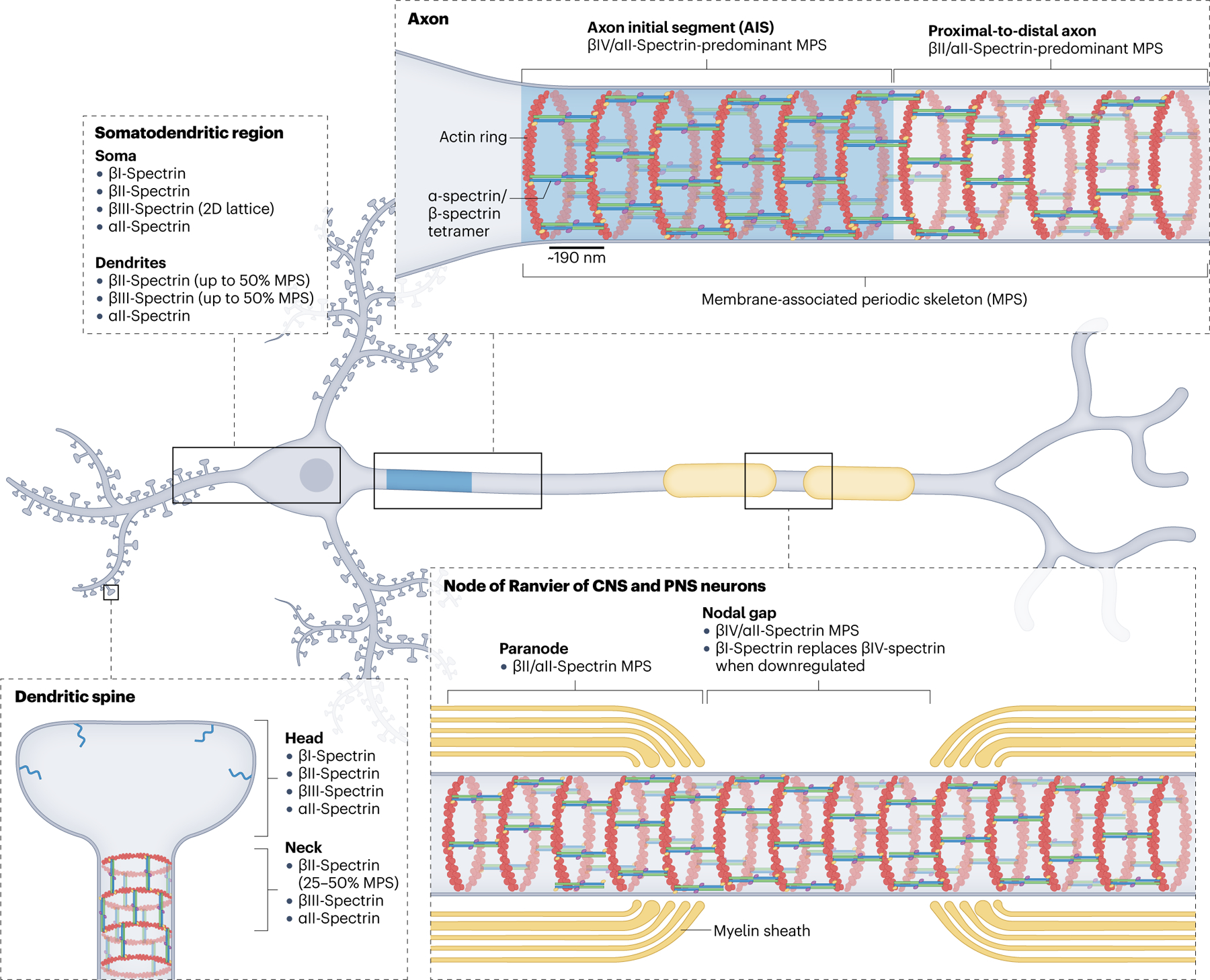
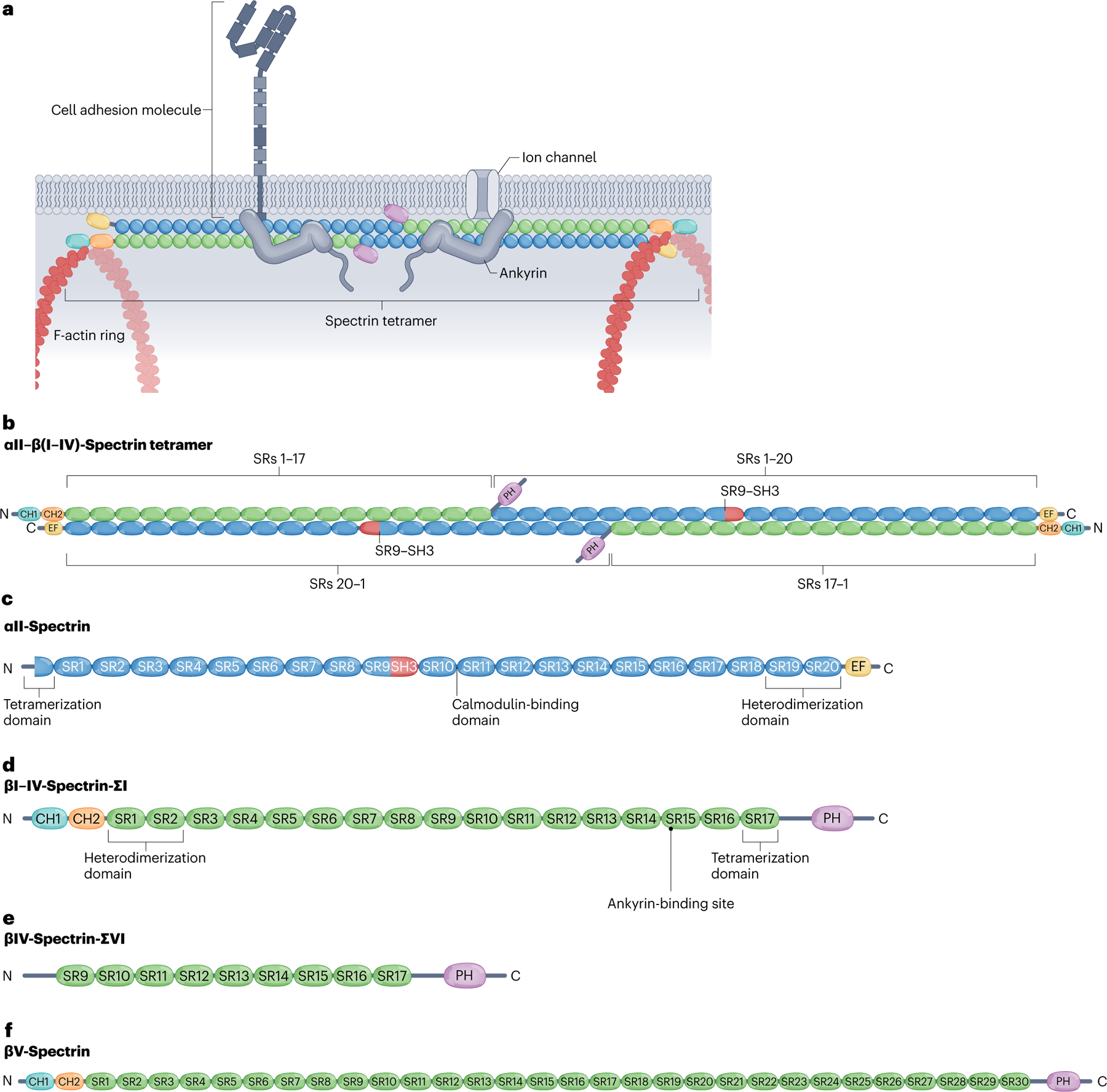
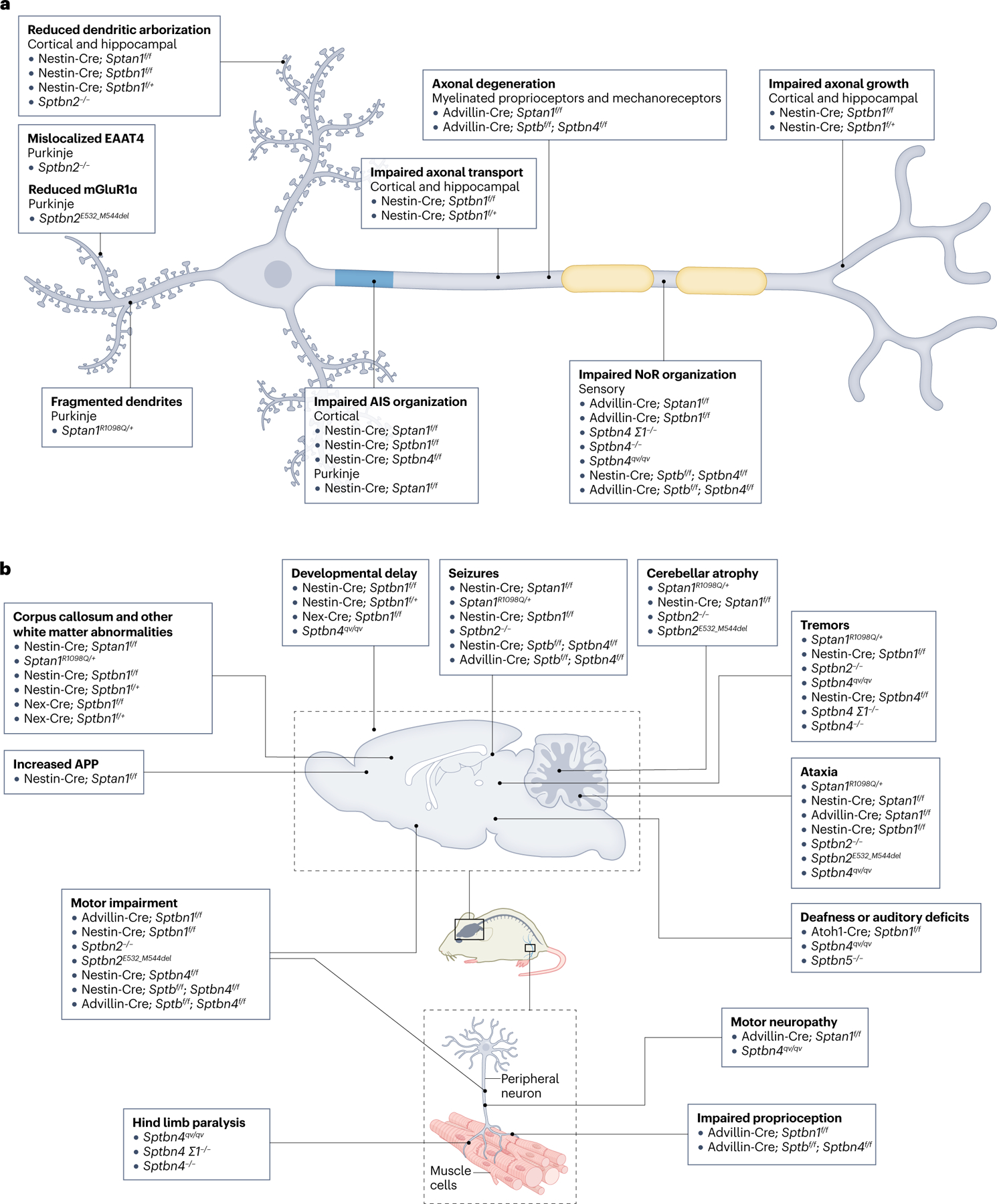
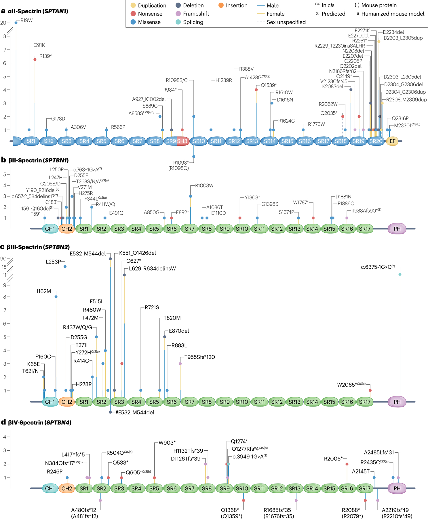
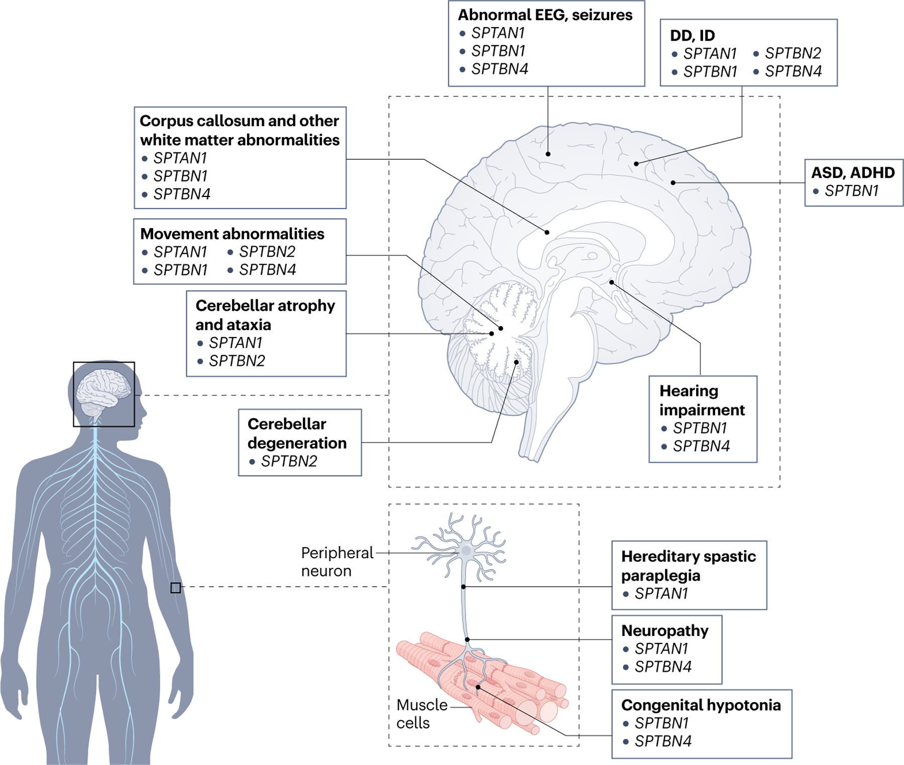
Spectrins are cytoskeletal proteins that are expressed ubiquitously in the mammalian nervous system. Pathogenic variants in SPTAN1, SPTBN1, SPTBN2 and SPTBN4, four of the six genes encoding neuronal spectrins, cause neurological disorders. Despite their structural similarity and shared role as molecular organizers at the cell membrane, spectrins vary in expression, subcellular localization and specialization in neurons, and this variation partly underlies non-overlapping disease presentations across spectrinopathies. Here, we summarize recent progress in discerning the local and long-range organization and diverse functions of neuronal spectrins. We provide an overview of functional studies using mouse models, which, together with growing human genetic and clinical data, are helping to illuminate the aetiology of neurological spectrinopathies. These approaches are all critical on the path to plausible therapeutic solutions.


Conflict of interest statement

Competing interests

The authors declare no competing interests.

Figures

Fig. 1 |
Fig. 1 |. Cellular localization and organization of neuronal spectrins.
Mammalian neurons express six of the seven spectrins, which follow a general pattern of domain localization and organization across different neuron types. βIV-spectrin is enriched at the axon initial segment (AIS), where, together with αII-spectrin, actin and other key molecules, it forms a membrane-associated periodic skeleton (MPS). The MPS is best characterized by actin rings enwrapping the circumference of neuronal processes with a ~190 nm periodicity, which is determined by their cross-linking by spectrin tetramers in their fully elongated conformation. The proximal-to-distal axon, including axonal branches, expresses βII-spectrin and αII-spectrin in high abundance together with relatively less abundant βIII-spectrin, all integrated into the MPS that spans the full axon. In myelinated axons of both the CNS and the PNS, βIV/αII-spectrins are localized and periodically organized in the nodal gap of nodes of Ranvier (NoR), flanked by βII/αII-spectrins in the paranode, also periodically distributed. Upon loss of βIV-spectrin, βI-spectrin localizes to NoR and rescues βIV-spectrin function. This redundancy is not available at the AIS, probably because βI-spectrin localization depends on its molecular partner ankyrin-R, which is not recruited to the AIS. Unlike in the axon, the probability of detecting the MPS in dendritic shafts of mature neurons, which includes βII-spectrin and βIII-spectrin, is about 50%. In addition to the quasi-1D organization of the MPS, βIII-spectrin can form 2D polygonal lattices in the soma and dendritic shaft. In dendritic spines, βII-spectrin and βIII-spectrin adopt MPS periodicity in the neck, but not in the head.
Fig. 2 |
Fig. 2 |. Tetrameric assembly and structural domains of neuronal spectrins.
a, Canonical spectrins form heterotetramers of two α-units and two β-units that crosslink F-actin rings along the neuronal membrane. Spectrins bind ankyrins, which in turn stabilize membrane-spanning proteins such as cell adhesion molecules and ion channels. b, Spectrin tetramers assemble by linking heterodimers head-to-head via non-covalent association between the partial spectrin repeats (SRs) in the N terminus adjacent to SR1 in the α-spectrin subunits (blue) and partial SR17 at the N terminus of the β-spectrin subunits (green). Complementary motifs in SR1 and SR2 of βI–IV spectrins and SR19 and SR20 of αII-spectrin bind covalently to enable the antiparallel lateral assembly of α–β-spectrin heterodimers. c, αII-Spectrin spans 20 modular SRs (blue), a calcium-binding EF hand domain (yellow) close to the C terminus, an Src-homology 3 (SH3) domain (red) in SR9 and a calmodulin (CaM)-binding loop in SR10. d, Canonical βI–βIV-spectrins contain 16 full SRs and a partial 17th SR (green), two N-terminal tandem calponin homology (CH) domains (teal and orange), an ankyrin-binding site in SR15 and a C-terminal pleckstrin homology (PH) domain (purple). The CH domains enable binding to actin and the PH domain binds membrane lipids. e, The alternatively spliced βIV-spectrin-ΣVI isoform, which is important for maintenance of the axon initial segment (AIS), lacks the CH domains and the first eight full SRs, but retains ankyrin-binding activity. f, Giant βV-spectrin contains 29 full SRs plus a partial 30th SR. Whether βV-spectrin associates with αII-spectrin is not clear.
Fig. 3 |
Fig. 3 |. Deficiencies in mouse models of neuronal spectrin dysfunction.
a, Loss, haploinsufficiency and mutations in spectrins in mice induce global, region and functional domain-specific neuronal defects in vivo and in vitro, including reduced dendritic arborization, axonal degeneration, protein mislocalization and reduced axonal transport. Neuron type and mouse model source (see Supplementary Table S1) indicated in parentheses. b, Major anatomical and functional phenotypes observed in mouse models of spectrin deficits in the CNS and PNS. Mouse model source (see Supplementary Table S1) indicated in parentheses. AIS, axon initial segment; APP, amyloid precursor protein; EAAT4, excitatory amino acid transporter 4; mGluR1α, metabotropic glutamate receptor type 1α; NoR, nodes of Ranvier.
Fig. 4 |
Fig. 4 |. Spectrin variants associated with neurological disorders.
a, Multiple reported αII-spectrin variants have been associated with neurological disorders. Variant types include missense (blue), nonsense (red), duplication (yellow), deletion (dark grey), splicing (teal), insertion (orange) and frameshift (violet). The cis superscript indicates compound heterozygous (in cis), with the letter in parentheses indicating the corresponding variant pair for a single individual. The sex of the reported individual is indicated by the lines below the dots (male, blue line; female, yellow line; unknown, discontinuous line). Number of individuals of each sex for each variant is indicated by the length of the corresponding line below the oval-shaped dot measured relative to the y-axis. Variants are distributed throughout the spectrin repeats (SRs; blue), with a cluster in the heterodimerization region (SRs 19–20). b, βII-Spectrin variants associated with a neurodevelopmental syndrome. These variants emerge largely de novo and are spread throughout the SRs (green), with a strong cluster in the second calponin homology (CH2; orange) domain. c, βIII-Spectrin variants associated with ataxia, developmental delay (DD) and intellectual disability (ID). d, Reported human βIV-spectrin variants associated with disorders of the CNS and PNS. Only homozygous and compound heterozygous carriers manifest clinical presentations. The carrier of the N384Qfs*17CIS(c) variant also bears a maternally inherited deletion with a breakpoint spanning [chr19.g.(?_41,001,394)_ (41,011,375_?)del (GRCh37)], which is predicted to delete exons 6–11 (ref. ). Knock-in mouse models are indicated in the lower part of the protein schematic, with the corresponding mutated site in the mouse spectrin homologue shown in parentheses. PH, pleckstrin homology; SH, Src-homology. Part b adapted from ref. , Springer Nature Limited.
Fig. 5 |
Fig. 5 |. Major phenotypes in humans with spectrinopathies of the nervous system.
Pathogenic variants in spectrins cause complex neurological syndromes in both the brain and periphery that have overlapping pathologies and clinical presentations across spectrin genes. Affected spectrin genes indicated in parentheses. ADHD, attention deficit hyperactivity disorder; ASD, autism spectrum disorder; DD, developmental delay; ID, intellectual disability.

References

    1. Bennett V & Lorenzo DN An adaptable spectrin/ankyrin-based mechanism for long-range organization of plasma membranes in vertebrate tissues. Curr. Top. Membr 77, 143–184 (2016). - PubMed
    1. Lorenzo DN Cargo hold and delivery: ankyrins, spectrins, and their functional patterning of neurons. Cytoskeleton 77, 129–148 (2020). - PMC - PubMed
    1. Bennett V & Lorenzo DN Spectrin- and ankyrin-based membrane domains and the evolution of vertebrates. Curr. Top. Membr 72, 1–37 (2013). - PubMed
    1. Zhou R, Han B, Xia C & Zhuang X Membrane-associated periodic skeleton is a signaling platform for RTK transactivation in neurons. Science 365, 929–934 (2019). - PMC - PubMed
    1. Marchesi VT & Steers EJ Selective solubilization of a protein component of the red cell membrane. Science 159, 203–204 (1968). - PubMed

Publication types