. 2023 Jan 25;20(1):4.
doi: 10.1186/s12950-023-00328-z.

Anti-inflammatory effects of β-FNA are sex-dependent in a pre-clinical model of LPS-induced inflammation

Affiliations

Anti-inflammatory effects of β-FNA are sex-dependent in a pre-clinical model of LPS-induced inflammation

Stephanie Myers et al. J Inflamm (Lond). .

Abstract

Background: Inflammation is present in neurological and peripheral disorders. Thus, targeting inflammation has emerged as a viable option for treating these disorders. Previous work indicated pretreatment with beta-funaltrexamine (β-FNA), a selective mu-opioid receptor (MOR) antagonist, inhibited inflammatory signaling in vitro in human astroglial cells, as well as lipopolysaccharide (LPS)-induced neuroinflammation and sickness-like-behavior in mice. This study explores the protective effects of β-FNA when treatment occurs 10 h after LPS administration and is the first-ever investigation of the sex-dependent effects of β-FNA on LPS-induced inflammation in the brain and peripheral tissues, including the intestines.

Results: Male and female C57BL/6J mice were administered LPS followed by treatment with β-FNA-immediately or 10 h post-LPS. Sickness- and anxiety-like behavior were assessed using an open-field test and an elevated-plus-maze test, followed by the collection of whole brain, hippocampus, prefrontal cortex, cerebellum/brain stem, plasma, spleen, liver, large intestine (colon), proximal small intestine, and distal small intestine. Levels of inflammatory chemokines/cytokines (interferon γ-induced-protein, IP-10 (CXCL10); monocyte-chemotactic-protein 1, MCP-1 (CCL2); interleukin-6, IL-6; interleukin-1β, IL-1β; and tumor necrosis factor-alpha, TNF-α) in tissues were measured using an enzyme-linked immunosorbent assay. Western blot analysis was used to assess nuclear factor-kappa B (NF-κB) expression. There were sex-dependent differences in LPS-induced inflammation across brain regions and peripheral tissues. Overall, LPS-induced CXCL10, CCL2, TNF-α, and NF-κB were most effectively downregulated by β-FNA; and β-FNA effects differed across brain regions, peripheral tissues, timing of the dose, and in some instances, in a sex-dependent manner. β-FNA reduced LPS-induced anxiety-like behavior most effectively in female mice.

Conclusion: These findings provide novel insights into the sex-dependent anti-inflammatory effects of β-FNA and advance this agent as a potential therapeutic option for reducing both neuroinflammation an intestinal inflammation.

Keywords: Chemokine; Cytokine; Neuroinflammation; Neuroprotective; Nuclear factor-κB; Opioid; Peripheral inflammation; β-funaltrexamine.


Conflict of interest statement

None.

Figures

Fig. 1
Fig. 1
β-FNA effects on LPS-induced behavioral deficits in male and female C57BL/6J mice. Mice (n = 11–12/group) were injected (i.p.) with saline (control), LPS (0.83 mg/kg), and LPS followed immediately by β-FNA treatment (50 mg/kg; i.p.; LPS + β-FNA), or LPS followed by β-FNA 10 h post-LPS (LPS + β-FNA 10 h). 24 h post-LPS, assessment of mice was conducted via A-C 10 min open-field test (OFT) and D 5 min elevated plus maze (EPM). Data are reported as mean ± SEM. Two-way ANOVA indicated a significant main effect of treatment (p < 0.0001) on distance moved in the OFT, but no significant effect of sex (p = 0.33) and no significant interaction of treatment and sex (p = 0.07). Two-way ANOVA indicated a significant main effect of sex (p < 0.01) on duration in the center but no main effect of treatment (p = 0.85) or interaction (p = 0.33). Two-way ANOVA indicated a significant main effect of sex (p < 0.01) on duration along the wall but no main effect of treatment (p = 0.84) or interaction (p = 0.31). Two-way ANOVA revealed significant main effects of sex (p < 0.03) and treatment (p < 0.01) on time in the open arms in the EPM, but no significant interaction (p = 0.52). Pairwise comparisons were assessed using a Fisher’s LSD test; * indicates p < 0.05 vs. saline group; # indicates p < 0.05 vs. LPS group. Δ indicates p < 0.05 vs. males LPS
Fig. 2
Fig. 2
β-FNAs effect on LPS-induced CXCL10 expression in the whole brain and brain regions (hippocampus, prefrontal cortex, and cerebellum/brain stem) of male and female C57BL/6J mice. Mice (n = 5–6/group) were injected (i.p.) with saline (control), LPS (0.83 mg/kg), LPS followed immediately by β-FNA treatment (50 mg/kg; i.p.; LPS + β-FNA), or LPS followed by β-FNA 10 h post-LPS (LPS + β-FNA 10 h). 24 h post-LPS, mice were terminated followed by tissue collection. Levels of CXCL10 in whole brain (A), hippocampus (B), prefrontal cortex (C), and cerebellum/brain stem (D) were measured by ELISA. Data are reported as mean ± SEM. Two-way ANOVA indicated a significant main effect of treatment (p < 0.0001) on CXCL10 levels in the whole brain; but no significant effect of sex (p = 0.27), nor a significant interaction (p = 0.88). Two-way ANOVA determined CXCL10 in the hippocampus had a significant main effect of sex (p < 0.01), treatment (p < 0.001), and interactions (p < 0.001). In the prefrontal cortex two-way ANOVA determined CXCL10 had a significant main effect of treatment (p < 0.0001) and interactions (p < 0.0001), but not sex (p = 0.94). Two-way ANOVA determined in the cerebellum/brain stem that CXCL10 had a significant main effect of treatment (p < 0.0001), and interactions (p < 0.01), but not sex (p = 0.73). Pairwise comparisons were assessed using a Fisher’s LSD test; * indicates p < 0.05 vs. saline group; # indicates p < 0.05 vs. LPS group
Fig. 3
Fig. 3
β-FNAs effect on LPS-induced CCL2 expression in the whole brain and brain regions (hippocampus, prefrontal cortex, and cerebellum/brain stem) of male and female C57BL/6J mice. Mice (n = 5–6/group) were injected (i.p.) with saline (control), LPS (0.83 mg/kg), LPS followed immediately by β-FNA treatment (50 mg/kg; i.p.; LPS + β-FNA), or LPS followed by β-FNA 10 h post-LPS (LPS + β-FNA 10 h). 24 h post-LPS, mice were terminated followed by tissue collection. Levels of CCL2 of whole brain (A), hippocampus (B), prefrontal cortex (C), and cerebellum/brain stem (D) of tissue homogenates were measured by ELISA. Data are reported as mean ± SEM. Two-way ANOVA indicated a significant main effect of treatment (p < 0.001) on CCL2 levels in the whole brain; but no significant effect of sex (p = 0.47), nor a significant interaction (p = 0.81). Two-way ANOVA determined CCL2 in the hippocampus had a no significant main effect of sex (p = 0.61), treatment (p = 0.14), or interaction (p = 0.79). In the prefrontal cortex two-way ANOVA determined CCL2 had a significant main effect of sex (p < 0.0001), treatment (p < 0.001), and an interaction (p < 0.005). Two-way ANOVA determined in the cerebellum/brain stem that CCL2 had a significant main effect of sex (p < 0.005), treatment (p < 0.005), and an interaction (p < 0.02). Pairwise comparisons were assessed using a Fisher’s LSD test; * indicates p < 0.05 vs. saline group; # indicates p < 0.05 vs. LPS group; Δ indicates p < 0.05 vs. males LPS
Fig. 4
Fig. 4
β-FNAs effect on LPS-induced IL-6 expression in the whole brain and brain regions (hippocampus, prefrontal cortex, and cerebellum/brain stem) of male and female C57BL/6J mice. Mice (n = 5–6/group) were injected (i.p.) with saline (control), LPS (0.83 mg/kg), LPS followed immediately by β-FNA treatment (50 mg/kg; i.p.; LPS + β-FNA), or LPS followed by β-FNA 10 h post-LPS (LPS + β-FNA 10 h). 24 h post-LPS, mice were terminated followed by tissue collection. Levels of IL-6 in whole brain (A), hippocampus (B), prefrontal cortex (C), and cerebellum/brain stem (D) of tissue homogenates were measured by ELISA. Data are reported as mean ± SEM. Two-way ANOVA indicated no significant main effect of treatment (p = 0.70), sex (p = 0.84), or interaction (p = 0.89) on IL-6 levels in the whole brain. Two-way ANOVA determined IL-6 in the hippocampus showed significant main effects of treatment (p < 0.0001), but not sex (p = 0.25), or interaction (p = 0.89). In the prefrontal cortex two-way ANOVA determined IL-6 showed significant main effects of sex (p < 0.001), but not treatment (p = 0.56), or interaction (p = 0.083). Two-way ANOVA determined in the cerebellum/brain stem that IL-6 showed significant main effects of sex (p < 0.001) and treatment (p < p < 0.04) but not interaction (p = 0.14). Pairwise comparisons were assessed using a Fisher’s LSD test; * indicates p < 0.05 vs. saline group; # indicates p < 0.05 vs. LPS group; Δ indicates p < 0.05 vs. males LPS
Fig. 5
Fig. 5
β-FNAs effect on LPS-induced IL-1β expression in the whole brain and brain regions (hippocampus, prefrontal cortex, and cerebellum/brain stem) of male and female C57BL/6J mice. Mice (n = 5–6/group) were injected (i.p.) with saline (control), LPS (0.83 mg/kg), LPS followed immediately by β-FNA treatment (50 mg/kg; i.p.; LPS + β-FNA), or LPS followed by β-FNA 10 h post-LPS (LPS + β-FNA 10 h). 24 h post-LPS, mice were terminated followed by tissue collection. Levels of IL-1β of whole brain (A), hippocampus (B), prefrontal cortex (C), and cerebellum/brain stem (D) of tissue homogenates were measured by ELISA. Data are reported as mean ± SEM. Two-way ANOVA indicated significant main effects of treatment (p < 0.001) and sex (p < 0.02) on IL-1β levels in the whole brain, but no significant effect of interaction (p = 0.39). Two-way ANOVA determined IL-1β in the hippocampus had a significant main effect of treatment (p < 0.05), but not sex (p = 0.32) or interaction (p = 0.86). In the prefrontal cortex two-way ANOVA determined IL-1β had a significant main effect of sex (p < 0.0001) and interaction (p < 0.01) but not treatment (p = 0.95). Two-way ANOVA determined in the cerebellum/brain stem that IL-1β had a significant main effect of sex (p < 0.001) and interaction (p = 0.04), but not treatment (p = 0.43). Pairwise comparisons were assessed using a Fisher’s LSD test; * indicates p < 0.05 vs. saline group; # indicates p < 0.05 vs. LPS group; Δ indicates p < 0.05 vs. males LPS
Fig. 6
Fig. 6
β-FNAs effect on LPS-induced TNF-α expression in the whole brain and brain regions (hippocampus, prefrontal cortex, and cerebellum/brain stem) of male and female C57BL/6J mice. Mice (n = 5–6/group) were injected (i.p.) with saline (control), LPS (0.83 mg/kg), LPS followed immediately by β-FNA treatment (50 mg/kg; i.p.; LPS + β-FNA), or LPS followed by β-FNA 10 h post-LPS (LPS + β-FNA 10 h). 24 h post-LPS, mice were terminated followed by tissue collection. Levels of TNF-α of whole brain (A), hippocampus (B), prefrontal cortex (C), and cerebellum/brain stem (D) of tissue homogenates were measured by ELISA. Data are reported as mean ± SEM. Two-way ANOVA indicated no significant main effect of treatment (p = 0.29) or sex (p = 0.19), but there was a significant interaction (p < 0.03) on TNF-α levels in the whole brain. Two-way ANOVA determined TNF-α in the hippocampus had a significant main effect of treatment (p < 0.001) but not sex (p = 0.06) and interaction (p = 0.70). In the prefrontal cortex two-way ANOVA determined TNF-α had a significant main effect of sex (p < 0.0001) and treatment (p < 0.01) but not interaction (p = 0.13). Two-way ANOVA determined in the cerebellum/brain stem that TNF-α had a significant main effect of sex (p < 0.0001), treatment (p < 0.01), and interaction (p < 0.04). Pairwise comparisons were assessed using a Fisher’s LSD test; * indicates p < 0.05 vs. saline group; # indicates p < 0.05 vs. LPS group; Δ indicates p < 0.05 vs. males LPS
Fig. 7
Fig. 7
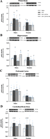
β-FNAs effect on LPS-induced NF-κB-p65 expression in the whole brain and brain regions (hippocampus, prefrontal cortex, and cerebellum/brain stem) of male and female C57BL/6J mice. Mice (n = 5–6/group) were injected (i.p.) with saline (control), LPS (0.83 mg/kg), LPS followed immediately by β-FNA treatment (50 mg/kg; i.p.; LPS + β-FNA), or LPS followed by β-FNA 10 h post-LPS (LPS + β-FNA 10 h). 24 h post-LPS, mice were terminated followed by tissue collection. Levels of NF-κB-p65 of whole brain (A), hippocampus (B), prefrontal cortex (C), and cerebellum/brain stem (D) of tissue homogenates were measured by western blot analysis (representative western blots are shown above each area analyzed). Data are reported as mean ± SEM. Two-way ANOVA indicated a significant main effect of treatment (p < 0.02) and sex (p < 0.0001) on levels of NF-κB-p65 in the whole brain; but no significant effect of interaction (p = 0.74). Two-way ANOVA determined NF-κB-p65 in the hippocampus had a significant main effect of sex (p < 0.01), treatment (p < 0.001), and interaction (p < 0.03). In the prefrontal cortex two-way ANOVA determined levels of NF-κB-p65 had a significant main effect of sex (p < 0.0001), but not treatment (p = 0.66) or interaction (p = 0.68). Two-way ANOVA determined in the cerebellum/brain stem that NF-κB-p65 had a no significant main effect of sex (p = 0.42), treatment (p = 0.08), or interaction (p = 0.62). Pairwise comparisons were assessed using a Fisher’s LSD test; * indicates p < 0.05 vs. saline group; # indicates p < 0.05 vs. LPS group; Δ indicates p < 0.05 vs. males LPS
Fig. 8
Fig. 8
β-FNAs effect on LPS-induced CXCL10, CCL2, IL-6, IL-1β, and TNF-α expression in the plasma of male and female C57BL/6J mice. Mice (n = 11–12/group) were injected (i.p.) with saline (control), LPS (0.83 mg/kg), LPS followed immediately by β-FNA treatment (50 mg/kg; i.p.; LPS + β-FNA), or LPS followed by β-FNA 10 h post-LPS (LPS + β-FNA 10 h). 24 h post-LPS, mice were terminated followed by plasma collection. Levels of CXCL10 (A), CCL2 (B), IL-6 (C), IL-1β (D), and TNF-α (E) of plasma were measured by ELISA. Data are reported as mean ± SEM. Two-way ANOVA indicated a significant main effect of treatment (p < 0.0001), sex (p < 0.0001), and interaction (p < 0.04) on CXCL10 levels in the plasma. Two-way ANOVA determined CCL2 had a significant main effect of sex (p < 0.0001), treatment (p < 0.0001), and interaction (p < 0.0001). Two-way ANOVA determined IL-6 had a significant main effect of sex (p < 0.0001) but not treatment (p = 0.14) and interaction (p = 0.06). Two-way ANOVA determined IL-1β had a significant main effect of sex (p < 0.0001) but not treatment (p = 0.28) or interaction (p = 0.07). Two-way ANOVA determined TNF-α had a significant main effect of sex (p < 0.04) and treatment (p < 0.0001) but not interaction (p = 0.08). Pairwise comparisons were assessed using a Fisher’s LSD test; * indicates p < 0.05 vs. saline group; # indicates p < 0.05 vs. LPS group; Δ indicates p < 0.05 vs. males LPS
Fig. 9
Fig. 9
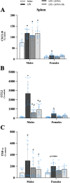
β-FNAs effect on LPS-induced CXCL10, CCL2, and TNF-α expression in the spleen of male and female C57BL/6J mice. Mice (n = 11–12/group) were injected (i.p.) with saline (control), LPS (0.83 mg/kg), LPS followed immediately by β-FNA treatment (50 mg/kg; i.p.; LPS + β-FNA), or LPS followed by β-FNA 10 h post-LPS (LPS + β-FNA 10 h). 24 h post-LPS, mice were terminated followed by tissue collection. Levels of CXCL10 (A), CCL2 (B), and TNF-α (C) of tissue homogenates were measured by ELISA. Data are reported as mean ± SEM. Two-way ANOVA indicated a significant main effect of treatment (p < 0.001), sex (p < 0.0001), and interaction (p < 0.001) on CXCL10 levels in the spleen. Two-way ANOVA determined CCL2 had a significant main effect of sex (p < 0.0001), treatment (p < 0.0001), and interaction (p < 0.0001). Two-way ANOVA determined TNF-α had a significant main effect of treatment (p < 0.003), but not sex (p = 0.25) or interaction (p = 0.54). Pairwise comparisons were assessed using a Fisher’s LSD test; * indicates p < 0.05 vs. saline group; # indicates p < 0.05 vs. LPS group; Δ indicates p < 0.05 vs. males LPS
Fig. 10
Fig. 10
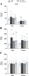
β-FNAs effect on LPS-induced CXCL10, CCL2, and TNF-α expression in the liver of male and female C57BL/6J mice. Mice (n = 11–12/group) were injected (i.p.) with saline (control), LPS (0.83 mg/kg), LPS followed immediately by β-FNA treatment (50 mg/kg; i.p.; LPS + β-FNA), or LPS followed by β-FNA 10 h post-LPS (LPS + β-FNA 10 h). 24 h post-LPS, mice were terminated followed by tissue collection. Levels of CXCL10 (A), CCL2 (B), and TNF-α (C) of tissue homogenates were measured by ELISA. Data are reported as mean ± SEM. Two-way ANOVA indicated a significant main effect of treatment (p < 0.001) and sex (p < 0.0001) on CXCL10 levels in the liver; but no significant effect on interaction (p = 0.61). Two-way ANOVA determined CCL2 had a significant main effect of treatment (p < 0.0001), but not of sex (p = 0.25) or interaction (p = 0.28). Two-way ANOVA determined TNF-α showed no main effects of sex (p = 0.63), treatment (p = 0.41), or interaction (p = 0.99). Pairwise comparisons were assessed using a Fisher’s LSD test; * indicates p < 0.05 vs. saline group; # indicates p < 0.05 vs. LPS group; Δ indicates p < 0.05 vs. males LPS
Fig. 11
Fig. 11
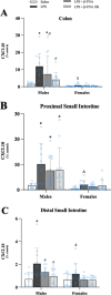
β-FNAs effect on LPS-induced CXCL10 expression in the colon, proximal small intestine (PSI), and distal small intestine (DSI) of male and female C57BL/6J mice. Mice (n = 11–12/group) were injected (i.p.) with saline (control), LPS (0.83 mg/kg), LPS followed immediately by β-FNA treatment (50 mg/kg; i.p.; LPS + β-FNA), or LPS followed by β-FNA 10 h post-LPS (LPS + β-FNA 10 h). 24 h post-LPS, mice were terminated followed by tissue collection. Levels of CXCL10 of the colon (A), PSI (B), and DSI (C) of tissue homogenates were measured by ELISA. Data are reported as mean ± SEM. Two-way ANOVA indicated a significant main effect of treatment (p < 0.01), sex (p < 0.0001), and interaction (p < 0.01) on CXCL10 levels in the colon. In the proximal small intestine two-way ANOVA determined CXCL10 showed main effects of sex (p < 0.0001) and treatment (p < 0.02), but not interaction (p = 0.14). In the distal small intestine two-way ANOVA determined CXCL10 showed main effects of sex (p < 0.005) and treatment (p < 0.001) but not an interaction (p = 0.28). Pairwise comparisons were assessed using a Fisher’s LSD test; * indicates p < 0.05 vs. saline group; # indicates p < 0.05 vs. LPS group; Δ indicates p < 0.05 vs. males LPS
Fig. 12
Fig. 12
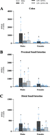
β-FNAs effect on LPS-induced CCL2 expression in the colon, proximal small intestine (PSI), and distal small intestine (DSI) of male and female C57BL/6J mice. Mice (n = 11–12/group) were injected (i.p.) with saline (control), LPS (0.83 mg/kg), LPS followed immediately by β-FNA treatment (50 mg/kg; i.p.; LPS + β-FNA), or LPS followed by β-FNA 10 h post-LPS (LPS + β-FNA 10 h). 24 h post-LPS, mice were terminated followed by tissue collection. Levels of CCL2 of the colon (A), PSI (B), and DSI (C) of tissue homogenates were measured by ELISA. Data are reported as mean ± SEM. Two-way ANOVA indicated a significant main effect of treatment (p < 0.0001), sex (p < 0.001), and an interaction (p < 0.002) on CCL2 levels in the colon. In the proximal small intestine two-way ANOVA determined CCL2 had a significant main effect of sex (p < 0.001) and treatment (p < 0.002) but not an interaction (p = 0.13). In the distal small intestine two-way ANOVA determined CCL2 had a significant main effect of treatment (p < 0.0001), but not sex (p = 0.37) or interaction (p = 0.42). Pairwise comparisons were assessed using a Fisher’s LSD test; * indicates p < 0.05 vs. saline group; # indicates p < 0.05 vs. LPS group; Δ indicates p < 0.05 vs. males LPS
Fig. 13
Fig. 13
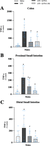
β-FNAs effect on LPS-induced TNF-α expression in the colon, proximal small intestine (PSI), and distal small intestine (DSI) of male C57BL/6J mice. Mice (n = 11–12/group) were injected (i.p.) with saline (control), LPS (0.83 mg/kg), LPS followed immediately by β-FNA treatment (50 mg/kg; i.p.; LPS + β-FNA), or LPS followed by β-FNA 10 h post-LPS (LPS + β-FNA 10 h). 24 h post-LPS, mice were terminated followed by tissue collection. Levels of TNF-α of the colon (A), PSI (B), and DSI (C) of tissue homogenates were measured by ELISA. Data are reported as mean ± SEM. One-way ANOVA indicated a significant main effect of treatment (p < 0.0001) on TNF-α levels in the colon, proximal small intestine (p < 0.0001), and distal small intestine (p < 0.01). Pairwise comparisons were assessed using a Fisher’s LSD test; * indicates p < 0.05 vs. saline group; # indicates p < 0.05 vs. LPS group; Δ indicates p < 0.05 vs. males LPS
Fig. 14
Fig. 14
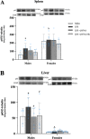
β-FNAs effect on LPS-induced NFκB-p65 expression in the spleen and liver of male and female C57BL/6J mice. Mice (n = 5–6/group) were injected (i.p.) with saline (control), LPS (0.83 mg/kg), LPS followed immediately by β-FNA treatment (50 mg/kg; i.p.; LPS + β-FNA), or LPS followed by β-FNA 10 h post-LPS (LPS + β-FNA 10 h). 24 h post-LPS, mice were terminated followed by tissue collection. Levels of NFκB-p65 of spleen A and liver B of tissue homogenates were measured by western blot analysis (representative western blots are shown above each area analyzed). Data are reported as mean ± SEM. Two-way ANOVA indicated a significant main effect of sex (p < 0.0001) on levels of NFκB-p65 in the spleen; but no significant effect of treatment (p = 0.16), nor a significant interaction (p = 0.62). In the liver two-way ANOVA determined levels of NFκB-p65 had a significant main effect of sex (p < 0.0001) but no significant effect of treatment (p = 0.28), nor a significant interaction (p = 0.26). Pairwise comparisons were assessed using a Fisher’s LSD test; * indicates p < 0.05 vs. saline group; # indicates p < 0.05 vs. LPS group; Δ indicates p < 0.05 vs. males LPS

Similar articles

Cited by

References

    1. Bai YM, et al. Comparison of pro-inflammatory cytokines among patients with bipolar disorder and unipolar depression and normal controls. Bipolar Disord. 2015;17(3):269–277. doi: 10.1111/bdi.12259. - DOI - PubMed
    1. Blank T, et al. Brain endothelial- and epithelial-specific interferon receptor chain 1 drives virus-induced sickness behavior and cognitive impairment. Immunity. 2016;44(4):901–912. doi: 10.1016/j.immuni.2016.04.005. - DOI - PubMed
    1. Burton MD, Sparkman NL, Johnson RW. Inhibition of interleukin-6 trans-signaling in the brain facilitates recovery from lipopolysaccharide-induced sickness behavior. J Neuroinflammation. 2011;8:54. doi: 10.1186/1742-2094-8-54. - DOI - PMC - PubMed
    1. Da Ré C, et al. Neuroinflammation induced by lipopolysaccharide leads to memory impairment and alterations in hippocampal leptin signaling. Behav Brain Res. 2020;379:112360. doi: 10.1016/j.bbr.2019.112360. - DOI - PubMed
    1. Dantzer R. Cytokine-induced sickness behavior: mechanisms and implications. Ann N Y Acad Sci. 2001;933:222–234. doi: 10.1111/j.1749-6632.2001.tb05827.x. - DOI - PubMed