

The utility of texture analysis based on quantitative synthetic magnetic resonance imaging in nasopharyngeal carcinoma: a preliminary study
- PMID: 36698156
- PMCID: PMC9875491
- DOI: 10.1186/s12880-023-00968-w
The utility of texture analysis based on quantitative synthetic magnetic resonance imaging in nasopharyngeal carcinoma: a preliminary study
Abstract
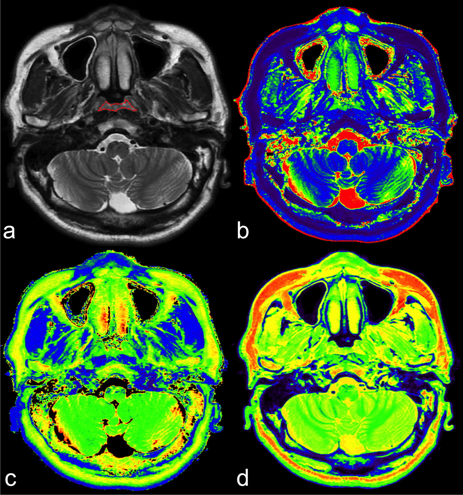
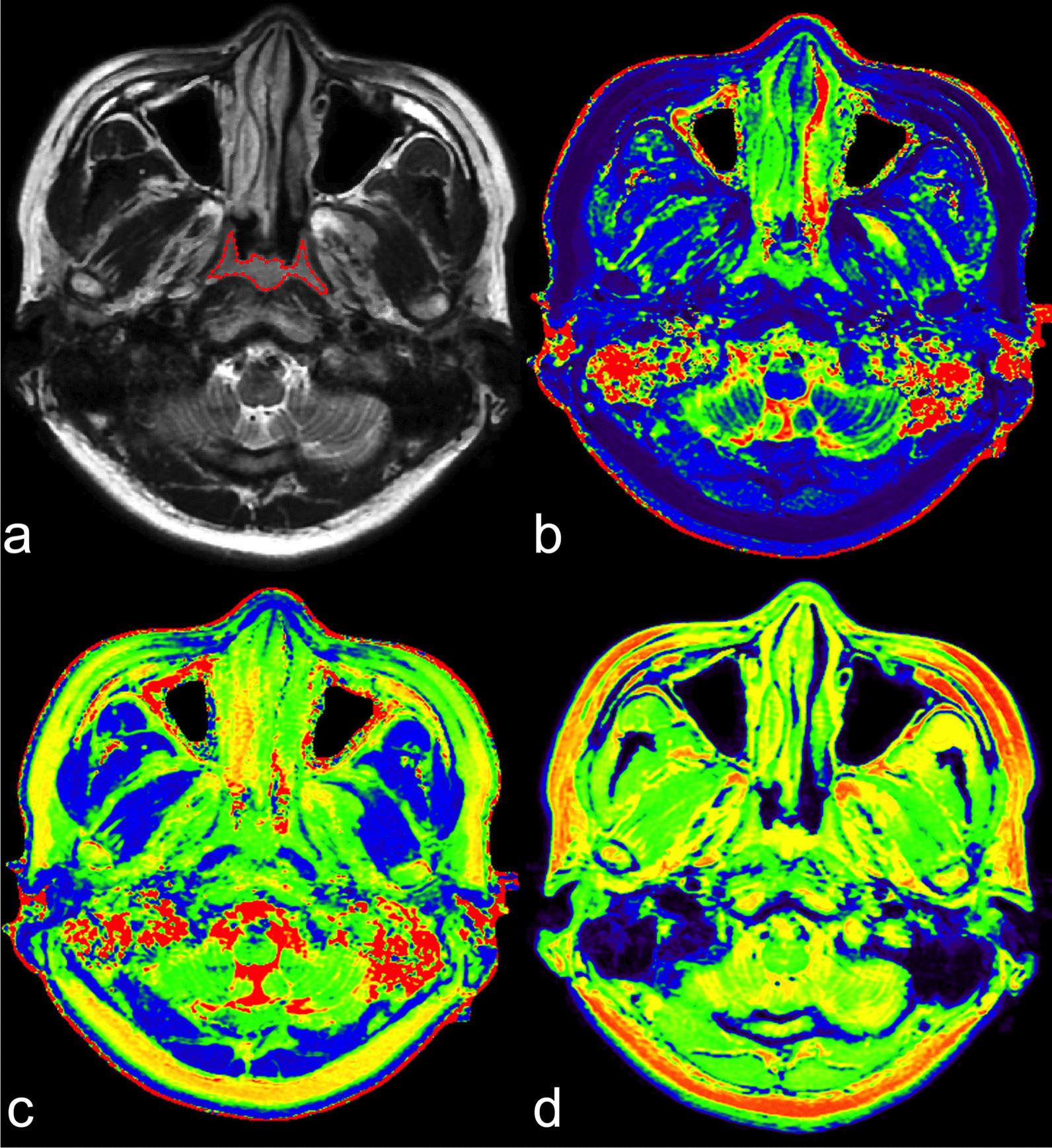
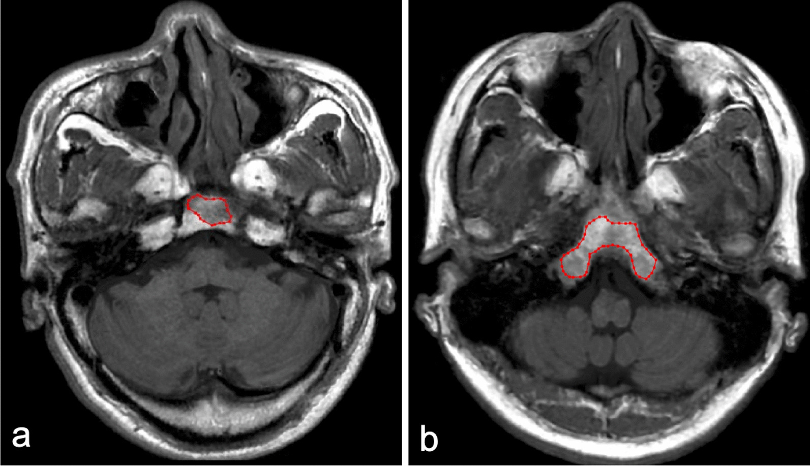
Background: Magnetic resonance imaging (MRI) is commonly used for the diagnosis of nasopharyngeal carcinoma (NPC) and occipital clivus (OC) invasion, but a proportion of lesions may be missed using non-enhanced MRI. The purpose of this study is to investigate the diagnostic performance of synthetic magnetic resonance imaging (SyMRI) in differentiating NPC from nasopharyngeal hyperplasia (NPH), as well as evaluating OC invasion.
Methods: Fifty-nine patients with NPC and 48 volunteers who underwent SyMRI examination were prospectively enrolled. Eighteen first-order features were extracted from VOIs (primary tumours, benign mucosa, and OC). Statistical comparisons were conducted between groups using the independent-samples t-test and the Mann-Whitney U test to select significant parameters. Multiple diagnostic models were then constructed using multivariate logistic analysis. The diagnostic performance of the models was calculated by receiver operating characteristics (ROC) curve analysis and compared using the DeLong test. Bootstrap and 5-folds cross-validation were applied to avoid overfitting.
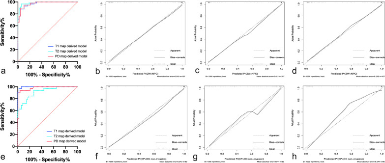
Results: The T1, T2 and PD map-derived models had excellent diagnostic performance in the discrimination between NPC and NPH in volunteers, with area under the curves (AUCs) of 0.975, 0.972 and 0.986, respectively. Besides, SyMRI models also showed excellent performance in distinguishing OC invasion from non-invasion (AUC: 0.913-0.997). Notably, the T1 map-derived model showed the highest diagnostic performance with an AUC, sensitivity, specificity, and accuracy of 0.997, 96.9%, 97.9% and 97.5%, respectively. By using 5-folds cross-validation, the bias-corrected AUCs were 0.965-0.984 in discriminating NPC from NPH and 0.889-0.975 in discriminating OC invasion from OC non-invasion.
Conclusions: SyMRI combined with first-order parameters showed excellent performance in differentiating NPC from NPH, as well as discriminating OC invasion from non-invasion.
Keywords: Differential diagnosis; Magnetic resonance imaging; Nasopharyngeal carcinoma.
© 2023. The Author(s).
Conflict of interest statement
The authors declare that they have no competing interests. And the author Lizhi Xie was the scientist of MR Research China of GE Healthcare who mainly contribute to manuscript editing and did not participate in study design, data collection, analysis or interpretation of this study.
Figures
Similar articles
-
Synthetic MRI and diffusion-weighted imaging for differentiating nasopharyngeal lymphoma from nasopharyngeal carcinoma: combination with morphological features.Br J Radiol. 2024 Jun 18;97(1159):1278-1285. doi: 10.1093/bjr/tqae095. Br J Radiol. 2024. PMID: 38733577 Free PMC article.
-
Investigation of the feasibility of synthetic MRI in the differential diagnosis of non-keratinising nasopharyngeal carcinoma and benign hyperplasia using different contoured methods for delineation of the region of interest.Clin Radiol. 2021 Mar;76(3):238.e9-238.e15. doi: 10.1016/j.crad.2020.10.010. Epub 2020 Nov 16. Clin Radiol. 2021. PMID: 33213835
-
Synthetic MRI quantitative parameters in discriminating stage T1 nasopharyngeal carcinoma and benign hyperplasia: Combination with morphological features.Eur J Radiol. 2024 Jan;170:111264. doi: 10.1016/j.ejrad.2023.111264. Epub 2023 Dec 15. Eur J Radiol. 2024. PMID: 38103492
-
Positron emission tomography/computed tomography outperforms MRI in the diagnosis of local recurrence and residue of nasopharyngeal carcinoma: An update evidence from 44 studies.Cancer Med. 2019 Jan;8(1):67-79. doi: 10.1002/cam4.1882. Epub 2018 Dec 21. Cancer Med. 2019. PMID: 30578604 Free PMC article.
-
A systematic review of the predictive value of radiomics for nasopharyngeal carcinoma prognosis.Medicine (Baltimore). 2024 Aug 30;103(35):e39302. doi: 10.1097/MD.0000000000039302. Medicine (Baltimore). 2024. PMID: 39213210 Free PMC article.
Cited by
-
Histogram analysis of quantitative parameters from synthetic MRI: correlations with prognostic factors in nasopharyngeal carcinoma.Eur Radiol. 2023 Aug;33(8):5344-5354. doi: 10.1007/s00330-023-09553-9. Epub 2023 Apr 10. Eur Radiol. 2023. PMID: 37036478
-
T1 and T2 mapping for identifying malignant lymph nodes in head and neck squamous cell carcinoma.Cancer Imaging. 2023 Dec 17;23(1):125. doi: 10.1186/s40644-023-00648-6. Cancer Imaging. 2023. PMID: 38105217 Free PMC article.
-
Technical aspects and clinical applications of synthetic MRI: a scoping review.Diagnosis (Berl). 2025 Feb 7;12(2):163-174. doi: 10.1515/dx-2024-0168. eCollection 2025 May 1. Diagnosis (Berl). 2025. PMID: 39913860
-
Synthetic MRI and diffusion-weighted imaging for differentiating nasopharyngeal lymphoma from nasopharyngeal carcinoma: combination with morphological features.Br J Radiol. 2024 Jun 18;97(1159):1278-1285. doi: 10.1093/bjr/tqae095. Br J Radiol. 2024. PMID: 38733577 Free PMC article.
-
Multiparametric approach with synthetic MR imaging for diagnosing salivary gland lesions.Jpn J Radiol. 2024 Sep;42(9):983-992. doi: 10.1007/s11604-024-01578-4. Epub 2024 May 11. Jpn J Radiol. 2024. PMID: 38733471 Free PMC article.
References
-
- Pan JJ, Ng WT, Zong JF, Chan LL, O'Sullivan B, Lin SJ, Sze HC, Chen YB, Choi HC, Guo QJ, et al. Proposal for the 8th edition of the AJCC/UICC staging system for nasopharyngeal cancer in the era of intensity-modulated radiotherapy. Cancer. 2016;122(4):546–558. doi: 10.1002/cncr.29795. - DOI - PMC - PubMed
-
- King AD, Wong LYS, Law BKH, Bhatia KS, Woo JKS, Ai QY, Tan TY, Goh J, Chuah KL, Mo FKF, et al. MR imaging criteria for the detection of nasopharyngeal carcinoma: discrimination of early-stage primary tumors from benign hyperplasia. Am J Neuroradiol. 2018;39(3):515–523. doi: 10.3174/ajnr.A5493. - DOI - PMC - PubMed
-
- Yoo MG, Kim J, Bae S, Ahn SS, Ahn SJ, Koh YW. Detection of clinically occult primary tumours in patients with cervical metastases of unknown primary tumours: comparison of three-dimensional THRIVE MRI, two-dimensional spin-echo MRI, and contrast-enhanced CT. Clin Radiol. 2018;73(4):410.e419–410.e415. doi: 10.1016/j.crad.2017.10.020. - DOI - PubMed
MeSH terms
LinkOut - more resources
Full Text Sources
