. 2023 Jan 25;16(1):30.
doi: 10.1186/s13071-022-05618-8.

Studies on the mechanism of Toxoplasma gondii Chinese 1 genotype Wh6 strain causing mice abnormal cognitive behavior

Affiliations

Studies on the mechanism of Toxoplasma gondii Chinese 1 genotype Wh6 strain causing mice abnormal cognitive behavior

Qing Tao et al. Parasit Vectors. .

Abstract

Background: Alzheimer's disease presents an abnormal cognitive behavior. TgCtwh6 is one of the predominant T. gondii strains prevalent in China. Although T. gondii type II strain infection can cause host cognitive behavioral abnormalities, we do not know whether TgCtwh6 could also cause host cognitive behavioral changes. So, in this study, we will focus on the effect of TgCtwh6 on mouse cognitive behavior and try in vivo and in vitro to explore the underlying mechanism by which TgCtwh6 give rise to mice cognitive behavior changes at the cellular and molecular level.

Methods: C57BL/6 mice were infected orally with TgCtwh6 cysts. From day 90 post-infection on, all mice were conducted through the open field test and then Morris water maze test to evaluate cognitive behavior. The morphology and number of cells in hippocampus were examined with hematoxylin-eosin (H&E) and Nissl staining; moreover, Aβ protein in hippocampus was determined with immunohistochemistry and thioflavin S plaque staining. Synaptotagmin 1, apoptosis-related proteins, BACE1 and APP proteins and genes from hippocampus were assessed by western blotting or qRT-PCR. Hippocampal neuronal cell line or mouse microglial cell line was challenged with TgCtwh6 tachyzoites and then separately cultured in a well or co-cultured in a transwell device. The target proteins and genes were analyzed by immunofluorescence staining, western blotting and qRT-PCR. In addition, mouse microglial cell line polarization state and hippocampal neuronal cell line apoptosis were estimated using flow cytometry assay.

Results: The OFT and MWMT indicated that infected mice had cognitive behavioral impairments. The hippocampal tissue assay showed abnormal neuron morphology and a decreased number in infected mice. Moreover, pro-apoptotic proteins, as well as BACE1, APP and Aβ proteins, increased in the infected mouse hippocampus. The experiments in vitro showed that pro-apoptotic proteins and p-NF-κBp65, NF-κBp65, BACE1, APP and Aβ proteins or genes were significantly increased in the infected HT22. In addition, CD80, pro-inflammatory factors, notch, hes1 proteins and genes were enhanced in the infected BV2. Interestingly, not only the APP and pro-apoptotic proteins in HT22, but also the apoptosis rate of HT22 increased after the infected BV2 were co-cultured with the HT22 in a transwell device.

Conclusions: Neuron apoptosis, Aβ deposition and neuroinflammatory response involved with microglia polarization are the molecular and cellular mechanisms by which TgCtwh6 causes mouse cognitive behavioral abnormalities.

Keywords: Apoptosis; Aβ; Cognitive behavior; Hippocampal neuron; Inflammatory response; Toxoplasma gondii Chinese 1 genotype Wh6 strain.


Conflict of interest statement

The authors declare that they have no competing interests.

Figures
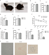
Fig. 1
Fig. 1
Effects of TgCtwh6 infection on mouse cognitive behavior. Mice were randomly divided into the infected and control groups. Each mouse in the infected group was infected orally with 30 TgCtwh6 cysts. TgCtwh6 infection caused mouse appearance and posture changes (A). The motor and exploration abilities were evaluated with OFT on the 90th day post-TgCtwh6 cyst infection (BE). The spatial learning ability was measured in MWMT for 5 consecutive days on the 6th day of the MWMT; spatial memory ability was assayed (FJ). Mice were killed on the 97th day post-infection, and brain cysts were assessed by tissue squash method with light microscopic examination at a magnification of 40 × 100 (Scale bars, 20 μm) (K). Data are represented as mean ± SEM and were analyzed by two-tailed Student’s t-test for comparing the difference between two groups (n = 24 each group). *P < 0. 05, **P < 0.01
Fig. 2
Fig. 2
TgCtwh6 infection leads to the apoptosis of hippocampus cells in mouse. The mouse brain tissues were cut into sections for staining. The hippocampus tissue section was first stained with hematoxylin and eosin, and then the neurons were observed under a light microscope at a magnification of 20 × 100 and 40 × 100 (scale bars, 50 μm and 20 μm, respectively) (A). Besides, the mouse hippocampus tissue section was stained with Nissl dye, and then the neurons were observed under a light microscope at a magnification of 20 × 100 and 40 × 100 (scale bars, 50 μm and 20 μm, respectively). The number of positive cells in each field of view were evaluated with semiquantitative analysis (B, C). The mouse right brain hippocampus tissues were lysed and proteins were extracted; then, the apoptosis-related proteins were assessed by western blotting and the protein expression was determined semiquantitatively (DG). Synaptotagmin 1 protein from hippocampus tissue was detected by western blotting and analyzed using semiquantitative method (H, I). Data are represented as mean ± SEM and were analyzed by two-tailed Student’s t-test for comparing the difference between the two groups (n = 3 each group). *P < 0. 05, **P < 0.01, ***P < 0.001
Fig. 3
Fig. 3
TgCtwh6 infection increases APP, BACE1 and Aβ production in mouse hippocampus tissue. First, the hippocampus tissue section was analyzed by immunhistochemistry method. The stained yellow positive areas were observed under a light microscope at a magnification of 20 × 100 and 40 × 100 (scale bars, 50 μm and 20 μm, respectively) (A). The Aβ protein expression in the mouse hippocampal zone was evaluated with semiquantitative method (B). Then, the hippocampus tissue section was stained with thioflavin S solution followed by counterstaining nuclei with DAPI. The stained bright green plaques were observed under a fluorescence microscope at a magnification of 20 × 100 and 40 × 100 (scale bars, 50 μm and 20 μm, respectively) (C). The Aβ protein expression in the mouse hippocampal zone was estimated using semiquantitative method (D). Third, the mouse right brain hippocampus tissues were lysed and proteins were extracted; APP and BACE1 protein were detected by western blotting and then analyzed semiquantitatively (EG). Finally, the RNA of the mouse right brain hippocampus tissue was extracted and then reversely transcribed to cDNA. APP and BACE1 gene expressions were assessed by qRT-PCR and then analyzed semiquantitatively (H, I). Data were represented as mean ± SEM and were analyzed by two-tailed Student’s t-test for comparing the difference between the two groups (n = 3 each group). *P < 0. 05, **P < 0.01, ***P < 0.001
Fig. 4
Fig. 4
TgCtwh6 tachyzoites affect apoptosis-related and synaptotagmin 1 protein expression in HT22. After TgCtwh6 tachyzoites infected HT22 cells for 24 h, the HT22 cells were lysed and proteins extracted. The expression of apoptosis-related proteins of HT22, such as Bax, Bcl-XL and caspase 3, was measured by western blotting and then analyzed by semiquantitative method (AD). Also, synaptotagmin 1 protein in the infected HT22 was detected by western blotting (E, F). Data were represented as mean ± SEM and were analyzed by two-tailed Student’s t-test for comparing the difference between the two groups (n = 3 each group). *P < 0. 05, **P < 0.01, ***P < 0.001
Fig. 5
Fig. 5
TgCtwh6 tachizoites polarize BV2 to M1 probably via Notch signaling pathway. After BV2 cells were infected by TgCtwh6 tachyzoites for 24 h, BV2 cells were collected and assessed quantitatively for polarization status using FCM (AC). Additionally, the infected BV2 cells were lysed and proteins extracted. The expression of Notch and Hes1 proteins was detected by western blotting and then analyzed semiquantitatively (DF). Data were represented as mean ± SEM and analyzed by two-tailed Student’s t-test for comparing the difference between the two groups (n = 3 each group). **P < 0.01, ***P < 0.001
Fig. 6
Fig. 6
TgCtwh6 tachyzoites indirectly induce HT22 apoptosis by polarizing BV2 into M1. BV2 cells were infected by TgCtwh6 tachyzoites for 24 h; then, RNA of the BV2 was extracted and reverse transcribed to cDNA. The gene expressions of IL-6, TNF-α, iNOS and TGF-β1 were assessed by qRT-PCR and then analyzed semiquantitatively (AD). After BV2 cells had been infected with TgCtwh6 tachyzoites for 24 h, the BV2 cells were co-cultured with HT22 for another 24 h in a transwell device. Then, HT22 cells were collected from the lower layer and apoptosis status was assessed using FCM (EG). Moreover, the HT22 cells were lysed and proteins were extracted. The expression of apoptosis-related proteins was detected by western blotting and then analyzed semiquantitatively (HK). Data were represented as mean ± SEM and were analyzed by two-tailed Student’s t-test for comparing the difference between the two groups (n = 3 each group). *P < 0. 05, **P < 0.01
Fig. 7
Fig. 7
TgCtwh6 tachyzoites upregulate APP, BACE1 and Aβ expression in HT22. After TgCtwh6 tachyzoites had been infected HT22 for 24 h, the HT22 were lysed and proteins were extracted. The expression of BACE1 and APP proteins was measured by western blotting and then analyzed with semiquantitative method (AC). Moreover, RNA in the infected HT22 was extracted and reversely transcribed to cDNA. The expressions of BACE1 and APP genes was assessed by qRT-PCR and then semiquantitatively evaluated (D, E). Furthermore, after the HT22 cells had been fixed and the cytomembranes penetrated by Triton, the BACE1 and Aβ proteins were visualized in situ by immunofluorescence staining and observed under a fluorescence microscope at a magnification of 40 × 100 (scale bars, 50 μm). The percentage of positively stained cells was calculated using morphometric software (FG). Data were represented as mean ± SEM and were analyzed by two-tailed Student’s t-test for comparing the difference between the two groups (n = 3 each group). *P < 0. 05, **P < 0.01, ***P < 0.001
Fig. 8
Fig. 8
TgCtwh6 tachyzoites indirectly induce APP protein production in HT22 by polarized BV2. BV2 cells infected with TgCtwh6 tachyzoites for 24 h were co-cultured with HT22 for another 24 h in a transwell system. Subsequently, the HT22 cells were collected from the lower layer of the co-culture system and then lysed. The proteins were extracted from the HT22. The expression of APP protein was detected by western blotting and analyzed by semiquantitative method (A, B). Data were represented as mean ± SEM and were analyzed by two-tailed Student’s t-test for comparing the difference between the two groups (n = 3 each group). *P < 0.05
Fig. 9
Fig. 9
TgCtwh6 tachyzoites induce Aβ production by activating NF-κB signaling pathway in HT22. HT22 cells pre-treated with or without PDTC for 12 h were challenged with or without TgCtwh6 tachyzoites for 24 h. Then, after the HT22 cells had been collected and lysed, the proteins in HT22 were extracted. The expressions of NF-κBp65, p-NF-κBp65, APP and BACE1 were detected by western blotting and then semiauantitative analyzed (AE). Additionally, after the HT22 cells had been fixed and the cytomembranes penetrated by Triton, the NF-κBp65, p-NF-κBp65, BACE1 and Aβ proteins were visualized in situ by immunofluorescence staining and were observed under a fluorescence microscope at a magnification of 40 × 100. The percentage of positively stained cells was calculated using morphometric software (FM). Data were represented as mean ± SEM and were analyzed by one-way ANOVA followed by Bonferroni’s post hoc test for multiple comparisons (n = 3 each group). *P < 0. 05, **P < 0.01, ***P < 0.001
Fig. 10
Fig. 10
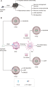
Mechanism with which TgCtwh6 infection induces mouse cognitive behavior disorder at the cellular and molecular level in our experiments. First, in vivo, each C57BL/6 mouse was infected orally with TgCtwh6 cysts. On day 90 post-infection, the infected mice manifested cognitive behavioral abnormalities. The infected mouse hippocampal tissue assay showed that neurons were disorganized in the arrangement, decreased in the number and abnormally stained in nuclei. Moreover, apoptotic cells and Aβ protein increased in the infected mouse hippocampus (A). TgCtwh6 tachyzoites led to HT22 apoptosis following HT22 infection for 24 h; meanwhile, TgCtwh6 tachyzoites promoted BACE1, APP and Aβ production by activating NF-κB signaling pathway in HT22. Additionally, after BV2 cells had been infected by TgCtwh6 tachyzoites for 24 h, they could drive polarization to M1 through Notch signaling pathway with production of pro-inflammatory factors, which provoked co-cultured HT22 apoptosis and APP expression (B)

Similar articles

Cited by

References

    1. Kinney JW, Bemiller SM, Murtishaw AS, Leisgang AM, Salazar AM, Lamb BT. Inflammation as a central mechanism in Alzheimer's disease. Alzheimers Dement (N Y) 2018;4:575–590. doi: 10.1016/j.trci.2018.06.014. - DOI - PMC - PubMed
    1. De Chiara G, Marcocci ME, Sgarbanti R, Civitelli L, Ripoli C, Piacentini R, et al. Infectious agents and neurodegeneration. Mol Neurobiol. 2012;46:614–638. doi: 10.1007/s12035-012-8320-7. - DOI - PMC - PubMed
    1. Postan M, Rottenberg ME. Editorial: Neurotropism of Parasites and the Immune Responses. Front Immunol. 2021;12:775666. doi: 10.3389/fimmu.2021.775666. - DOI - PMC - PubMed
    1. Milne G, Webster JP, Walker M. Toxoplasma gondii: An Underestimated Threat? Trends Parasitol. 2020;36:959–969. doi: 10.1016/j.pt.2020.08.005. - DOI - PubMed
    1. Molan A, Nosaka K, Hunter M, Wang W. Global status of Toxoplasma gondii infection: systematic review and prevalence snapshots. Trop Biomed. 2019;36:898–925. - PubMed

Substances

LinkOut - more resources