. 2023 Jan 16:2023:5915988.
doi: 10.1155/2023/5915988. eCollection 2023.

Matricellular Protein SMOC2 Potentiates BMP9-Induced Osteogenic Differentiation in Mesenchymal Stem Cells through the Enhancement of FAK/PI3K/AKT Signaling

Affiliations

Matricellular Protein SMOC2 Potentiates BMP9-Induced Osteogenic Differentiation in Mesenchymal Stem Cells through the Enhancement of FAK/PI3K/AKT Signaling

Wen-Ge He et al. Stem Cells Int. .

Abstract

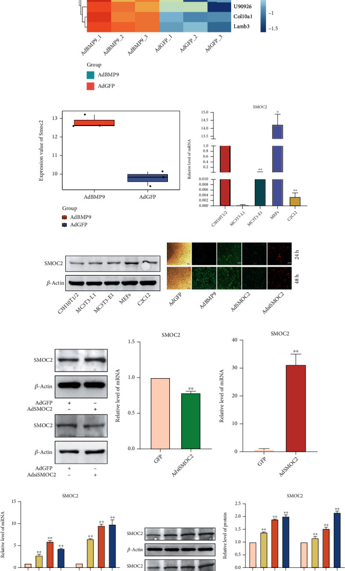
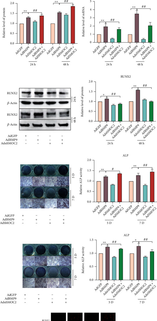
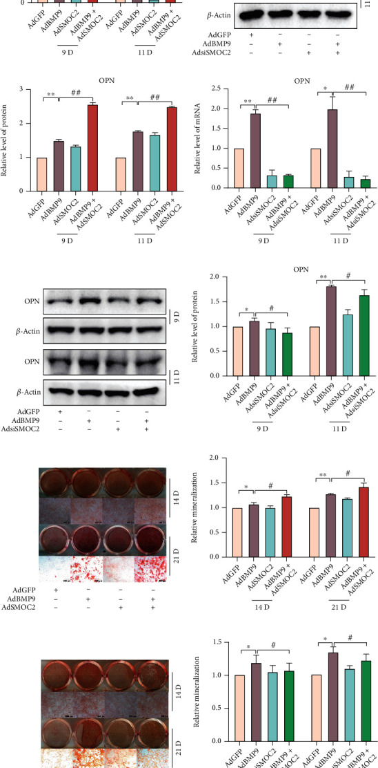
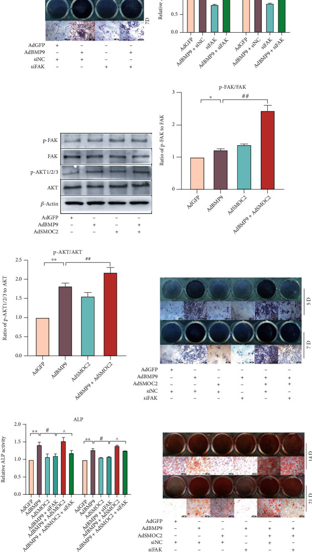
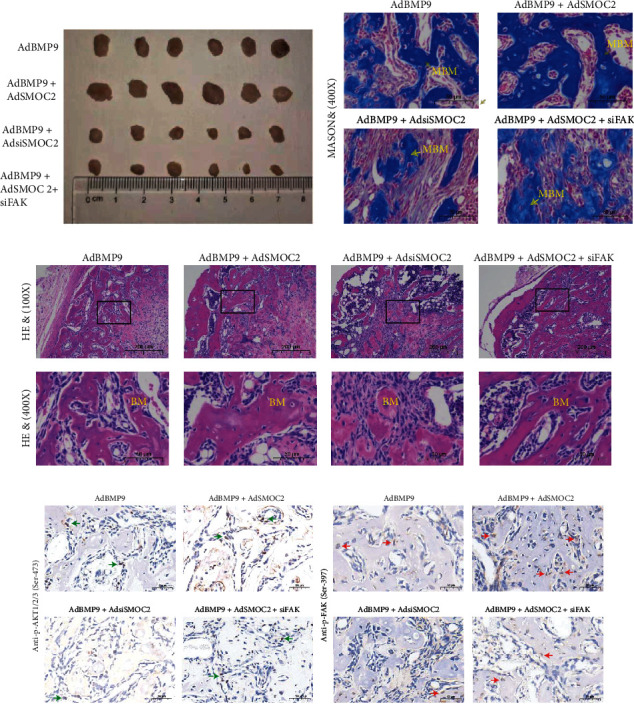
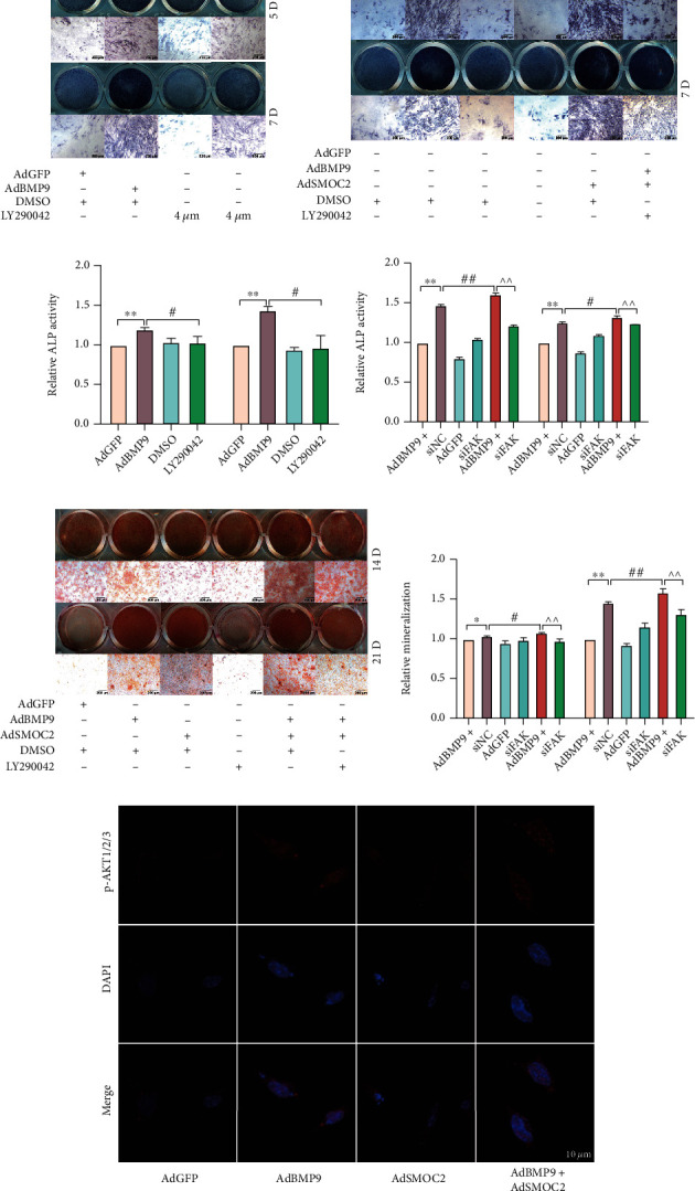
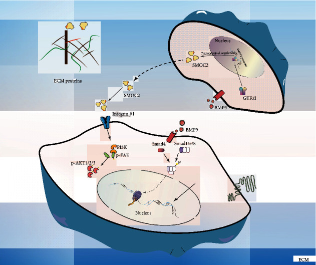
Mesenchymal stem cells (MSCs) can self-renew and differentiate into multiple lineages, making MSC transplantation a promising option for bone regeneration. Both matricellular proteins and growth factors play an important role in regulating stem cell fate. In this study, we investigated the effects of matricellular protein SMOC2 (secreted modular calcium-binding protein 2) on bone morphogenetic protein 9 (BMP9) in mouse embryonic fibroblasts (MEFs) and revealed a possible molecular mechanism underlying this process. We found that SMOC2 was detectable in MEFs and that exogenous SMOC2 expression potentiated BMP9-induced osteogenic markers, matrix mineralization, and ectopic bone formation, whereas SMOC2 knockdown inhibited these effects. BMP9 increased the levels of p-FAK and p-AKT, which were either enhanced or reduced by SMOC2 and FAK silencing, respectively. BMP9-induced osteogenic markers were increased by SMOC2, and this increase was partially abolished by silencing FAK or LY290042. Furthermore, we found that general transcription factor 2I (GTF2I) was enriched at the promoter region of SMOC2 and that integrin β1 interacted with SMOC2 in BMP9-treated MEFs. Our findings demonstrate that SMOC2 can promote BMP9-induced osteogenic differentiation by enhancing the FAK/PI3K/AKT pathway, which may be triggered by facilitating the interaction between SMOC2 and integrin β1.


Conflict of interest statement

The authors declare that they have no competing interests.

Figures

Figure 1
Figure 1
BMP9 upregulates the expression of SMOC2 in MSCs. (a) Heatmap of the top 10 upregulated and downregulated mRNAs between the control (AdGFP) and experimental groups (AdBMP9). (b) RNA-seq analysis showed the expression value of smoc2 between AdGFP and AdBMP9. (c) Real-time PCR assay results showed endogenous mRNA expression of smoc2 in mesenchymal progenitor cells. (d) Western blot assay results showed endogenous protein expression in mesenchymal progenitor cells; β-actin was used as an internal reference. (e) Efficient transduction of MEFs by the recombinant adenoviruses (AdGFP, AdBMP9, AdSMOC2, and AdsiSMOC2) at 24 h. The expression of the marker genes monomeric RFP and GFP was detected at 24 h after infection in both bright and fluorescence fields. (f–h) Western blot and real-time PCR assays showed the expression of SMOC2 in MEFs at 24 h after infection by recombinant adenoviruses overexpressing SMOC2 and knocking down SMOC2. (i) Real-time PCR assay results showed the effect of BMP9 on mRNA expression of smoc2 in MEFs (p < 0.05, ∗∗p < 0.01 vs. control). (j) Western blot assays showed the effect of BMP9 on SMOC2 protein expression in MEFs. (k) The quantification results of the Western blot assay showed the effect of BMP9 on the protein expression of SMOC2 in MEFs (p < 0.05, ∗∗p < 0.01 vs. control).
Figure 2
Figure 2
The effect of SMOC2 on BMP9-induced early osteogenic marker levels. (a) Real-time PCR assay results showed the effect of SMOC2 on the mRNA expression of RUNX2 induced by BMP9. (b) Western blot results showed the effect of SMOC2 on the protein expression of RUNX2 induced by BMP9 at 24 h and 48 h. (c) The quantification results of the Western blot results showed the effect of SMOC2 on the protein expression of RUNX2 induced by BMP9. (d) Real-time PCR assay results showed the effect of SMOC2 knockdown on the mRNA expression of RUNX2 induced by BMP9. (e–f) Western blot assay and quantification results showed the effect of SMOC2 knockdown on the protein expression of RUNX2 induced by BMP9 at 24 h and 48 h. (g–h) ALP assay and quantification results showed the effect of overexpression or knockdown of SMOC2 on the activity of ALP induced by BMP9 in MEFs. (k) Immunofluorescence staining assay results showed the effect of SMOC2 on the expression of RUNX2 induced by BMP9. Compared with the control group, p < 0.05; ∗∗p < 0.01; compared with the AdBMP9 group, #p < 0.05, ##p < 0.01.
Figure 3
Figure 3
Exogenous SMOC2 expression enhances BMP-9-induced late osteogenic marker expression and matrix mineralization. (a) Real-time PCR assay results showed the effect of SMOC2 on the mRNA expression of OPN promoted by BMP9 in MEFs. (b) Western blot assay results showed the effect of SMOC2 on the protein expression of OPN promoted by BMP9 in MEFs. (c) Quantification of the western blot results showed the effect of SMOC2 on OPN protein expression promoted by BMP9 in MEFs. (d) Real-time PCR assay results showed the effect of SMOC2 knockdown on the mRNA expression of OPN promoted by BMP9 in MEFs. (e) Western blotting assay results showed the effect of SMOC2 knockdown on OPN protein expression induced by BMP9 in MEFs. (f) Quantification of the western blot results showed the effect of SMOC2 on OPN protein expression promoted by BMP9 in MEFs. (g) Alizarin Red S staining results showed the effect of SMOC2 on BMP9-induced mineralization in MEFs. (h) Quantification results of Alizarin Red S staining showed the effect of SMOC2 on BMP9-induced mineralization in MEFs. (i) Alizarin Red S staining results showed the effect of SMOC2 knockdown on BMP9-induced mineralization in MEFs. (j) Quantification results of Alizarin Red S staining showed the effect of SMOC2 knockdown on BMP9-induced mineralization in MEFs. Compared with the control group, p < 0.05; ∗p < 0.01; compared with the AdBMP9 group, #p < 0.05, ##p < 0.01.
Figure 4
Figure 4
The activation of FAK signaling in the enhancement effect of SMOC2 on BMP9-induced osteogenic differentiation in MEFs. (a) Western blot assay results showed that the levels of p-FAK and FAK were affected by BMP9 in MEFs at 30 h. (b) Quantification of the Western blot results showed that the levels of p-FAK and FAK were affected by BMP9. (c) ALP assay results showed the effect of FAK knockdown on the activity of ALP induced by BMP9 in MEFs. (d) Quantification results of the ALP assay showed the effect of FAK knockdown on the activity of ALP induced by BMP9. (e) Western blot assay results showed the effect of SMOC2 on the expression of p-FAK and p-AKT promoted by BMP9 in MEFs. (f–g) The quantification results of western blotting showed the effect of SMOC2 on the expression of p-FAK and p-AKT promoted by BMP9 in MEFs. (h) ALP assay results showed the reverse effect of SMOC2 on the effect of FAK knockdown on the BMP9-induced activity of ALP. (i) Quantification results of the ALP assay showed the reversal effect of SMOC2 on the effect of FAK knockdown on the BMP9-induced activity of ALP in MEFs. (j) Alizarin Red S staining showed the reversal effect of SMOC2 on the effect of FAK knockdown on BMP9-induced mineralization in MEFs. (k) Quantification results of Alizarin Red S staining showed the effect of SMOC2 on the effect of FAK knockdown on BMP9-induced mineralization in MEFs. Compared with the control group, p < 0.05; ∗∗p < 0.01; compared with the AdBMP9 group, #p < 0.05, ##p < 0.01; compared with the AdBMP9 and siFAK group, ^p < 0.05, ^^p < 0.01.
Figure 5
Figure 5
SMOC2 potentiates BMP9-induced ectopic bone formation in MEF implantation in vivo, while knockdown of FAK inhibits bone formation. (a) Macrographic images of ectopic bone mass. MEFs were implanted subcutaneously after infection with the designed adenoviruses. Ectopic osseous masses were retrieved at 4 weeks. Representative images are shown. (b) Masson's trichrome staining results showed the effect of SMOC2 and FAK knockdown on BMP9-induced ectopic bone masses in MEFs (scale bar is 50 μm for the panel). (c) Hematoxylin-eosin (H&E) staining results showed that the osteogenesis ability was affected by SMOC2 and/or FAK knockdown in MEFs (scale bar is 200 μm for upper panel and 50 μm for lower panel). (d) Immunohistochemical staining results showed the expression relationship between p-FAK and p-AKT1/2/3. Ectopic osseous masses were retrieved at 4 weeks. The expression of p-FAK and p-AKT1/2/3 was assessed by immunohistochemical staining analysis. Representative images are shown (scale bar is 50 μm for the panel).
Figure 6
Figure 6
The activation of FAK/AKT signaling may be triggered by facilitating the interaction of SMOC2 and integrin β1 in MEFs. (a) Western blot assay results showed the effect of SMOC2 and/or siFAK on the phosphorylation levels of FAK and AKT promoted by BMP9 in MEFs at 30 h. (b–c) Quantification of the western blot results showed the effect of SMOC2 and/or siFAK on the phosphorylation levels of FAK and AKT promoted by BMP9 in MEFs. (d) ALP assay results showed the effect of LY290042 on the activity of ALP induced by BMP9 in MEFs. (e) ALP assay results showed the effect of SMOC2 and/or LY290042 on BMP9-induced ALP activity in MEFs. (f) Quantification results of the ALP assay showed the effect of LY290042 on the activity of ALP induced by BMP9 in MEFs. (g) Quantification results of the ALP assay showed the effect of SMOC2 and/or LY290042 on BMP9-induced ALP activity in MEFs. (h) Alizarin Red S staining showed the effect of SMOC2 and/or LY290042 on BMP9-induced mineralization in MEFs. (i) Quantification results of Alizarin Red S staining showed the effect of SMOC2 and/or Ly294002 on the effect of BMP9-induced mineralization in MEFs. (j) Confocal laser scanning immunofluorescence staining results showed the effect of SMOC2 on the phosphorylation of AKT induced by BMP9 in MEFs at 30 h. (k) ChIP assay shows the enrichment of GTF2I at the promoter region of smoc2 in MEFs. (l) Immunoprecipitation assay (IP) results showed that SMOC2 may interact with integrin β1 in MEFs. Compared with the control group, p < 0.05; ∗∗p < 0.01; compared with the AdBMP9 group, #p < 0.05, ##p < 0.01; compared with the AdBMP9 and LY290042 group, ^p < 0.05, ^^p < 0.01.
Figure 7
Figure 7
Mechanisms of action of SMOC2 in BMP-induced osteogenic differentiation. The activation of FAK/AKT signaling may be triggered by facilitating the interaction of SMOC2 and integrin β1.

Similar articles

Cited by

References

    1. Grafe I., Alexander S., Peterson J. R., et al. TGF-β Family Signaling in Mesenchymal Differentiation. Cold Spring Harbor Perspectives in Biology . 2018;10(5, article a022202) doi: 10.1101/cshperspect.a022202. - DOI - PMC - PubMed
    1. Mostafa S., Pakvasa M., Coalson E., et al. The wonders of BMP9: from mesenchymal stem cell differentiation, angiogenesis, neurogenesis, tumorigenesis, and metabolism to regenerative medicine. Genes & Diseases . 2019;6(3):201–223. doi: 10.1016/j.gendis.2019.07.003. - DOI - PMC - PubMed
    1. Eiraku N., Chiba N., Nakamura T., et al. BMP9 directly induces rapid GSK3-β phosphorylation in a Wnt-independent manner through class I PI3K-Akt axis in osteoblasts. The FASEB Journal . 2019;33(11):12124–12134. doi: 10.1096/fj.201900733RR. - DOI - PMC - PubMed
    1. Deng Y., Li L., Zhu J. H., et al. COX-2 promotes the osteogenic potential of BMP9 through TGF-β1/p38 signaling in mesenchymal stem cells. Aging (Albany NY) . 2021;13(8):11336–11351. doi: 10.18632/aging.202825. - DOI - PMC - PubMed
    1. Kang Q., Sun M. H., Cheng H., et al. Characterization of the distinct orthotopic bone-forming activity of 14 BMPs using recombinant adenovirus-mediated gene delivery. Gene Therapy . 2004;11(17):1312–1320. doi: 10.1038/sj.gt.3302298. - DOI - PubMed

LinkOut - more resources