

Isolated Adductor Magnus Injuries in Athletes: A Case Series
- PMID: 36698789
- PMCID: PMC9869219
- DOI: 10.1177/23259671221138806
Isolated Adductor Magnus Injuries in Athletes: A Case Series
Abstract
Background: Little is known about injuries to the adductor magnus (AM) muscle and how to manage them.
Purpose: To describe the injury mechanisms of the AM and its histoarchitecture, clinical characteristics, and imaging features in elite athletes.
Study design: Case series; Level of evidence, 4.
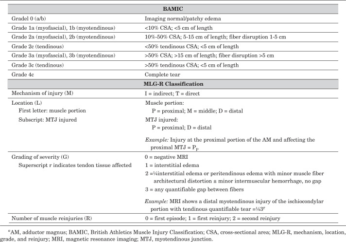
Methods: A total of 11 competitive athletes with an AM injury were included in the study. Each case was clinically assessed, and the diagnosis and classification were made by magnetic resonance imaging (MRI) according to the British Athletics Muscle Injury Classification (BAMIC) and mechanism, location, grade, and reinjury (MLG-R) classification. A 1-year follow-up was performed, and return-to-play (RTP) time was recorded.
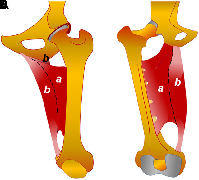
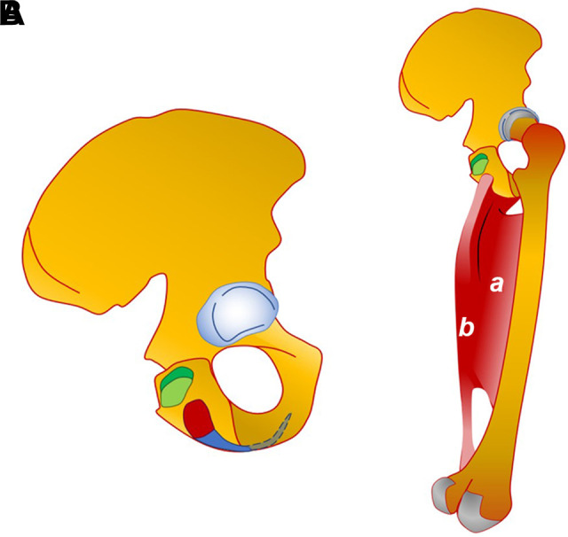
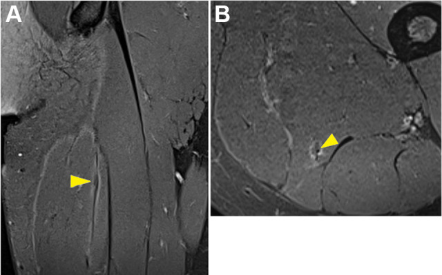
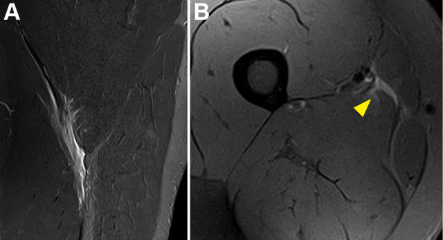
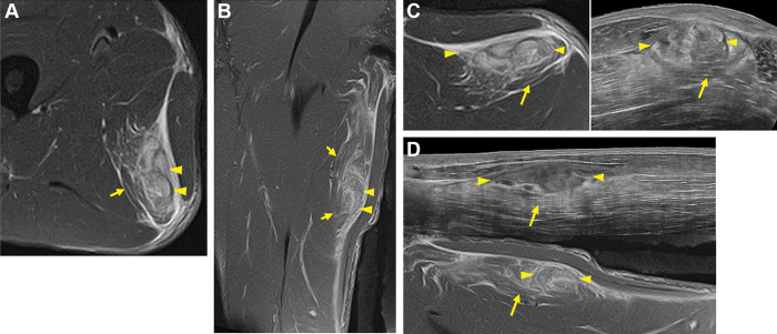
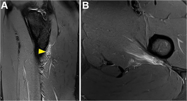
Results: Different mechanisms of injury were found; most of the athletes (10/11) had flexion and internal rotation of the hip with extension or slight flexion of the knee. Symptoms consisted of pain in the posteromedial (7/11) or medial (4/11) thigh during adduction and flexion of the knee. Clinically, there was a suspicion of an injury to the AM in only 3 athletes. According to MRI, 5 lesions were located in the ischiocondylar portion (3 in the proximal and 2 in the distal myoconnective junction) and 6 in the pubofemoral portion (4 in the distal and 2 in the proximal myoconnective junction). Most of the ischiocondylar lesions were myotendinous (3/5), and most of the pubofemoral lesions were myofascial (5/6). The BAMIC and MLG-R classification coincided in distinguishing injuries of moderate and mild severity. The management was nonoperative in all cases. The mean RTP time was 14 days (range, 0-35 days) and was longer in the ischiocondylar cases than in the pubofemoral cases (21 vs 8 days, respectively). Only 1 recurrence, at <10 months, was recorded.
Conclusion: Posteromedial thigh pain after an eccentric contraction during forced adduction of the thigh from hip internal rotation should raise a suspicion of AM lesions. The identification of the affected portion was possible on MRI. An injury in the ischiocondylar portion entailed a longer RTP time than an injury in the pubofemoral portion.
Keywords: BAMIC and MLG-R classification; MRI; adductor magnus injury; hip; pelvis; thigh.
© The Author(s) 2023.
Conflict of interest statement
The authors declared that there are no conflicts of interest in the authorship and publication of this contribution. AOSSM checks author disclosures against the Open Payments Database (OPD). AOSSM has not conducted an independent investigation on the OPD and disclaims any liability or responsibility relating thereto.
Figures
References
-
- Attarian DE. Isolated acute hip adductor brevis strain. J South Orthop Assoc. 2000;9(3):213–215. - PubMed
-
- Ballestero E, Duran C, Planas A, López Bedoya J, Vernetta M. Fuerza y dominancia lateral. Apunts Sports Med. 1997;1(47):74–80.
-
- Broski SM, Murthy NS, Krych AJ, Obey MR, Collins MS. The adductor magnus “mini-hamstring”: MRI appearance and potential pitfalls. Skeletal Radiol. 2016;45(2):213–219. - PubMed
-
- Chopra A, Robinson P. Imaging athletic groin pain. Radiol Clin North Am. 2016;54(5):865–873. - PubMed
-
- Delahunt E, Kennelly C, McEntee BL, Coughlan GF, Green BS. The thigh adductor squeeze test: 45° of hip flexion as the optimal test position for eliciting adductor muscle activity and maximum pressure values. Man Ther. 2011;16(5):476–480. - PubMed
LinkOut - more resources
Full Text Sources
