

Peripheral Blood Gene Expression Profile of Infants with Atopic Dermatitis
- PMID: 36699197
- PMCID: PMC9868882
- DOI: 10.1016/j.xjidi.2022.100165
Peripheral Blood Gene Expression Profile of Infants with Atopic Dermatitis
Abstract
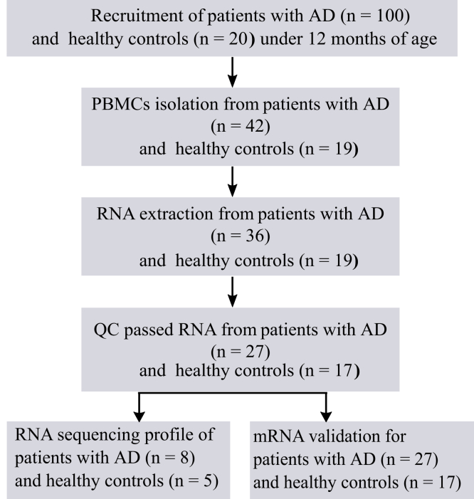
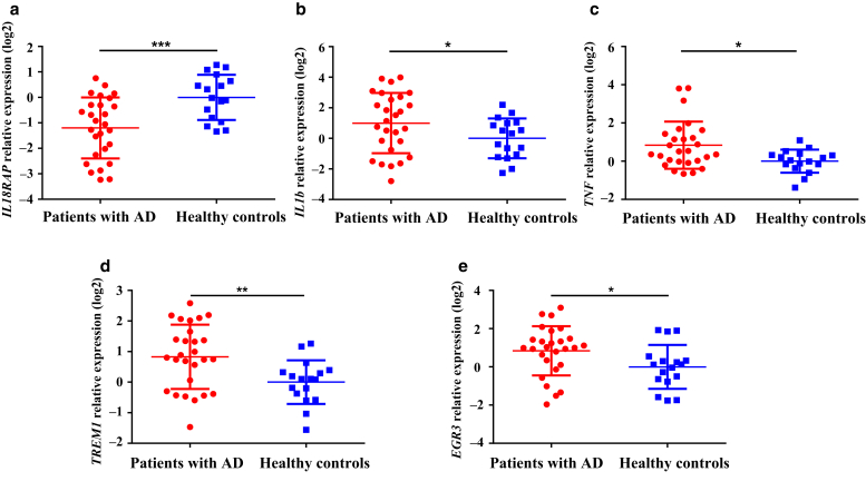
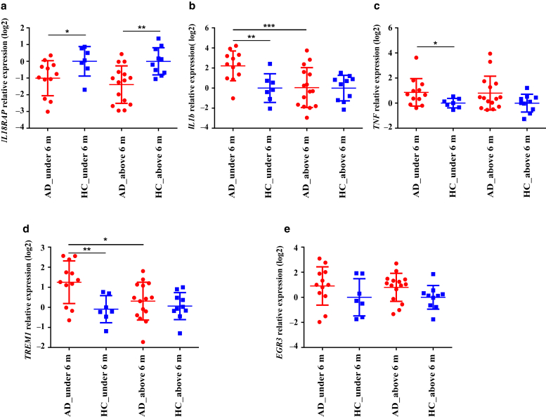
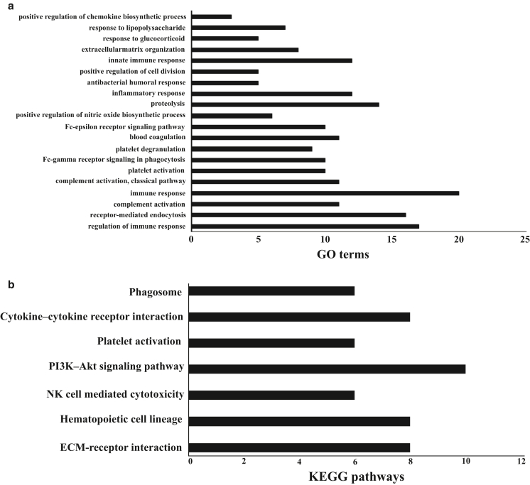
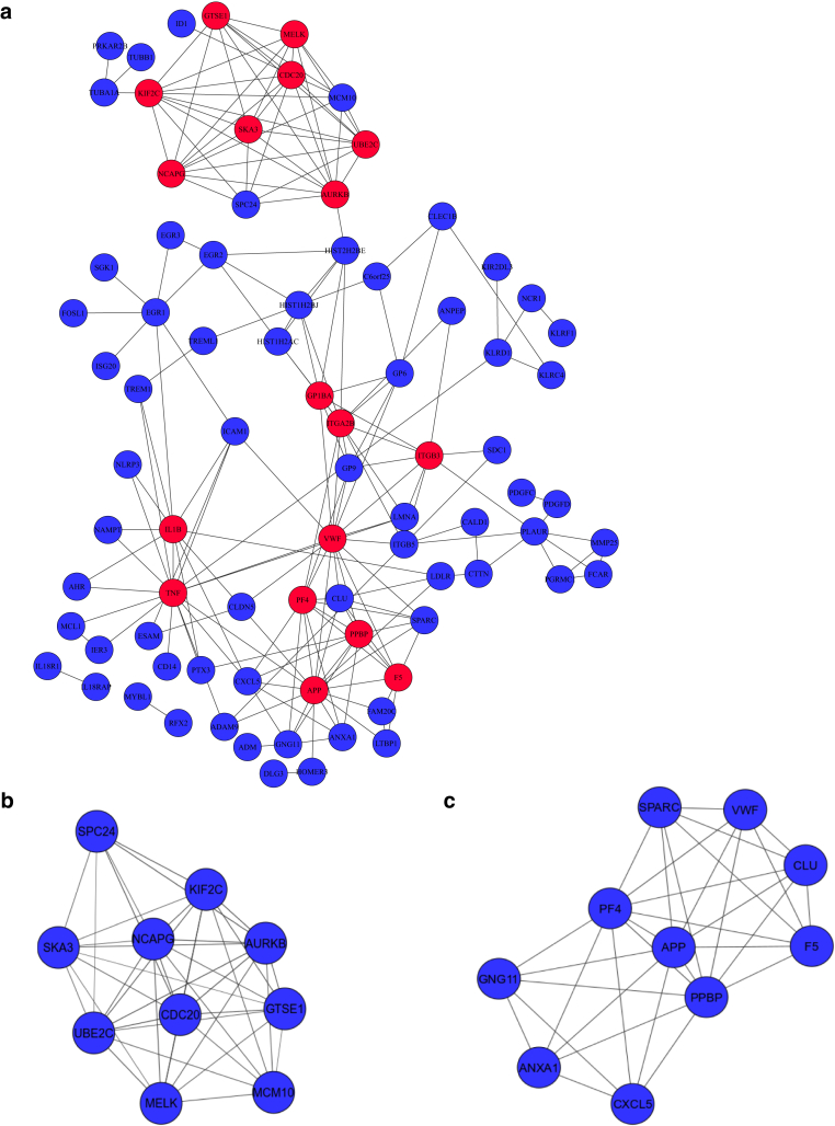
To enhance the understanding of molecular mechanisms and mine previously unidentified biomarkers of pediatric atopic dermatitis, PBMC gene expression profiles were generated by RNA sequencing in infants with atopic dermatitis and age-matched controls. A total of 178 significantly differentially expressed genes (DEGs) (115 upregulations and 63 downregulations) were seen, compared with those in healthy controls. The DEGs identified included IL1β, TNF, TREM1, IL18R1, and IL18RAP. DEGs were validated by real-time RT- qPCR in a larger number of samples from PBMCs of infants with atopic dermatitis aged <12 months. Using the DAVID (Database for Annotation, Visualization and Integrated Discovery) database, functional and pathway enrichment analyses of DEGs were performed. Gene ontology enrichment analysis showed that DEGs were associated with immune responses, inflammatory responses, regulation of immune responses, and platelet activation. Pathway analysis indicated that DEGs were enriched in cytokine‒cytokine receptor interaction, immunoregulatory interactions between lymphoid and nonlymphoid cells, hematopoietic cell lineage, phosphoinositide 3-kinase‒protein kinase B signaling pathway, NK cell‒mediated cytotoxicity, and platelet activation. Furthermore, the protein‒protein interaction network was predicted using the STRING (Search Tool for the Retrieval of Interacting Genes/Proteins) database and visualized with Cytoscape software. Finally, on the basis of the protein‒protein interaction network, 18 hub genes were selected, and two significant modules were obtained. In conclusion, this study sheds light on the molecular mechanisms of pediatric atopic dermatitis and may provide diagnostic biomarkers and therapeutic targets.
Keywords: AD, atopic dermatitis; BP, biological process; CC, cellular component; DEG, differentially expressed gene; GO, gene ontology; MF, molecular function; PIP, protein‒protein interaction; RNA-seq, RNA sequencing.
© 2023 The Authors.
Figures
Similar articles
-
Identification of candidate biomarkers and pathways associated with SCLC by bioinformatics analysis.Mol Med Rep. 2018 Aug;18(2):1538-1550. doi: 10.3892/mmr.2018.9095. Epub 2018 May 29. Mol Med Rep. 2018. PMID: 29845250 Free PMC article.
-
Bioinformatics analysis of gene expression profile data to screen key genes involved in intracranial aneurysms.Mol Med Rep. 2019 Nov;20(5):4415-4424. doi: 10.3892/mmr.2019.10696. Epub 2019 Sep 23. Mol Med Rep. 2019. PMID: 31545495 Free PMC article.
-
Identification of Effective Diagnostic Biomarkers and Immune Cell Infiltration in Atopic Dermatitis by Comprehensive Bioinformatics Analysis.Front Mol Biosci. 2022 Jul 14;9:917077. doi: 10.3389/fmolb.2022.917077. eCollection 2022. Front Mol Biosci. 2022. PMID: 35911963 Free PMC article.
-
Bioinformatic analysis of key pathways and genes involved in pediatric atopic dermatitis.Biosci Rep. 2021 Jan 29;41(1):BSR20193517. doi: 10.1042/BSR20193517. Biosci Rep. 2021. PMID: 33289509 Free PMC article.
-
Common gene signatures and key pathways in hypopharyngeal and esophageal squamous cell carcinoma: Evidence from bioinformatic analysis.Medicine (Baltimore). 2020 Oct 16;99(42):e22434. doi: 10.1097/MD.0000000000022434. Medicine (Baltimore). 2020. PMID: 33080677 Free PMC article.
Cited by
-
Associations of 2923 plasma proteins with incident atopic dermatitis in a prospective cohort study and genetic analysis.Medicine (Baltimore). 2025 Jul 18;104(29):e43447. doi: 10.1097/MD.0000000000043447. Medicine (Baltimore). 2025. PMID: 40696606 Free PMC article.
-
Deregulated Long Non-Coding RNAs (lncRNA) as Promising Biomarkers in Hidradenitis Suppurativa.J Clin Med. 2024 May 20;13(10):3016. doi: 10.3390/jcm13103016. J Clin Med. 2024. PMID: 38792557 Free PMC article.
-
Genome-wide identification of dysregulated alternative splicing and RNA-binding proteins involved in atopic dermatitis.Front Genet. 2024 Mar 1;15:1287111. doi: 10.3389/fgene.2024.1287111. eCollection 2024. Front Genet. 2024. PMID: 38495671 Free PMC article.
-
Effect of Topical Corticosteroid Treatment on microRNA Expression in Infants with Atopic Dermatitis.JID Innov. 2025 Jun 10;5(5):100388. doi: 10.1016/j.xjidi.2025.100388. eCollection 2025 Sep. JID Innov. 2025. PMID: 40686936 Free PMC article.
-
Use of Transcriptional Signatures to Differentiate Pathogen-Specific and Treatment-Specific Host Responses in Patients With Bacterial Bloodstream Infections.J Infect Dis. 2024 May 15;229(5):1535-1545. doi: 10.1093/infdis/jiad498. J Infect Dis. 2024. PMID: 38001039 Free PMC article.
References
-
- Bernard M., Carrasco C., Laoubi L., Guiraud B., Rozières A., Goujon C., et al. IL-1β induces thymic stromal lymphopoietin and an atopic dermatitis-like phenotype in reconstructed healthy human epidermis. J Pathol. 2017;242:234–245. - PubMed
-
- Bieber T. Atopic dermatitis. N Engl J Med. 2008;358:1483–1494. - PubMed
-
- Brunner P.M., Israel A., Leonard A., Pavel A.B., Kim H.J., Zhang N., et al. Distinct transcriptomic profiles of early-onset atopic dermatitis in blood and skin of pediatric patients. Ann Allergy Asthma Immunol. 2019;122:318–330.e3. - PubMed
-
- Brunner P.M., Israel A., Zhang N., Leonard A., Wen H.C., Huynh T., et al. Early-onset pediatric atopic dermatitis is characterized by TH2/TH17/TH22-centered inflammation and lipid alterations. J Allergy Clin Immunol. 2018;141:2094–2106. - PubMed
LinkOut - more resources
Full Text Sources
