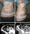
Absorbable Barbed Continuous versus Nonabsorbable Nonbarbed Interrupted Suturing Methods for Donor-site Closure of the Rectus Abdominis Myocutaneous Flap
- PMID: 36699207
- PMCID: PMC9848532
- DOI: 10.1097/GOX.0000000000004742
Absorbable Barbed Continuous versus Nonabsorbable Nonbarbed Interrupted Suturing Methods for Donor-site Closure of the Rectus Abdominis Myocutaneous Flap
Abstract
Abdominal incisional hernia is a complication of the rectus abdominis myocutaneous (RAMC) flap harvest. This study aimed to compare the incidence of abdominal incisional hernia and donor-site closure time between absorbable barbed continuous (ABC) and non-absorbable non-barbed interrupted (nAnBI) methods.
Methods: This study included 145 patients who underwent free RAMC flap reconstruction after head and neck cancer surgery at Kobe University Hospital between January 2012 and March 2020. The nAnBI method was selected between January 2012 and August 2016, and the ABC method was selected between September 2016 and March 2020. The incidence of abdominal incisional hernia and the average time required for donor-site closure were compared between the two groups.
Results: Of the 145 patients surveyed, 116 (57 and 59 in the nAnBI and ABC groups, respectively) were followed-up for at least 90 days after the surgery. The incidence rates of abdominal incisional hernia were 0% and 5.1% (n = 3) in the nAnBI and ABC groups, respectively, with no significant differences (p = 0.244). The average donor-site closure times were 127.6 and 111.3 minutes in the nAnBI and ABC groups, respectively, with no significant differences (p = 0.122).
Conclusions: No significant differences in the incidence of abdominal incisional hernia and donor-site closure time were observed between the nAnBI and ABC groups. However, there was a tendency for increased hernia occurrence and shorter wound closure time in the ABC group. A randomized prospective multicenter study is warranted to validate our findings of the ABC method.
Copyright © 2023 The Authors. Published by Wolters Kluwer Health, Inc. on behalf of The American Society of Plastic Surgeons.
Conflict of interest statement
Disclosure: The authors have no financial interest to declare in relation to the content of this article.
Figures


Similar articles
-
Double-blind randomized controlled trial of collagen mesh for the prevention of abdominal incisional hernia in patients having a vertical rectus abdominis myocutaneus flap during surgery for advanced pelvic malignancy.Colorectal Dis. 2017 May;19(5):491-500. doi: 10.1111/codi.13552. Colorectal Dis. 2017. PMID: 27805791 Clinical Trial.
-
The use of mesh versus primary fascial closure of the abdominal donor site when using a transverse rectus abdominis myocutaneous flap for breast reconstruction: a cost-utility analysis.Plast Reconstr Surg. 2015 Mar;135(3):682-689. doi: 10.1097/PRS.0000000000000957. Plast Reconstr Surg. 2015. PMID: 25719690 Review.
-
Donor Site Morbidity of Patients Receiving Vertical Rectus Abdominis Myocutaneous Flap for Perineal, Vaginal or Inguinal Reconstruction.World J Surg. 2021 Jan;45(1):132-140. doi: 10.1007/s00268-020-05788-5. Epub 2020 Sep 29. World J Surg. 2021. PMID: 32995931 Free PMC article.
-
Contralateral Component Separation Technique for Abdominal Wall Closure in Patients Undergoing Vertical Rectus Abdominis Myocutaneous Flap Transposition for Pelvic Exenteration Reconstruction.Ann Plast Surg. 2016 Jan;77(1):90-2. doi: 10.1097/SAP.0000000000000327. Ann Plast Surg. 2016. PMID: 25188251
-
Vertical rectus abdominis flap (VRAM) for perineal reconstruction following pelvic surgery: A systematic review.J Plast Reconstr Aesthet Surg. 2021 Mar;74(3):523-529. doi: 10.1016/j.bjps.2020.10.100. Epub 2020 Nov 9. J Plast Reconstr Aesthet Surg. 2021. PMID: 33317983
Cited by
-
Initial Experience with Unidirectional Barbed Suture for Abdominal Donor Site Closure in Deep Inferior Epigastric Perforator Flap Breast Reconstruction.Plast Reconstr Surg Glob Open. 2024 Mar 25;12(3):e5681. doi: 10.1097/GOX.0000000000005681. eCollection 2024 Mar. Plast Reconstr Surg Glob Open. 2024. PMID: 38528844 Free PMC article.
-
Assessing the application of barbed sutures in comparison to conventional sutures for surgical applications: a global systematic review and meta-analysis of preclinical animal studies.Int J Surg. 2024 May 1;110(5):3060-3071. doi: 10.1097/JS9.0000000000001230. Int J Surg. 2024. PMID: 38445518 Free PMC article.
References
-
- Kroll SS, Baldwin BJ. Head and neck reconstruction with the rectus abdominis free flap. Clin Plast Surg. 1994;21:97–105. - PubMed
-
- Aki FE, Besteiro JM, Ferreira MC. Immediate reconstruction of extensive defects in head and neck with the rectus abdominis musculocutaneous free flap. Rev Soc Bras Cir Plast. 1997;12:37–54.
-
- Mortensen AR, Grossmann I, Rosenkilde M, et al. . Double-blind randomized controlled trial of collagen mesh for the prevention of abdominal incisional hernia in patients having a vertical rectus abdominis myocutaneus flap during surgery for advanced pelvic malignancy. Colorectal Dis. 2017;19:491–500. - PubMed
-
- Rosen A, Hartman T. Repair of the midline fascial defect in abdominoplasty with long-acting barbed and smooth absorbable sutures. Aesthet Surg J 2011;31:668–673. - PubMed
LinkOut - more resources
Full Text Sources
