

Epidermal growth factor regulates autophagy activity and endocytosis of yak cumulus cells in a concentration-dependent manner
- PMID: 36699336
- PMCID: PMC9868291
- DOI: 10.3389/fvets.2022.1081643
Epidermal growth factor regulates autophagy activity and endocytosis of yak cumulus cells in a concentration-dependent manner
Abstract
Introduction: Autophagy and endocytosis are crucial biological activities in mammalian follicle development and oocyte maturation, which are easily affected by external environmental factors. Epidermal growth factor (EGF), as an important component of follicular fluid, regulates the growth and apoptosis of follicular cells. However, its regulatory mechanism of autophagy and endocytosis in mammals, especially in large domestic animals such as plateau yak, remains unclear. This study aimed to investigate the regulatory mechanism of EGF on autophagy and endocytosis in yak cumulus cells.
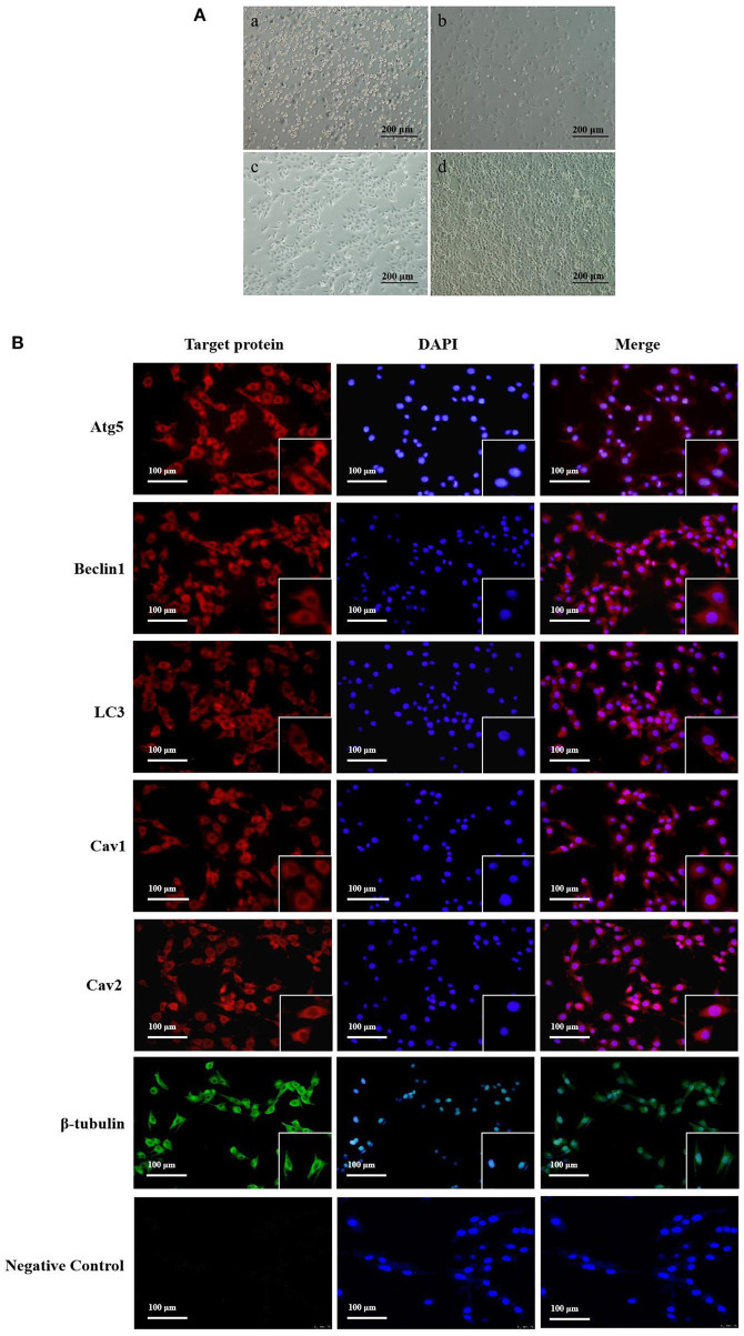
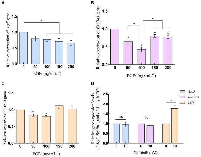
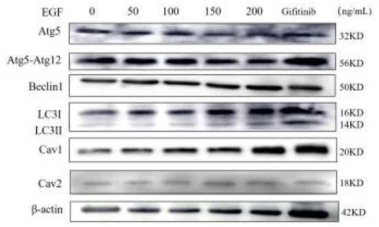
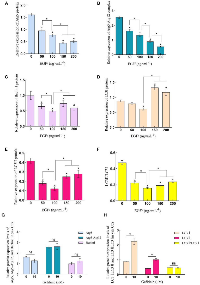
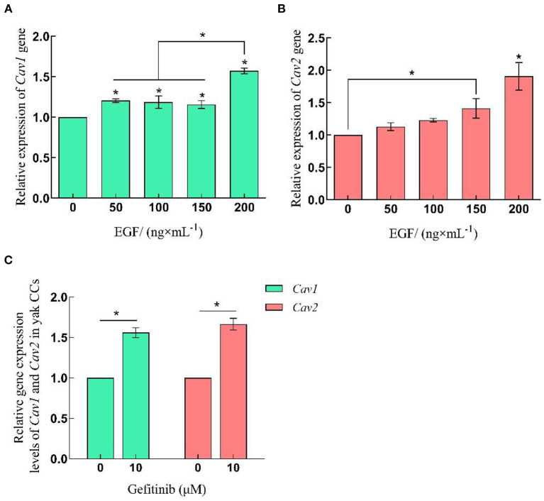
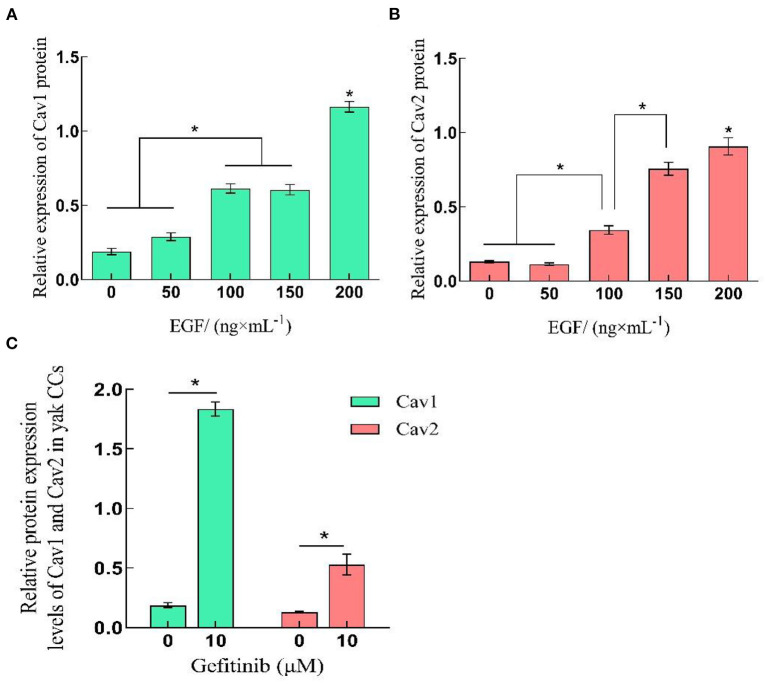
Methods: Yak cumulus cells were treated with different concentrations of EGF and appropriate concentrations of EGFR inhibitor gefitinib (10 μM). The dynamic expression levels of Atg5, Beclin1, LC3, Cav1 and Cav2 were detected by immunofluorescence staining, qRT-PCR and Western-blot.
Results: EGF inhibited autophagy in yak cumulus cells by down-regulating the expression of Atg5, Beclin1, and LC3. The level of autophagy varied with the concentration of ligands, and the inhibition was most significant at 100 ng/mL. Noteworthy, EGF can promote endocytosis by regulating the expression of Cav1 and Cav2, but the EGFR-mediated signaling pathway is not the main way to regulate the expression of these proteins.
Discussion: These results provide a reference for further exploring the effects of growth factors on livestock germ cells and the regulatory role of autophagy-endocytosis crosstalk mechanism in follicle development and oocyte maturation, to improve the fecundity of yaks.
Keywords: EGF; autophagy; cumulus cells; endocytosis; yak.
Copyright © 2023 Ma, Yu, Cui, Pan, Wang, Wang, Wang, Zhao and Zhang.
Conflict of interest statement
The authors declare that the research was conducted in the absence of any commercial or financial relationships that could be construed as a potential conflict of interest.
Figures
Similar articles
-
Hypoxia-Inducible Factor 1α Affects Yak Oocyte Maturation and Early Embryonic Development by Regulating Autophagy.Antioxidants (Basel). 2024 Jul 14;13(7):840. doi: 10.3390/antiox13070840. Antioxidants (Basel). 2024. PMID: 39061908 Free PMC article.
-
Exosomes Derived from Yak Follicular Fluid Increase 2-Hydroxyestradiol Secretion by Activating Autophagy in Cumulus Cells.Animals (Basel). 2022 Nov 16;12(22):3174. doi: 10.3390/ani12223174. Animals (Basel). 2022. PMID: 36428401 Free PMC article.
-
Proteomics and Expression of HIF2α/BNIP3L Signaling in Yak Brains at Different Altitudes.Int J Mol Sci. 2025 Feb 16;26(4):1675. doi: 10.3390/ijms26041675. Int J Mol Sci. 2025. PMID: 40004139 Free PMC article.
-
Epidermal growth factor-like growth factors in the follicular fluid: role in oocyte development and maturation.Semin Reprod Med. 2009 Jan;27(1):52-61. doi: 10.1055/s-0028-1108010. Epub 2009 Feb 5. Semin Reprod Med. 2009. PMID: 19197805 Free PMC article. Review.
-
The epidermal growth factor network: role in oocyte growth, maturation and developmental competence.Hum Reprod Update. 2018 Jan 1;24(1):1-14. doi: 10.1093/humupd/dmx029. Hum Reprod Update. 2018. PMID: 29029246 Review.
References
-
- Ge H, Zhang F, Duan P, Zhu N, Zhang J, Ye F, et al. . Mitochondrial uncoupling protein 2 in human cumulus cells is associated with regulating autophagy and apoptosis, maintaining gap junction integrity and progesterone synthesis. Mol Cell Endocrinol. (2017) 443:128–37. 10.1016/j.mce.2017.01.020 - DOI - PubMed
-
- Miao J-K, Liu Y-H, Liu S, Liu X-M, Wang P-C, Du Z-Q, et al. . Lysosomal dysfunction disturbs porcine oocyte maturation and developmental capacity by disorganizing chromosome/cytoskeleton and activating autophagy/apoptosis. Theriogenology. (2019) 140:44–51. 10.1016/j.theriogenology.2019.08.019 - DOI - PubMed
LinkOut - more resources
Full Text Sources
Research Materials
Miscellaneous