

Alterations of degree centrality and functional connectivity in classic trigeminal neuralgia
- PMID: 36699513
- PMCID: PMC9870176
- DOI: 10.3389/fnins.2022.1090462
Alterations of degree centrality and functional connectivity in classic trigeminal neuralgia
Abstract
Objectives: Recent neuroimaging studies have indicated a wide range of structural and regional functional alterations in patients with classic trigeminal neuralgia (CTN). However, few studies have focused on the intrinsic functional characteristics of network organization in the whole brain. Therefore, the present study aimed to characterize the potential intrinsic dysconnectivity pattern of the whole brain functional networks at the voxel level using the degree centrality (DC) analysis in CTN patients.
Methods: Thirty-four patients with CTN and twenty-nine well-matched healthy controls (HCs) participated in this study. All subjects underwent resting-state functional magnetic resonance imaging (rs-MRI) examination and clinical and neuropsychologic assessments. DC is a graph theory-based measurement that represents the overall functional connectivity (FC) numbers between one voxel and other brain voxels. We first investigated brain regions exhibiting abnormal DC, and further identified their perturbation on FC with other brain regions using a seed-based FC analysis in patients with CTN. In addition, correlation analyses were performed to evaluate the relationship between the abnormal DC value and clinical variables in CTN patients.
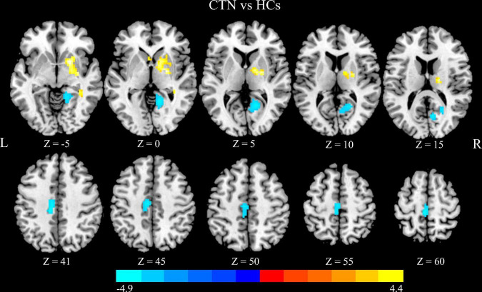
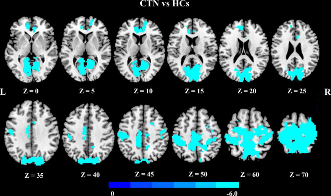
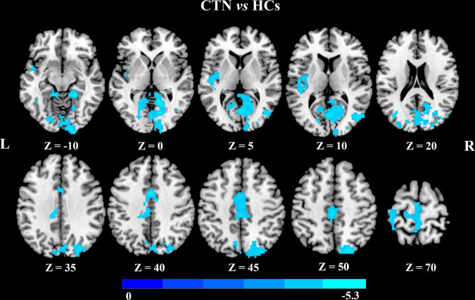
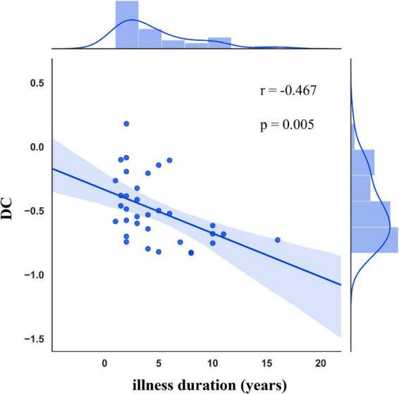
Results: Compared with the HCs, the patients with CTN exhibited significantly greater DC values in the right pallidum and right putamen, and lower DC values in the right lingual gyrus, right calcarine sulcus, left paracentral lobule, and left midcingulate cortex. A further seed-based FC analysis revealed that the right lingual gyrus showed decreased FC within the visual network and with other core brain networks, including the sensorimotor network, default mode network, and salience network, relative to HCs. Additionally, the left midcingulate cortex exhibited decreased FC within the middle cingulate cortex and the visual network in CTN patients. Moreover, the DC value in the left midcingulate cortex was negatively correlated with the illness duration.
Conclusion: The present study shows that CTN patients exhibited specific functional connectivity network alterations in the basal ganglia, visual network, and salience network, which may reflect the aberrant neural network communication in pain processing and modulation. These findings may provide novel insight for understanding the mechanisms of pain chronicity in CTN patients.
Keywords: classic trigeminal neuralgia; degree centrality; functional connectivity; neuropathic pain; resting-state functional MRI.
Copyright © 2023 Liu, Zheng, Zhang, Zhang, Hou, Cheng and Han.
Conflict of interest statement
The authors declare that the research was conducted in the absence of any commercial or financial relationships that could be construed as a potential conflict of interest.
Figures
Similar articles
-
Structural and Functional Brain Changes in Patients With Classic Trigeminal Neuralgia: A Combination of Voxel-Based Morphometry and Resting-State Functional MRI Study.Front Neurosci. 2022 Jun 29;16:930765. doi: 10.3389/fnins.2022.930765. eCollection 2022. Front Neurosci. 2022. PMID: 35844235 Free PMC article.
-
Altered brain functional network dynamics in classic trigeminal neuralgia: a resting-state functional magnetic resonance imaging study.J Headache Pain. 2021 Dec 11;22(1):147. doi: 10.1186/s10194-021-01354-z. J Headache Pain. 2021. PMID: 34895135 Free PMC article.
-
Altered Structural and Functional Connectivity of Salience Network in Patients with Classic Trigeminal Neuralgia.J Pain. 2022 Aug;23(8):1389-1399. doi: 10.1016/j.jpain.2022.02.012. Epub 2022 Apr 3. J Pain. 2022. PMID: 35381362
-
Alterations in degree centrality and functional connectivity in tension-type headache: a resting-state fMRI study.Brain Imaging Behav. 2024 Aug;18(4):819-829. doi: 10.1007/s11682-024-00875-w. Epub 2024 Mar 21. Brain Imaging Behav. 2024. PMID: 38512647 Review.
-
Functional changes of default mode network and structural alterations of gray matter in patients with irritable bowel syndrome: a meta-analysis of whole-brain studies.Front Neurosci. 2023 Oct 24;17:1236069. doi: 10.3389/fnins.2023.1236069. eCollection 2023. Front Neurosci. 2023. PMID: 37942144 Free PMC article.
Cited by
-
Brain entropy changes in classical trigeminal neuralgia.Front Neurol. 2023 Nov 23;14:1273336. doi: 10.3389/fneur.2023.1273336. eCollection 2023. Front Neurol. 2023. PMID: 38073647 Free PMC article.
-
Abnormal Degree Centrality in Zoster-Associated Pain with or Without Psychiatric Comorbidities: A Resting-State Functional MRI Study.J Pain Res. 2024 Aug 12;17:2629-2638. doi: 10.2147/JPR.S465018. eCollection 2024. J Pain Res. 2024. PMID: 39155954 Free PMC article.
-
Disruption of functional and structural topological organization in specific subnetwork among patients with classical trigeminal neuralgia: a graph theory-based magnetic resonance imaging study.Brain Imaging Behav. 2025 Jun 24. doi: 10.1007/s11682-025-01035-4. Online ahead of print. Brain Imaging Behav. 2025. PMID: 40551062 No abstract available.
-
Brain functional connectivity patterns associated with symptoms of vestibular migraine.Front Neurosci. 2023 Dec 14;17:1231273. doi: 10.3389/fnins.2023.1231273. eCollection 2023. Front Neurosci. 2023. PMID: 38156263 Free PMC article.
-
Hypoconnectivity of the Amygdala in Patients with Low-Back-Related Leg Pain Linked to Individual Mechanical Pain Sensitivity: A Resting-State Functional MRI Study.J Pain Res. 2023 Nov 8;16:3775-3784. doi: 10.2147/JPR.S425874. eCollection 2023. J Pain Res. 2023. PMID: 38026465 Free PMC article.
References
-
- Bennetto L., Patel N. K., Fuller G. (2007). Trigeminal neuralgia and its management. BMJ 334 201–205. 10.1136/bmj.39085.614792.BE - DOI - PMC - PubMed
-
- Buckner R. L., Sepulcre J., Talukdar T., Krienen F. M., Liu H., Hedden T., et al. (2009). Cortical hubs revealed by intrinsic functional connectivity: Mapping, assessment of stability, and relation to Alzheimer’s disease. J. Neurosci. 29 1860–1873. 10.1523/jneurosci.5062-08.2009 - DOI - PMC - PubMed
LinkOut - more resources
Full Text Sources