. 2023 Jan 9:16:1067493.
doi: 10.3389/fnins.2022.1067493. eCollection 2022.

Chronic Δ9-tetrahydrocannabinol impact on plasticity, and differential activation requirement for CB1-dependent long-term depression in ventral tegmental area GABA neurons in adult versus young mice

Affiliations

Chronic Δ9-tetrahydrocannabinol impact on plasticity, and differential activation requirement for CB1-dependent long-term depression in ventral tegmental area GABA neurons in adult versus young mice

Isaac Ostlund et al. Front Neurosci. .

Abstract

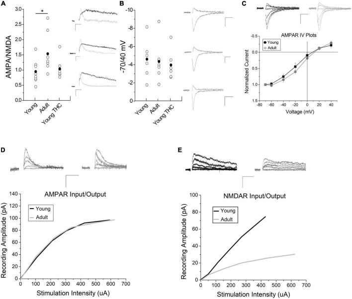
The ventral tegmental area (VTA) mediates incentive salience and reward prediction error through dopamine (DA) neurons that are regulated by local VTA GABA neurons. In young mice, VTA GABA cells exhibit a form of synaptic plasticity known as long-term depression (LTD) that is dependent on cannabinoid 1 (CB1) receptors preceded by metabotropic glutamate receptor 5 (mGluR5) signaling to induce endocannabinoid production. This LTD was eliminated following chronic (7-10 consecutive days) exposure to the marijuana derived cannabinoid Δ9 -tetrahydrocannabinol (THC). We now examine the mechanism behind THC-induced elimination of LTD in adolescents as well as plasticity induction ability in adult versus young male and female mice using whole-cell electrophysiology experiments of VTA GABA cells. Chronic THC injections in adolescents resulted in a loss of CB1 agonist-mediated depression, illustrating chronic THC likely desensitizes or removes synaptic CB1. We noted that seven days withdrawal from chronic THC restored LTD and CB1 agonist-induced depression, suggesting reversibility of THC-induced changes. Adult mice continue to express functional mGluR5 and CB1, but require a doubling of the synaptic stimulation compared to young mice to induce LTD, suggesting a quantitative difference in CB1-dependent plasticity between young and adult mice. One potential rationale for this difference is changes in AMPA and NMDA glutamate receptors. Indeed, AMPA/NMDA ratios were increased in in adults compared to young mice. Lastly, we performed quantitative reverse-transcription PCR and identified that CB1, DAGLα, and GluA1 levels increased following chronic THC exposure. Collectively, our data demonstrate the first age-dependent GABA neuron plasticity in the VTA, which could have implications for decreased THC dependence capacity in adults, as well as the mechanism behind chronic THC-induced synaptic alterations in young mice.

Keywords: CB1; GABA; LTD; THC; VTA; age; development.


Conflict of interest statement

The authors declare that the research was conducted in the absence of any commercial or financial relationships that could be construed as a potential conflict of interest.

Figures

FIGURE 1
FIGURE 1
Chronic THC impact on glutamate inputs and plasticity of VTA GABA neurons. (A) Within naïve young mice (P15-40) a high frequency stimulus (HFS) induces LTD (33 ± 7.1% reduction) 15–20 min post stimulus compared to baseline (n = 7, p < 0.001), confirming previously reported LTD in mice (Friend et al., 2017). (B) Young mice with chronic (7–10 consecutive days) THC intraperitoneal (IP) injections do not exhibit WIN55,212-2-induce depression compared to baseline (n = 8, p > 0.1). (C) In contrast, young mice chronically injected with vehicle (0.1% EtOH saline) exhibit significant WIN55,212-2-induced depression compared to baseline (38.3% ± 6.3% depression, p < 0.001). (D) Seven days withdrawal following chronic exposure to THC in young mice, WIN55,212-2 again induced depression of excitatory currents (by 26.3 ± 7.3%; n = 6, p < 0.001), which is significantly different than the chronic THC group in panel (B) at the same time points (p < 0.001; 15–20 min post-drug application). (E) Chronic THC elimination of HFS-induced LTD is reversed in young mice by seven days withdrawal as they undergo LTD (38.1 ± 5.7%; n = 6, p < 0.001). This is not significantly different (p > 0.1) than naïve LTD in panel (A). (F) HFS continues to induce significant LTD within age-match control mice chronically injected with vehicle after 7 days withdrawal (29.3 ± 7.3%; n = 4, p < 0.001). Here and throughout horizontal gray bars indicate duration of drug application (10 min) and whole-cell plots are normalized excitatory post-synaptic currents (EPSC) amplitude means with bars indicating standard error of the mean (SEM). All recordings were excitatory glutamate currents recorded from GAD67/GFP + neurons. N values are number of individual cells, with one cell per animal recorded. Arrows indicate time of HFS. Example traces representing 12–14 averaged traces taken before (black) and 15–20 min after (gray) conditioning or drug application. Note that all stimulation artifacts were removed from example traces for clarity. Scale bars here and throughout represent 50 pA, 5 ms. Supplementary Table 1 is a list of statistics and significance values of all the experiments in these figures.
FIGURE 2
FIGURE 2
Adult mice exhibit increased LTD induction thresholds of excitatory inputs to VTA GABA neurons. (A) HFS does not induce LTD in adult mice compared to baseline (1.4 ± 5.7%, n = 8, p > 0.1, P70-150) and is significantly different than HFS young mice at the same time points compared to Figure 1A (p < 0.001). (B) WIN55,212-2 significantly depresses excitatory currents in adult mice compared to baseline (by 31.0 ± 10.1%; n = 5, p < 0.001) and is significantly different than the naïve adult HFS group at the same time points (p < 0.001). (C) DHPG significantly depresses excitatory postsynaptic currents (EPSCs) in adult mice compared to baseline (by 41.7 ± 3.1%; n = 6, p < 0.001) and is significantly different than the naïve adult HFS group at the same time points (p < 0.001). Note that WIN55,212-2 and DHPG-induced depression in adults are not significantly (p > 0.1) different from depression induced by these agonists in adolescents, comparing to data in our prior publication (Friend et al., 2017). (D) Doubling the HFS protocol induces LTD in adult mice compared to baseline (32.3 ± 4.4%; n = 6, p < 0.001) and is significantly different than the naïve adult HFS group at the same time points (p < 0.001). (E) The CB1 antagonist AM-251 (2 μM) blocked 2× HFS LTD (n = 6, 4.3 ± 2.2% above baseline, p > 0.1), which was not significantly different (p > 0.1) than control HFS in panel (A). (F) Chronic (7–10 days) THC exposure results in a loss of 2× HFS-induced LTD in adult mice (n = 6, p > 0.1) and is significantly different than naïve adults with double HFS (p < 0.001). This is comparable to chronic THC elimination of HFS-induced LTD seen in young mice (see Friend et al., 2017). (G) Application of THC (1 μM) to the bath ACSF induces significant depression (p < 0.001) compared to baseline (35% depression), which was not significantly (p > 0.1) different from THC-induced depression in adolescents (20% depression (Friend et al., 2017). (H) The coefficient of variance (1/CV2) in adult experimental groups showed a significant decrease following 2× HFS LTD (n = 6, p < 0.05), WIN55,212-2-induced depression (n = 5, p < 0.05), and DHPG-induced depression (n = 6, p < 0.05). (I) The paired pulse ratio (PPR) for adult experimental groups increased when comparing baseline to post 2× HFS LTD (n = 6, p < 0.05) and WIN55,212-2-induced depression (n = 5, p < 0.05). No significant change was noted following DHPG depression, but trend only (n = 6, 0.1 > p > 0.05).
FIGURE 3
FIGURE 3
Impact of development and chronic THC on AMPA/NMDA ratios. (A) AMPA/NMDA ratios are significantly higher in adult mice than young (n = 8 young, n = 7 adults, p < 0.05). Chronic THC injection in young mice has no significant effect on AMPA/NMDA ratios when compared to naive young mice (n = 7 young chronic THC, p > 0.1). Insets represent AMPA currents and NMDA currents at +40 mV, the latter being a subtraction of AMPA from total current (see methods for details). (B) The -70/40 ratio remains unchanged from naive young mice to adults (n = 6 young and n = 7 adult, p > 0.1), and from naïve young mice to chronic THC injected young mice (n = 6 chronic THC young, p > 0.1). Insets represent AMPA currents taken at -70 and +40 mV. (C) No significant difference is noted in IV plots between young and adult mice (n = 7 for both groups, p > 0.1; ANCOVA). Rectification between the two groups at positive potentials is unchanged (p > 0.1). Insets represent AMPA currents taken at +40, +20, 0, -20, -40, -60, and -70 mV. (D) Input/output curves of evoked AMPA currents as correlated with stimulation intensity were unchanged between age groups (n = 15 adults, n = 13 young, p > 0.1, ANCOVA). Curves were created by post hoc analysis of raw data using two-minute averages of EPSCs comparing the amount of current evoked in an AMPA or NMDA only EPSCs at +40 mV compared to current injected into the stimulation electrode, which were then best fit to a curve. The data points used to create the curves were taken at various points throughout the curve range. The curves were compared statistically between young and adult mice for AMPA (D) and NMDA (E) currents using an ANCOVA. Raw data was that attained in Figure 3A at +40 mV in order to measure NMDA currents. (E) Input/output curves of evoked NMDA currents were significantly higher in young mice compared to adults (n = 9 adults, n = 8 young mice, p < 0.05, ANCOVA). In both panels (D,E) inset AMPA and NMDA current traces were measured at +40 mV at various stimulus intensities. Scale bars represent 20 pA, 5 ms. *p < 0.05.
FIGURE 4
FIGURE 4
Expression of mRNA for CB1, GluA1, DAGLα and FAAH is modified following chronic THC injection or development. (A) Glutamate receptor subunits are largely unchanged across all groups, with the GluA1 subunit being a notable exception. GluA1 gene expression is increased following chronic THC exposure compared to naive young mice (p < 0.05). GluA1 gene expression is also increased in adults compared to naive young mice (p < 0.001), vehicle controls (p < 0.05), and THC withdrawal groups (p < 0.05). (B) Endocannabinoid components exhibit significant changes or trends following chronic THC injection. Gene expression for the CB1 receptor is significantly increased following chronic THC exposure compared to naive young mice (p < 0.05). DAGLα shows significant (p < 0.05) increased gene expression following chronic THC exposure, while FAAH demonstrates a trend in Naive adults compared to naïve young mouse levels (0.1 > p < 0.05). MAGL is increased in chronic THC exposed mice compared to vehicle control mice (p < 0.05). (C) Various genetic transcription or epigenetic modifiers were also examined, where HDAC3 gene expression decreased (p < 0.05) in young THC withdrawal groups when compared to naive young mice. HDAC3 gene expression is also increased in adults compared to THC withdrawal group (p < 0.05). (D) Proper amplification of each cellular target was examined using 4% agarose gel electrophoresis to verify amplicon size. See Table 1 for base pair (BP) lengths. Targets not shown in the gel were published previously (Merrill et al., 2015). The n value ranged from 7 to 10 for each group with at least 3 females and 3 males per group, and each n represents one mouse. The range was the result of some failed PCR experiments in some groups, indicated by qPCR results > 20 cycles from the control target, illustrating non-specificity, likely resulting from experimental error. *p < 0.05, **p < 0.001.
FIGURE 5
FIGURE 5
Working model of CB1-dependent LTD of VTA GABA neurons and increased induction thresholds in adults. (A) HFS (2 × 100 Hz) within young mice causes glutamate release sufficient to activate both mGluR5 and NMDA receptors, causing Ca2+ influx (1). Ca2+ activates DAGLα (2), creating the retrograde signaling endocannabinoid, 2-AG (3). 2-AG leaves the postsynaptic neuron and binds presynaptic CB1 receptors (4). CB1 activation depresses glutamate transmission resulting in LTD (5). (B) HFS within young mice chronically exposed to THC does not induce LTD. This loss of LTD is potentially at the CB1 receptor through internalization following constitutive activation by THC or impediments to its signal cascade. Following withdrawal this synapse reverses back to plasticity induction capacity similar to THC naïve (see “Withdrawal reversal”). (C) Within adults, HFS is not strong enough to induced LTD, likely due to a potential decrease in NMDA receptor levels reducing Ca2+ influx (1). Lower Ca2+ levels are insufficient to activate DAGLα (2). No 2-AG is produced (3), CB1 is not activated (4), and there is no LTD (5). (D) Double HFS within adults causes glutamate release sufficient to activate both mGluR5 and NMDA receptors more intensely, causing increased Ca2+ influx (1). Ca2+ activates DAGLα (2), creating the retrograde signaling endocannabinoid, 2-AG (3) that binds the CB1 receptor (4) to induce LTD (5).

Similar articles

Cited by

References

    1. Aitta-Aho T., Möykkynen T. P., Panhelainen A. E., Vekovischeva O. Y., Bäckström P., Korpi E. R. (2012). Importance of GluA1 subunit-containing ampa glutamate receptors for morphine state-dependency. PLoS One 7:e38325. 10.1371/journal.pone.0038325 - DOI - PMC - PubMed
    1. Albertella L., Le Pelley M. E., Yucel M., Copeland J. (2018). Age moderates the association between frequent cannabis use and negative schizotypy over time. Addict. Behav. 87 183–189. 10.1016/j.addbeh.2018.07.016 - DOI - PubMed
    1. Al-Hasani R., Gowrishankar R., Schmitz G. P., Pedersen C. E., Marcus D. J., Shirley S. E., et al. (2021). Ventral tegmental area Gabaergic inhibition of cholinergic interneurons in the ventral nucleus accumbens shell promotes reward reinforcement. Nat. Neurosci. 24:1501. 10.1038/s41593-021-00928-z - DOI - PubMed
    1. Allen M. T., Matthews K. A. (1997). Hemodynamic responses to laboratory stressors in children and adolescents: The influences of age, race, and gender. Psychophysiology 34 329–339. 10.1111/j.1469-8986.1997.tb02403.x - DOI - PubMed
    1. Authement M. E., Langlois L. D., Kassis H., Gouty S., Dacher M., Shepard R. D., et al. (2016). Morphine-induced synaptic plasticity in the Vta is reversed by Hdac inhibition. J. Neurophysiol. 116 1093–1103. 10.1152/jn.00238.2016 - DOI - PMC - PubMed

LinkOut - more resources