. 2023 Jan 9:13:1074107.
doi: 10.3389/fpls.2022.1074107. eCollection 2022.

The COPII subunit MoSec24B is involved in development, pathogenicity and autophagy in the rice blast fungus

Affiliations

The COPII subunit MoSec24B is involved in development, pathogenicity and autophagy in the rice blast fungus

Hui Qian et al. Front Plant Sci. .

Abstract

The endoplasmic reticulum (ER) acts as the starting point of the secretory pathway, where approximately one-third of the proteins are correctly folded and modified, loaded into vesicles, and transported to the Golgi for further processing and modification. In this process, COPII vesicles are responsible for transporting cargo proteins from the ER to the Golgi. Here, we identified the inner shell subunit of COPII vesicles (MoSec24B) and explored the importance of MoSec24B in the rice blast fungus. The targeted disruption of MoSec24B led to decreased growth, reduced conidiation, restricted glycogen and lipids utilization, sensitivity to the cell wall and hypertonic stress, the failure of septin-mediated repolarization of appressorium, impaired appressorium turgor pressure, and decreased ability to infect, which resulted in reduced pathogenicity to the host plant. Furthermore, MoSec24B functions in the three mitogen-activated protein kinase (MAPK) signaling pathways by acting with MoMst50. Deletion of MoSec24B caused reduced lipidation of MoAtg8, accelerated degradation of exogenously introduced GFP-MoAtg8, and increased lipidation of MoAtg8 upon treatment with a late inhibitor of autophagy (BafA1), suggesting that MoSec24B regulates the fusion of late autophagosomes with vacuoles. Together, these results suggest that MoSec24B exerts a significant role in fungal development, the pathogenesis of filamentous fungi and autophagy.

Keywords: COPII; Magnaporthe oryzae; Sec24; autophagy; pathogenicity; signaling pathway.


Conflict of interest statement

The authors declare that the research was conducted in the absence of any commercial or financial relationships that could be construed as a potential conflict of interest.

Figures

Figure 1
Figure 1
Identification of MoSec24 in M. oryzae and interactions within the COPII complex. (A) Structural domain analysis of the Sec24 homologous gene in M. oryzae. (B) Co-localization of MoSec24A-GFP and MoSec24B-mCherry in conidia and mycelium observed by fluorescence microscopy images. Scale Bar = 10 μm. (C) Analysis of the interactions between different subunits within the COPII complex by yeast-two-hybrid.
Figure 2
Figure 2
Subcellular localization of MoSec24B in M. oryzae. (A) Confocal fluorescence microscopy images observed the co-localization of MoSec24B-mCherry with ER-localized marker MoSlp1-GFP and Golgi-localized marker MoSft2-GFP, respectively, in untreated conidia (Fv3000, 60 × oil). (B) Confocal fluorescence microscopy images observed the co-localization of MoSec24B-mCherry with ER-localized marker MoSlp1-GFP and Golgi-localized marker MoSft2-GFP, respectively, in 0.1% DMSO-treated conidia (Fv3000, 60 × oil). (C) Confocal fluorescence microscopy images observed the co-localization of MoSec24B-mCherry with ER-localized marker MoSlp1-GFP and Golgi-localized marker MoSft2-GFP, respectively, in BFA-treated conidia (Fv3000, 60 × oil). Scale Bar = 5 μm. Fifteen spores were selected under each treatment (Normal, DMSO, BFA), the reticular or spot-like structures representing the ER and Golgi in each spore were labeled as number A, and the MoSec24-mCherry, which co-located with green fluorescence in each spore, was labeled as number B. (B/A) *100 was calculated as the co-localization percentage. The bar graphs indicate the percentage of localization of MoSec24B-mCherry in ER and Golgi, respectively, under different treatment conditions. Error bars represent the standard deviation. An analysis of the data was carried out using an unpaired two-tailed Student’s t-test. Asterisks represent statistically significant differences in the data (**P < 0.01).
Figure 3
Figure 3
MoSec24B is involved in growth and conidiation. (A) WT, ΔMosec24B, and ΔMosec24B::MoSEC24B strains were cultured on CM solid medium for 8 days. Scale Bar = 1 cm. (B) Mycelial diameters of WT, ΔMosec24B, and ΔMosec24B::MoSEC24B strains. (C) Colony side views of aerial hyphae growth in WT, ΔMosec24B, and ΔMosec24B::MoSEC24B strains. (D) Conidiation of WT, ΔMosec24B, and ΔMosec24B::MoSEC24B strains. (E) Conidiophores of WT, ΔMosec24B, and ΔMosec24B::MoSEC24B strains were observed under a light microscope. Scale Bar = 50 μm. Error bars represent the standard deviation. An analysis of the data was carried out using an unpaired two-tailed Student’s t-test. Asterisks represent statistically significant differences in the data (**P < 0.01).
Figure 4
Figure 4
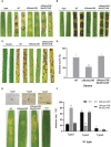
MoSec24B is important for pathogenicity. (A) Mycelial plugs from WT, ΔMosec24B, and ΔMosec24B::MoSEC24B strains were inoculated on detached barley leaves. (B) Mycelial plugs from WT, ΔMosec24B, and ΔMosec24B::MoSEC24B strains were inoculated on detached rice leaves. (C) Conidial suspensions (5 × 10⁴ conidia/ml) from WT, ΔMosec24B, and ΔMosec24B::MoSEC24B strains were sprayed on rice seedlings. (D) Disease areas of rice leaves whose length is 5 cm were measured using Photoshop CS6. (E) Conidial suspensions (5 × 10⁴ conidia/ml) from WT, ΔMosec24B, and ΔMosec24B::MoSEC24B strains were sprayed on barley leaves for 4 days, and IHs on barley cells were divided into three types. Scale Bar = 20 μm. (F) Statistical analysis of three IH types from WT, ΔMosec24B, and ΔMosec24B::MoSEC24B strains. Error bars represent the standard deviation. An analysis of the data was carried out using an unpaired two-tailed Student’s t-test. Asterisks represent statistically significant differences in the data (**P < 0.01, *P < 0.05).
Figure 5
Figure 5
MoSec24B affects appressorium turgor and the mobilization of glycogen and lipid droplets. (A) Treatment with 1.0 M glycerol for appressoria collapse. Scale Bar = 10 μm. (B) Appressorium collapse rates of WT, ΔMosec24B, and ΔMosec24B::MoSEC24B strains under 0.5–2.0 M glycerol treatment. (C) Cellular distribution of glycogen in conidia and appressoria was observed at 0–24 (h) The staining of the samples with I2/KI solution showed a dark brown color of glycogen under a white light microscope. Scale Bar = 10 μm. (D) The number of conidia containing glycogen as a percentage. (E) Cellular distribution of lipid droplets in conidia and appressoria was observed at 0–24 (h) Samples were stained with BODIPY solution, and lipid droplets displayed green fluorescence under a fluorescence microscope. Scale Bar = 10 μm. (F) The number of conidia containing lipid droplets as a percentage. (G) The number of appressoria containing lipid droplets as a percentage. Error bars represent the standard deviation. An analysis of the data was carried out using an unpaired two-tailed Student’s t-test. Asterisks represent statistically significant differences in the data (**P < 0.01, *P < 0.05).
Figure 6
Figure 6
(A) Interaction between MoSec24B and MoRas1 was detected using a yeast-two-hybrid assay. pGBKT7-53 and pGADT7-T were used as positive controls, and pGBKT7 and pGADT7 were used as negative controls. (B) Interaction between MoSec24B and MoRas1 was examined using a pull-down assay. GST-MoSec24B and His-MoRas1, empty GST- and His-MoRas1 were incubated sequentially with glutathione agarose gel beads with GST labels for two hours, respectively. The final eluate obtained was detected by immunoprecipitation. (C) Interaction between MoSec24B and MoRas1 was detected using a co-IP assay. MoRas1-GFP and MoSec24B-Flag, empty GFP and MoSec24B-Flag were incubated with anti-GFP beads for four hours, respectively. The final eluate obtained was detected by immunoprecipitation. (D) Subcellular localization of MoRas1 in the wild-type and the ΔMosec24B mutant mycelium. Scale Bar = 10 μm.
Figure 7
Figure 7
(A) Interaction between MoSec24B and MoSep4 was detected using a yeast-two-hybrid assay. (B) Interaction between MoSec24B and MoSep4 was examined using a pull-down assay. (C) Interaction between MoSec24B and MoSep4 was detected using a co-IP assay. (D) Subcellular localization of MoSep4 in the wild-type and ΔMosec24B mutant appressoria (Fv3000, 60 × oil). Scale Bar = 5 μm.
Figure 8
Figure 8
(A) Interaction between MoSec24B and MoMst50 was detected using a yeast-two-hybrid assay. (B) Interaction between MoSec24B and MoMst50 was examined using a pull-down assay. (C) Interaction between MoSec24B and MoMst50 was detected using a co-IP assay. (D) Phosphorylation analysis of Pmk1 in WT and ΔMosec24B. (E) Mycelial plugs from WT, ΔMosec24B, and ΔMosec24B::MoSEC24B strains were grown on CM medium supplemented with 100 μg/ml CFW, 800 μg/ml CR, and 0.004% SDS for 8 days. Scale Bar = 1 cm. (F) The growth inhibition rates of WT, ΔMosec24B, and ΔMosec24B::MoSEC24B strains on CFW, CR, and SDS. Error bars represent the standard deviation. (G) Phosphorylation analysis of Mps1 in WT and ΔMosec24B under treatment with 100 μg/ml CFW. (H) Mycelial plugs from WT, ΔMosec24B, and ΔMosec24B::MoSEC24B strains were grown on CM medium containing 0.6 M NaCl, 0.6 M KCl, and 1 M sorbitol for 8 days. Scale Bar = 1 cm. (I) The growth inhibition rates of WT, ΔMosec24B, and ΔMosec24B::MoSEC24B strains on NaCl, KCl, and sorbitol. Error bars represent the standard deviation. An analysis of the data was carried out using an unpaired two-tailed Student’s t-test. Asterisks represent statistically significant differences in the data (**P < 0.01). (J) Phosphorylation analysis of Osm1 in WT and ΔMosec24B under treatment with NaCl using the protein content of GAPDH as a control.
Figure 9
Figure 9
MoSec24B is engaged in the process of autophagy. (A) Analysis of MoAtg8 lipidation to form MoAtg8-PE in WT and ΔMosec24B. To assess the conversion rate of MoAtg8 lipidation to form MoAtg8-PE, the ratio of total MoAtg8-PE to total GAPDH was calculated. (B) Analysis of MoAtg8 protein detection in WT and ΔMosec24B. To assess the content of MoAtg8, the ratio of MoAtg8 to total GAPDH was calculated. (C) Analysis of MoAtg8 lipidation to form MoAtg8-PE in WT and ΔMosec24B after BafA1 treatment for 4 h. (D) Analysis of MoAtg8 protein detection in WT and ΔMosec24B after BafA1 treatment for 4 h. (E) Subcellular localization of GFP-MoAtg8 in WT and ΔMosec24B under fluorescence microscopy. Scale Bar = 10 μm. Mycelia were stained with CMAC, which is used to stain vacuoles. (F) Autophagy flux analysis of GFP-MoAtg8 in WT and ΔMosec24B. Total GFP-MoAtg8 and free GFP protein were detected by western blotting. The proportion of the amount of free GFP to the total amount of intact GFP-MoAtg8 and free GFP was calculated for the purpose of assessing the extent of autophagy. The protein content of GAPDH as a control. ImageJ software was used to analyze the grayscale value of each independent band.
Figure 10
Figure 10
The model of MoSec24B in signaling pathways and autophagy. MoSec24B interacts with both MoRas1 and MoMst50, participating in the Pmk1, Mps1, and Osm1 pathways. Additionally, MoSec24B is involved in autophagy process, which affects the pathogenicity of M. oryzae. Autophagy, double membrane structure is autophagosome and single membrane structure is vacuole.

Similar articles

Cited by

References

    1. Aboelfotoh Hendy A., Xing J., Chen X., Chen X. L. (2019). The farnesyltransferase beta-subunit RAM1 regulates localization of RAS proteins and appressorium-mediated infection in Magnaporthe oryzae . Mol. Plant Pathol. 20, 1264–1278. doi: 10.1111/mpp.12838 - DOI - PMC - PubMed
    1. Ammanathan V., Mishra P., Chavalmane A. K., Muthusamy S., Jadhav V., Siddamadappa C., et al. . (2020). Restriction of intracellular Salmonella replication by restoring TFEB-mediated xenophagy. Autophagy 16, 1584–1597. doi: 10.1080/15548627.2019.1689770 - DOI - PMC - PubMed
    1. Barlowe C. K., Miller E. A. (2013). Secretory protein biogenesis and traffic in the early secretory pathway. Genetics 193, 383–410. doi: 10.1534/genetics.112.142810 - DOI - PMC - PubMed
    1. Beckerman J. L., Ebbole D. J. (1996). MPG1, a gene encoding a fungal hydrophobin of Magnaporthe grisea, is involved in surface recognition. Mol. Plant Microbe Interact. 9, 450–456. doi: 10.1094/mpmi-9-0450 - DOI - PubMed
    1. Bonifacino J. S., Glick B. S. (2004). The mechanisms of vesicle budding and fusion. Cell 116, 153–166. doi: 10.1016/s0092-8674(03)01079-1 - DOI - PubMed