Review
. 2022 Aug 9;1(2):103-119.
doi: 10.1093/lifemedi/lnac030. eCollection 2022 Oct.

Targeting senescent cells for a healthier longevity: the roadmap for an era of global aging

Affiliations
Review

Targeting senescent cells for a healthier longevity: the roadmap for an era of global aging

Yu Sun et al. Life Med. .

Abstract

Aging is a natural but relentless process of physiological decline, leading to physical frailty, reduced ability to respond to physical stresses (resilience) and, ultimately, organismal death. Cellular senescence, a self-defensive mechanism activated in response to intrinsic stimuli and/or exogenous stress, is one of the central hallmarks of aging. Senescent cells cease to proliferate, while remaining metabolically active and secreting numerous extracellular factors, a feature known as the senescence-associated secretory phenotype. Senescence is physiologically important for embryonic development, tissue repair, and wound healing, and prevents carcinogenesis. However, chronic accumulation of persisting senescent cells contributes to a host of pathologies including age-related morbidities. By paracrine and endocrine mechanisms, senescent cells can induce inflammation locally and systemically, thereby causing tissue dysfunction, and organ degeneration. Agents including those targeting damaging components of the senescence-associated secretory phenotype or inducing apoptosis of senescent cells exhibit remarkable benefits in both preclinical models and early clinical trials for geriatric conditions. Here we summarize features of senescent cells and outline strategies holding the potential to be developed as clinical interventions. In the long run, there is an increasing demand for safe, effective, and clinically translatable senotherapeutics to address healthcare needs in current settings of global aging.

Keywords: aging; clinical trial; senescence-associated secretory phenotype; senescent cell; senotherapeutics.


Conflict of interest statement

J.L.K. has a financial interest related to this research including patents and pending patents covering senolytic drugs and their uses that are held by Mayo Clinic. This research has been reviewed by the Mayo Clinic Conflict of Interest Review Board and was conducted in compliance with Mayo Clinic conflict of interest policies. The other authors declare no conflict of interest.

Figures

Figure 1.
Figure 1.
Major timelines depicting historic milestones in cellular senescence research. The first theory defining inherent limitations of tissue self-renewal, cell proliferation and organismal developmental potential was proposed in 1891 by August Weissman, interpreting the distinction between the heritable germline and the perishable soma. In 1961, Hayflick et al. Moorhead reported the finite proliferation potential of human primary fibroblasts, which can be observed after serial passaging in culture and was termed “cellular senescence.” Years afterwards, numerous scientists not only confirmed this finding, but made continued and significant efforts to reveal mechanisms underlying senescence and to identify biological properties of senescent cells. As one of the critical breakthroughs in aging biology, several groups demonstrated that clearance of senescent cells via targeted agents in animal models and, more recently, in aged humans who typically develop chronic pathologies, can delay age-related dysfunction, and alleviate age-related diseases. The key breakthroughs are individually presented in the order of the name of major scientist, a brief description of the central finding or concept, as well as the year of the work published. “D + Q,” a combination of dasatinib and quercetin.
Figure 2.
Figure 2.
The impact of senescent cells on human aging and age-related pathologies and emerging interventions. During the lifespan senescent cells can accumulate, which secret a plethora of extracellular pro-inflammatory factors and/or other factors, collectively referred to as the SASP. Notably, some SASP components can not only reinforce cellular senescence per se, but induce passive senescence of adjacent and distant cells, together resulting in tissue remodeling and causing age-related organ dysfunction, such as nephropathy (presented as graphic symbols of the kidney, affected vs. healthy). Age-associated degeneration in several key organs contributes to organismal aging, affecting healthspan, and lifespan. As part of these recent advances, small molecule compounds that specifically target senescent cells (namely senotherapeutics, including senomorphics and senolytics) and minimize their detrimental effects in vivo are being intensively developed. Such agents with a potential to promote tissue rejuvenation, provide new therapeutic opportunities for intervening against multiple age-associated conditions. The clock at the center represents circadian rhythm or time relapse in the course of human aging.
Figure 3.
Figure 3.
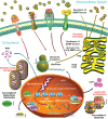
Major characteristics and distinct markers of senescent cells. In response to endogenous stimuli and/or environmental stress, proliferating cells can be forced to into a stable and usually irreversible state of cell cycle arrest, termed cellular senescence. Of note, senescent cells are typically enlarged, flattened, exhibit an irregular cell shape, and develop a secretory phenotype, the SASP, which can be pro-inflammatory and pro-fibrotic. Their nuclear integrity is compromised due to the loss of Lamin B1 and nuclear exclusion of the HMGB1, with the appearance of extranuclear DNA species, particularly CCFs. Senescent cells display increased lysosomal content and enhanced SA-β-Gal activity, with an increased number of large but dysfunctional mitochondria producing high levels of ROS. There is often an elevated level of lipofuscin in the cytoplasm, up-regulated expression of p16INK4a/p21CIP1 and aberrant histone modifications in the nucleus. Biosynthesis and extracellular release of a number of pro-inflammatory factors as part of the SASP can be a prominent hallmark or feature of senescent cells. CCF, cytoplasmic chromatin fragments; HMGB1, high mobility group box 1. Dark red arrows, signal transduction initiated upon binding of extracellular ligands to their corresponding receptors at the plasma membrane. Blue arrows, generation and transportation of ROS in the senescent cell. Purple arrows, transition within and release of molecules from the nucleus. Yellow arrows, protein translation, posttranslational process, and extracellular release of SASP factors. White and black arrows beside molecules or organelles, up- or down-regulation of expression level or biological activity of indicated molecules in the nucleus (white) or the cytoplasm (black), individually.

References

    1. Lopez-Otin C, Blasco MA, Partridge L, et al. . The hallmarks of aging. Cell 2013;153:1194–217. - PMC - PubMed
    1. Chan ASL, Narita M.. Short-term gain, long-term pain: the senescence life cycle and cancer. Genes Dev 2019;33:127–43. - PMC - PubMed
    1. Gorgoulis V, Adams PD, Alimonti A, et al. . Cellular senescence: defining a path forward. Cell 2019;179:813–27. - PubMed
    1. Munoz-Espin D, Serrano M.. Cellular senescence: from physiology to pathology. Nat Rev Mol Cell Biol 2014;15:482–96. - PubMed
    1. Paez-Ribes M, Gonzalez-Gualda E, Doherty GJ, et al. . Targeting senescent cells in translational medicine. EMBO Mol Med 2019;11:e10234. - PMC - PubMed

LinkOut - more resources