Review
. 2023 Jan 12;9(2):e1434.
doi: 10.1097/TXD.0000000000001434. eCollection 2023 Feb.

Overview of Corneal Transplantation for the Nonophthalmologist

Affiliations
Review

Overview of Corneal Transplantation for the Nonophthalmologist

Yujia Zhou et al. Transplant Direct. .

Abstract

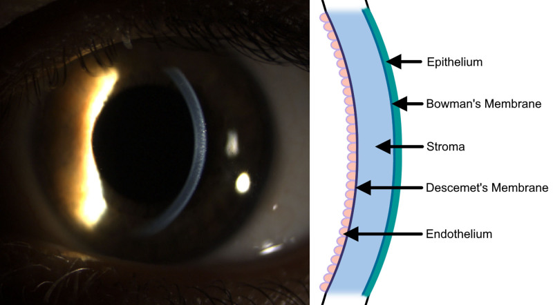
Corneal transplant is a procedure that aims to replace dysfunctional corneal tissue with a transparent graft and is one of the most widely performed transplant surgeries, but its public and professional awareness is low outside of ophthalmology. Corneal tissue consists of 5 major layers that serve to maintain its structural integrity and refractive shape: the epithelium, Bowman's layer, the stroma, Descemet's membrane, and the endothelium. Failure or irreversible damage to any layer of the cornea may be an indication for corneal transplant, and variants of this procedure may be full thickness or selectively lamellar. Complications related to corneal transplantation may occur anywhere from during surgery to years afterward, including rejection, dehiscence, cataract, and glaucoma. Complications should be managed by an ophthalmologist, but other physicians should be aware of prophylactic medications. Topical immunosuppressants and steroids are effective for preventing and treating rejection episodes, whereas there is little evidence to support the use of systemic immunosuppression. Eye protection is recommended for any corneal transplant recipient. Physicians should counsel patients on corneal donation, especially if outside the United States, where donor tissue is in short supply.


Conflict of interest statement

The authors declare no funding or conflicts of interest.

Figures

FIGURE 1.
FIGURE 1.
Cornea location and anatomy. The cornea is the anterior refractive structure of the eye. A slit-lamp photograph with oblique lighting is shown with a schematic detailing layers of the cornea. In order from anterior to posterior, the layers are the epithelium, Bowman’s membrane, corneal stroma, Descemet’s membrane, and endothelium.
FIGURE 2.
FIGURE 2.
Cross sections and photos of corneal transplant procedures. Transplanted tissue is colored, and the recipient eye is grayed out in schematics. A, PKP replaces the entire corneal thickness, with a photo several months after transplant with several sutures removed. B, ALK replaces the anterior stroma, Bowman’s membrane, and epithelium. Photograph shows an intact graft with all sutures removed several months after transplant. C, DALK replaces the entire stroma, Bowman’s membrane, and epithelium while leaving Descemet’s membrane intact. Photograph shows an intact graft several months after transplant with selected sutures removed to correct astigmatism. D, DSEK replaces Descemet’s membrane and endothelium, supported by a thin layer of donor stroma. E, DMEK replaces Descemet’s membrane and endothelium without any donor stroma. An air bubble is used to flatten the graft onto the cornea, and an “S” is used to orient the graft. F, Keratoprosthesis replaces the entire corneal thickness, with artificial optical surfaces displayed in red on the schematic. ALK, anterior lamellar keratoplasty; DALK, deep anterior lamellar keratoplasty; DMEK, Descemet membrane endothelial keratoplasty; DSEK, Descemet stripping endothelial keratoplasty; PKP, penetrating keratoplasty.
FIGURE 3.
FIGURE 3.
Cross sections comparing automated and traditional endothelial keratoplasty. Transplanted tissue is colored, and the recipient eye is grayed out. A, Traditional DSEK grafts include manually resected layers of stroma, Descemet’s membrane, and endothelium. B, DSAEK grafts are produced by microkeratome or femtolaser incisions and may reduce the stromal thickness compared with DSEK. C, Traditional DMEK grafts include manually resected Descemet’s membrane and endothelium with no stromal tissue. D, DMAEK grafts are produced by microkeratome or femtolaser incisions and may include a peripheral rim of stromal tissue not included in DMEK. DMEK, Descemet membrane endothelial keratoplasty; DMAEK, Descemet membrane automated endothelial keratoplasty; DMEK, Descemet membrane endothelial keratoplasty; DSAEK, Descemet stripping automated endothelial keratoplasty; DSEK, Descemet stripping endothelial keratoplasty.

References

    1. Zirm E. Successful total keratoplasty. Graefes Arch Klin Exp Ophthalmol. 1906;64:581.
    1. Albert DM, Edwards DD. The History of Ophthalmology. Vol 7. Blackwell Science Cambridge; 1996.
    1. Lee SH, Cortina MS, Cruz Jdl. History of the artificial cornea. In: Keratoprostheses and Artificial Corneas. Springer; 2015:13–16.
    1. Tan DTH, Dart JKG, Holland EJ, et al. . Corneal transplantation. Lancet. 2012;379:1749–1761. - PubMed
    1. George AJT, Larkin DFP. Corneal transplantation: the forgotten graft. Am J Transplant. 2004;4:678–685. - PubMed