Review
. 2023 Apr;40(2):159-167.
doi: 10.1007/s10719-023-10101-2. Epub 2023 Jan 26.

Gangliosides as Siglec ligands

Affiliations
Review

Gangliosides as Siglec ligands

Ronald L Schnaar. Glycoconj J. 2023 Apr.

Abstract

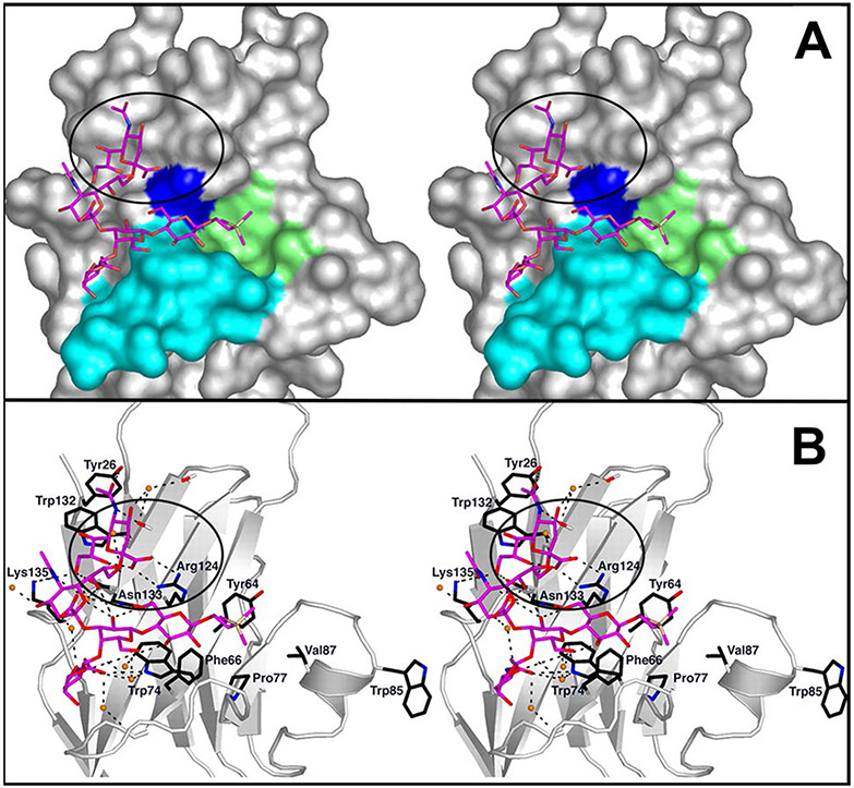
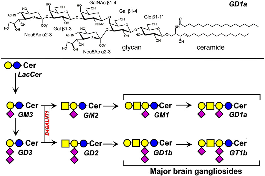
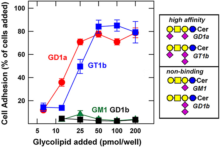
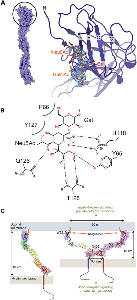
The structure of a sialoglycan can be translated into to a biological response when it binds to a specific endogenous lectin. Among endogenous sialic acid-binding lectins in humans are those comprising the 15-member Siglec family, most of which are expressed on overlapping sets of immune cells. Endogenous Siglec ligands are sialoglycolipids (gangliosides) and/or sialoglycoproteins, on cell surfaces or in the extracellular milieu, that bind to and initiate signaling by cell surface Siglecs. In the nervous system, where gangliosides are the predominant sialoglycans, Siglec-4 (myelin-associated glycoprotein) on myelinating cells binds to gangliosides GD1a and GT1b on nerve cell axons to ensure stable and productive axon-myelin interactions. In the immune system, Siglec-7 on natural killer cells binds to gangliosides GD3 and GD2 to inhibit immune signaling. Expression of GD3 and GD2 on cancer cells can lead to tumor immune evasion. Siglec-1 (sialoadhesin, CD169) on macrophages binds to gangliosides on tumors and enveloped viruses. This may enhance antigen presentation in some cases, or increase viral distribution in others. Several other Siglecs bind to gangliosides in vitro, the biological significance of which has yet to be fully established. Gangliosides, which are found on all human cells and tissues in cell-specific distributions, are functional Siglec ligands with varied roles driving Siglec-mediated signaling.

Keywords: CD33; Macrophages; Myelin-associated glycoprotein; Natural killer cells; Sialic acid; Sialoadhesin.


Conflict of interest statement

Conflicts of interest The authors declare that they have no conflicts of interest.

Figures

Fig. 1
Fig. 1
Binding of a ganglioside GT1b analog to Siglec7. A Stereo image of the outermost Ig-like domain of Siglec-7 bound to a synthetic GT1b analog. Five of the seven sugars of GT1b are resolved at the binding site in the crystal structure (Neu5Acα2-8Neu5Acα2–3[GalNAcβ1-4Galβ1-4Glcβ-R). The terminal sialic acid (Neu5Ac) binds to the conserved Arg residue, R124 (blue). The circled area shows the terminal sialic acid binding via its carboxylate to R124. A convex shelf (marine blue), forms the base of the binding site over which the rest of the glycan lies. The synthetic trimethylsilyl aglycone (yellow) lies in a hydrophobic cup (green). B Stereo image of the network of potential hydrogen bonds (black-dashed lines) at the binding site. Stably associated water molecules are shown as orange spheres. Adapted from reference [12]
Fig. 2
Fig. 2
Ganglioside structure and biosynthesis. The structure of disialo ganglioside GD1a (top). Biosynthesis of major human gangliosides (bottom) using symbol nomenclature for glycans [27]. The biosynthetic gene B4GALNT1 discussed in the text is boxed. Cer = ceramide
Fig. 3
Fig. 3
MAG binding to major brain gangliosides. Fibroblasts were transfected to express full-length MAG on their surface, then were placed in microwells adsorbed with the indicated concentrations of the indicated gangliosides. MAG-mediated cell adhesion is expressed as a percent of the MAG-transfected cells added to each well. Ganglioside structures are shown using symbol nomenclature for glycans [27]. Image adapted from reference [24]
Fig. 4
Fig. 4
MAG-ganglioside binding and a model for myelin-axon engagement. A crystal structure of MAG and its terminal Ig-like domain binding to the “3-O” trisaccharide (Neu5Acα2,3Galβ1-3GalNAc, orange). B Protein-ligand interactions with hydrogen bonds indicated by dashes and Van der Waals’ contacts by curved blue lines, C Model for MAG-mediated myelin-axon engagement and signaling. Dimerization of MAG restricts the distance between the innermost myelin sheath and axon membrane to 10 nm. Reproduced from reference [34]
Fig. 5
Fig. 5
Ganglioside-liposomes bind Siglec-1 on THP-1 human monocyte/macrophage cells and are internalized. Fluorescently-labeled ganglioside-liposomes were incubated with THP1 cells overexpressing Siglec-1, and binding at 4 °C or uptake at 37 °C determined by flow cytometry. Binding or uptake of ganglioside-liposomes at different concentrations are shown. Data are from reference [48]
Fig. 6
Fig. 6
Binding of select Siglecs to major gangliosides. Gangliosides were adsorbed to microwell plates and overlaid with soluble expressed Siglec-Fc chimeras precomplexed to alkaline-phosphatase(AP)-labeled anti-Fc antibody. After incubation and washing, bound Siglec was determined by measuring AP colorimetrically. Data are from references [52] and [53]

Similar articles

Cited by

References

    1. Varki A, Schnaar RL, Schauer R: Sialic acids and other nonulosonic acids. In: Varki A, Cummings RD, Esko JD, Stanley P, Hart GW, Aebi M, Darvill AG, Kinoshita T, Packer NH, Prestegard JH, Schnaar RL, Seeberger PH (eds.) Essentials of glycobiology, 3rd edn., pp. 179–195. Cold Spring Harbor Laboratory Press, Cold Spring Harbor; (2017). https://www.ncbi.nlm.nih.gov/books/NBK579918/
    1. Lundblad A: Gunnar Blix and his discovery of sialic acids. Fascinating molecules in glycobiology. Ups J. Med. Sci 120(2), 104–112 (2015). 10.3109/03009734.2015.1027429 - DOI - PMC - PubMed
    1. Schauer R, Kamerling JP: Exploration of the sialic acid world. Sialic Acids, Pt I: Historical Background and Development, and Chemical Synthesis 75, 1–213 (2018). 10.1016/bs.accb.2018.09.001 - DOI - PMC - PubMed
    1. Blix G: Über die Kohlenhydratgruppen des Submaxillarismucins [Concerning the carbohydrate groups of submaxillary mucin]. Hoppe Seylers Z. Physiol. Chem 240, 43–54 (1936). 10.1515/bchm2.1936.240.1-2.43 - DOI
    1. Gottschalk A, Lind PE: Product of interaction between influenza virus enzyme and ovomucin. Nature 164(4162), 232 (1949). 10.1038/164232a0 - DOI - PubMed

Publication types