. 2023 Feb 8;15(5):6312-6325.
doi: 10.1021/acsami.2c17214. Epub 2023 Jan 26.

Targeted Lymphoma Therapy Using a Gold Nanoframework-Based Drug Delivery System

Affiliations

Targeted Lymphoma Therapy Using a Gold Nanoframework-Based Drug Delivery System

Manpreet Bariana et al. ACS Appl Mater Interfaces. .

Abstract

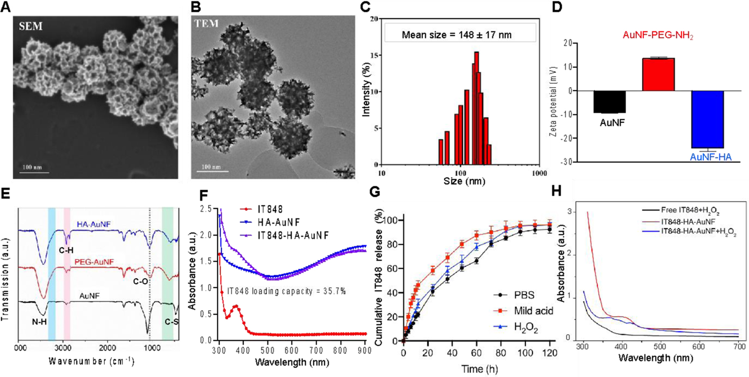
Precision nanomedicine can be employed as an alternative to chemo- or radiotherapy to overcome challenges associated with the often narrow therapeutic window of traditional treatment approaches, while safely inducing effective, targeted antitumor responses. Herein, we report the formulation of a therapeutic nanocomposite comprising a hyaluronic acid (HA)-coated gold nanoframework (AuNF) delivery system and encapsulated IT848, a small molecule with potent antilymphoma and -myeloma properties that targets the transcriptional activity of nuclear factor kappa B (NF-κB). The porous AuNFs fabricated via a liposome-templated approach were loaded with IT848 and surface-functionalized with HA to formulate the nanotherapeutics that were able to efficiently deliver the payload with high specificity to myeloma and lymphoma cell lines in vitro. In vivo studies characterized biodistribution, pharmacokinetics, and safety of HA-AuNFs, and we demonstrated superior efficacy of HA-AuNF-formulated IT848 vs free IT848 in lymphoma mouse models. Both in vitro and in vivo results affirm that the AuNF system can be adopted for targeted cancer therapy, improving the drug safety profile, and enhancing its efficacy with minimal dosing. HA-AuNF-formulated IT848 therefore has strong potential for clinical translation.

Keywords: CD44; gold nanoparticles; hyaluronic acid; lymphoma; nuclear factor-κB inhibitor; targeted drug delivery.


Figures

Figure 1.
Figure 1.
Engineering and pharmacodynamics of IT848-loaded mesoporous gold nanoframeworks (AuNFs). A) Depiction of the expected biodistribution of IT8484-AuNFs in the tumor microenvironment and major organs (liver, spleen, kidneys, lungs, bone marrow and heart) after intravenous injection into the lymphoma-bearing mice, along with selective target/uptake by HA receptor (CD44)-overexpressing cancer cells. Upon uptake by target cells, IT848 released from AuNFs will mediate the inhibition of NF-kB transcriptional activity; B) Schematic illustration of preparation of IT848-HA-AuNFs following 1) AuNF fabrication via the liposome templating approach, 2) IT848 encapsulation and 3) HA surface functionalization of porous AuNFs. The chemical structure of IT848 is also shown.
Figure 2.
Figure 2.
In vitro characterization of AuNFs and IT848-HA-AuNFs. A and B) SEM and TEM images of HA-AuNFs with large mesopores for drug loading; C and D) Size distribution (hydrodynamic radius) (C) and zeta potential (D) of AuNFs determined by DLS; E) Chemical modifications of AuNFs confirmed by FTIR; F) IT848 loading capacity (35.7%) into AuNFs measured using UV spectrometry; G) Cumulative release of IT848 from IT848-HA-AuNFs for 5 days in PBS, pH=6 acidic solution or PBS containing 100 μM H2O2. H) Peroxide-based oxidative degradation of IT848-HA-AuNFs measured by UV-vis absorption spectra as described in the M&M section.
Figure 3.
Figure 3.
HA-AuNF formulation enhances the specificity and potency of IT848 delivery to target cells in vitro. A) CD44 expression levels in EL4 cells, MM.1S cells and control splenocytes from B6 mice were measured by flow cytometry and are presented as mean florescence intensity (MFI), B+C) Selective cellular uptake of Cy7-AuNFs without coating or coated with polyethylene glycol (PEG), hyaluronic acid (HA) (MW 10K) or HA (MW 1000K). AuNFs were incubated for 4 h with EL4 cells, MM.1S cells, and control splenocytes and AuNF cellular uptake was analyzed by flow cytometry. Representative scatter plots (B) and the Mean ± SEM of Cy7 MFI (C) are presented, D +E) Jurkat/GFP/NF-κB transcriptional reporter cells were stimulated with TNF-α and incubated in presence of empty vehicle or 2, 4 and 6 μM of free IT848 (Free-IT848) and equivalent amounts of IT848-loaded AuNFs for up to 96 h. NF-κB transcriptional activity was analyzed at 6, 24, 30, 48, 72 and 96 h by measuring GFP florescence (D) and by flow cytometric analysis of GFP negative cells at 24 and 48 h timepoints (E). Data presented as Mean ± SEM were normalized as percentage of the vehicle treated baseline activity. One of two independent experiments is shown. F-H) Luciferase-expressing EL4 cells were incubated with free or AuNF-encapsulated IT848 (2, 4 μM) or empty vehicle or control particles; F) apoptosis of EL4 cells after 24 h was analyzed by Annexin V/7AAD staining, G) EL4 growth over 72 h was quantified by luciferase assay, H) EL4 metabolic viability for up to 96 h was measured by MTS assay. Data were presented as Mean ± SEM.
Figure 4.
Figure 4.
HA coating enhances the uptake of AuNFs by liver and lymphoma cells. A+B) B6 naïve mice or mice bearing EL4 subcutaneous tumors were sacrificed and hepatic cells were analyzed by flow cytometry (n=3–5), A) Monocytic (M)-MDSC (CD11b+Ly6G-Ly6C+) and polymorphonuclear (PMN)-MDSC (CD11b+Ly6G+Ly6C-) frequencies of hepatic CD45+ cells are presented (Mean ± SEM); B) Expression of CD44 in the hepatic hematopoietic cells (CD45+) and epithelial cells (EpCAM+) was determined via flow cytometry; C-H) Subcutaneous EL4 lymphoma nodules were established in B6 mice and groups of four mice each were treated with Cy7-labled control AuNFs or HA-coated AuNFs. C) Biodistribution of Cy-AuNFs and Cy7-HA-AuNFs was analyzed at different timepoints (10 min, 3 h, 6 h, 9 h and 24 h) by in vivo NIR fluorescence imaging; D) Ex vivo fluorescence imaging-based quantitative analysis of various organs at 12 h and 24 h post i.v. injection of control and HA-coated AuNFs; E) ICP-OES analysis to detect the trace amounts of gold nanoparticles in explanted organs at 12 h and 24 h post-AuNF injection. Data is presented as Mean ± SEM; F) The normalized HA-AuNF quantity in the whole blood of mice (percentage of the injected dose (ID) remaining in the blood) at various time points after HA-AuNF injection (15 min, 30 min, 1 h, 4 h, 12 h, 1 day, 2 day, 4 day, 7 day) was used to determine the pharmacokinetics of intravenously administered HA-AuNFs in mice (n=5); G) Co-localization of fluorescence signal showing that HA-coated AuNFs (red) accumulate in EL4 tumor (blue) nodules in vivo 9 h after i.v. HA-AuNF administration; an image of one representative mouse is presented; H) AuNFs in EL4 lymphoma nodules were quantified via ICP-OES for up to 7 days post i.v. injection of control and HA-coated AuNFs. Nonlinear regression curves and Mean ± SEM of individual measurements are presented.
Figure 5.
Figure 5.
In vitro and in vivo toxicology testing of IT848-loaded AuNFs. A+B) Human PBMCs were resuspended in 100 mL of a hypotonic solution (water) or an isotonic solution (PBS). PBS samples were supplemented with empty HA-AuNFs or IT848-loaded HA-AuNFs (30, 62, 100 mg/mL) or no HA-AuNFs (control). Samples were incubated at room temperature for 4 h. Photographs showing the presence or absence of hemolysis are presented in panel A. Panel B shows the quantitative analysis of the presence or absence of hemolysis by measuring absorbance at 541 nm using a microplate spectrophotometer. Data is presented as Mean ± SEM. C-F) B6 mice were injected with 100 mL of PBS (control) or IT848-loaded HA-AuNFs (5 mg of IT848/kg) via retroorbital sinus injection (n=5). C) Time-dependent body weight changes are presented as Mean ± SEM; D) Complete blood counts (CBC) were measured 2-, 4-, 6-, and 8-weeks post-injection. White blood cell counts (WBC), neutrophils, lymphocytes, red blood cells (RBC) and platelets are presented as Mean ± SEM; E+F) Serum samples were collected from control and IT848-AuNF-injected mice at week 4 and 8 post-injection and levels of blood urea nitrogen (BUN), Creatinine, Albumin, total Bilirubin and Alanine Transaminase (ALT) were measured. Data is presented as Mean ± SEM; G) Histological images of the major organs of the control and IT848 treated mice, 8 weeks post-injection with HA-AuNFs. Images were acquired at 10× magnification.
Figure 6.
Figure 6.
HA-AuNFs loaded with IT848 inhibit lymphoma progression with infrequent intravenous dosing in subcutaneous and orthotopic immunocompetent mouse models. B6 mice received 2×10 EL4 lymphoma cell via subcutaneous injection. One week after lymphoma cell inoculation, mice were assigned to treatment (free IT848 5mg/kg i.v. once a week; IT848-HA-AuNF 5mg/kg i.v. once a week) and control (empty HA-AuNF i.v. once a week) groups. A) Schematic representation of subcutaneous EL4 tumor inoculation followed by IT848-AuNF intervention once a week; B) Mean and SEM of longitudinal tumor volume measurements are presented (n=5). The inset shows representative tumor images at the end of treatment (day 20); C-E) Lymphoma nodules on day 20 post-treatment were harvested for histological and flow cytometric studies. C) Representative images of TUNEL- labeled tumor sections of different treatment groups. Apoptotic cell nuclei were stained dark brown by TUNEL assay and the sections were counter-stained with methyl green. D) Semi-quantification of TUNEL-positive cells out of the total cells. Values are expressed as mean ± SEM. E) Homogenized lymphoma nodules were analyzed via multiparameter flow cytometry for the presence of regulatory T cells (Tregs). Mean and SEM of the Treg frequency of CD45+ cells in the tumor microenvironment are presented (n=5). F) Dissemination patterns of lymphoma progression after intravenous injection of 2 × 10⁶ luciferase-expressing EL4 cells was analyzed longitudinally by in vivo bioluminescence imaging at the indicated time points. Pseudocolor images of a representative mouse superimposed on conventional photographs is presented. G) Schematic representation of intravenous EL4 tumor inoculation followed by IT848-AuNF administration once a week; H) The curves show the probability of overall survival curve in the orthotopic lymphoma model and the comparison between the following groups: control (empty AuNFs i.v. once a week); free IT848 5mg/kg i.v. once a week; IT848-AuNF (5mg/kg) i.v. once a week. Differences between groups were analyzed by Log-rank test. Combined data from three independent experiments are presented (n=10–20).

Similar articles

Cited by

References

    1. Samal P; Begum S Drug Loaded Nanomaterials for Hematological Malignancies Diagnosis and Enhanced Targeted Therapy. In Advanced Nanomaterials for Point of Care Diagnosis and Therapy; Elsevier: 2022; pp 383–398.
    1. Powsner EH; Harris JC; Day ES Biomimetic Nanoparticles for the Treatment of Hematologic Malignancies. Advanced NanoBiomed Research 2021, 1 (4), 2000047.
    1. Bromma K; Chithrani DB Advances in Gold Nanoparticle-based Combined Cancer Therapy. Nanomaterials 2020, 10 (9), 1671. - PMC - PubMed
    1. Jain S; Hirst D; O’Sullivan J Gold Nanoparticles as Novel Agents for Cancer Therapy. The British Journal of Radiology 2012, 85 (1010), 101–113. - PMC - PubMed
    1. Scott AM; Allison JP; Wolchok JD Monoclonal Antibodies in Cancer Therapy. Cancer Immunity Archive 2012, 12 (1), 14–22. - PMC - PubMed