Friction Blisters of the Feet: A New Paradigm to Explain Causation
- PMID: 36701751
- PMCID: PMC10783477
- DOI: 10.4085/1062-6050-0309.22
Friction Blisters of the Feet: A New Paradigm to Explain Causation
Abstract
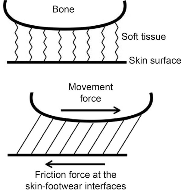
Friction blisters on the feet commonly occur when individuals engage in active pursuits such as running, hiking, and military training. The high prevalence of blisters in active individuals underscores the fact that the pathomechanics of this condition are not fully understood. The traditional blister causation paradigm revolves around heat, moisture, and friction. In reality, foot friction blisters are caused by repetitive shear deformation. The 3 fundamental elements of blister-inducing shear deformation are (1) motion of bone, (2) high friction force, and (3) repetition of the resulting shear events. Rubbing at the skin surface is not a mechanism for friction blister formation. To that end, prevention of the friction blister continues to be an elusive quest for both the patient and the treating clinician. In this article, we aimed to highlight the limitations of the long-held blister-causation paradigm and offer a new explanation.
Keywords: foot injury; shear; skin injury.
© by the National Athletic Trainers’ Association, Inc.
Figures
Similar articles
-
Friction Blisters of the Feet: A Critical Assessment of Current Prevention Strategies.J Athl Train. 2024 Jan 1;59(1):8-21. doi: 10.4085/1062-6050-0341.22. J Athl Train. 2024. PMID: 36701678 Free PMC article.
-
Influence of an antiperspirant on foot blister incidence during cross-country hiking.J Am Acad Dermatol. 1998 Aug;39(2 Pt 1):202-6. doi: 10.1016/s0190-9622(98)70075-1. J Am Acad Dermatol. 1998. PMID: 9704829
-
Blisters on the battlefield: the prevalence of and factors associated with foot friction blisters during Operation Iraqi Freedom I.Mil Med. 2012 Feb;177(2):157-62. doi: 10.7205/milmed-d-11-00325. Mil Med. 2012. PMID: 22360060
-
Friction blisters. Pathophysiology, prevention and treatment.Sports Med. 1995 Sep;20(3):136-47. doi: 10.2165/00007256-199520030-00002. Sports Med. 1995. PMID: 8570998 Review.
-
Etiological Foundation for Practical Strategies to Prevent Exercise-Related Foot Blisters.Curr Sports Med Rep. 2016 Sep-Oct;15(5):330-5. doi: 10.1249/JSR.0000000000000297. Curr Sports Med Rep. 2016. PMID: 27618242 Review.
Cited by
-
Influence of changes in foot morphology and temperature on bruised toenail injury risk during running.Sci Rep. 2024 Jan 21;14(1):1826. doi: 10.1038/s41598-024-51826-w. Sci Rep. 2024. PMID: 38246957 Free PMC article.
-
Influence of skin hydration level on the occurrence of blisters on the foot during hiking.Int Wound J. 2024 Dec;21(12):e70024. doi: 10.1111/iwj.70024. Int Wound J. 2024. PMID: 39675818 Free PMC article.
References
MeSH terms
LinkOut - more resources
Full Text Sources
Medical