. 2023 May:637:193-206.
doi: 10.1016/j.jcis.2023.01.086. Epub 2023 Jan 21.

Cancer cells inhibition by cationic carbon dots targeting the cellular nucleus

Affiliations

Cancer cells inhibition by cationic carbon dots targeting the cellular nucleus

Jiuyan Chen et al. J Colloid Interface Sci. 2023 May.

Abstract

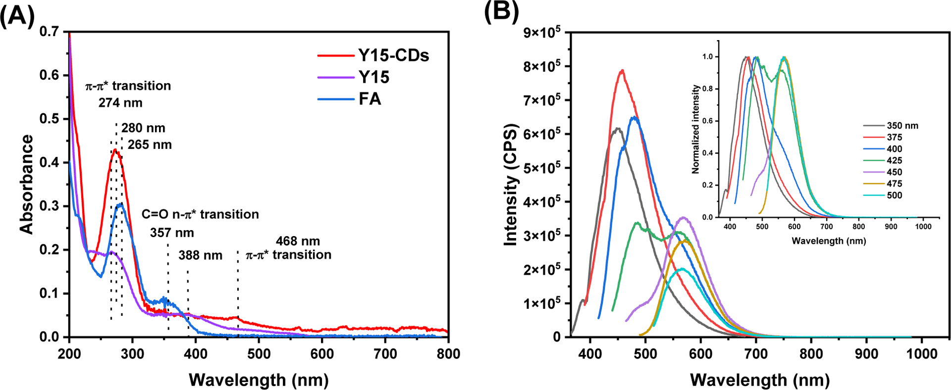
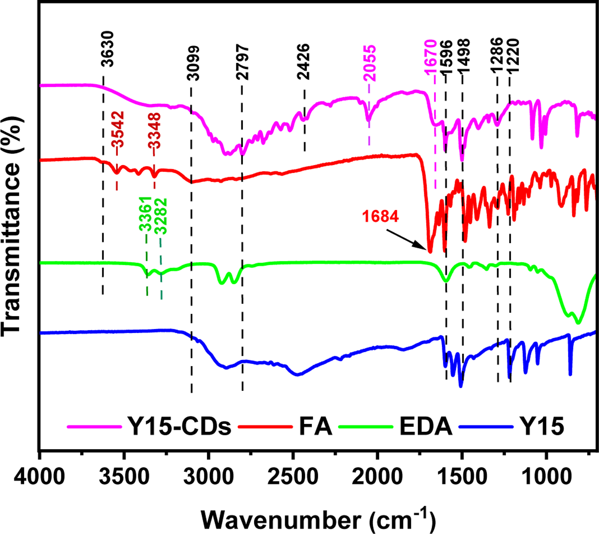
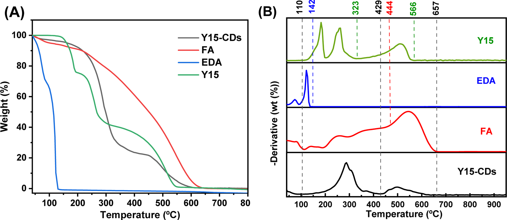
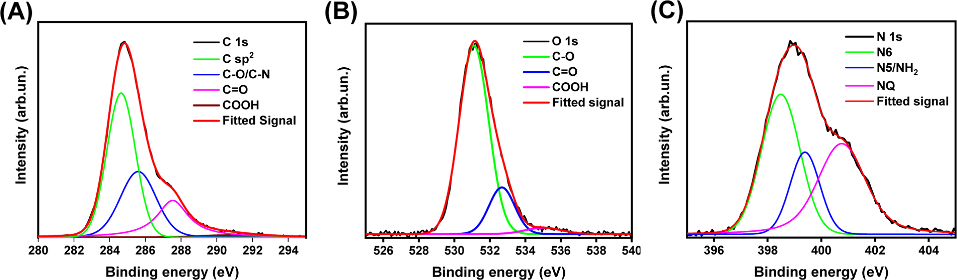
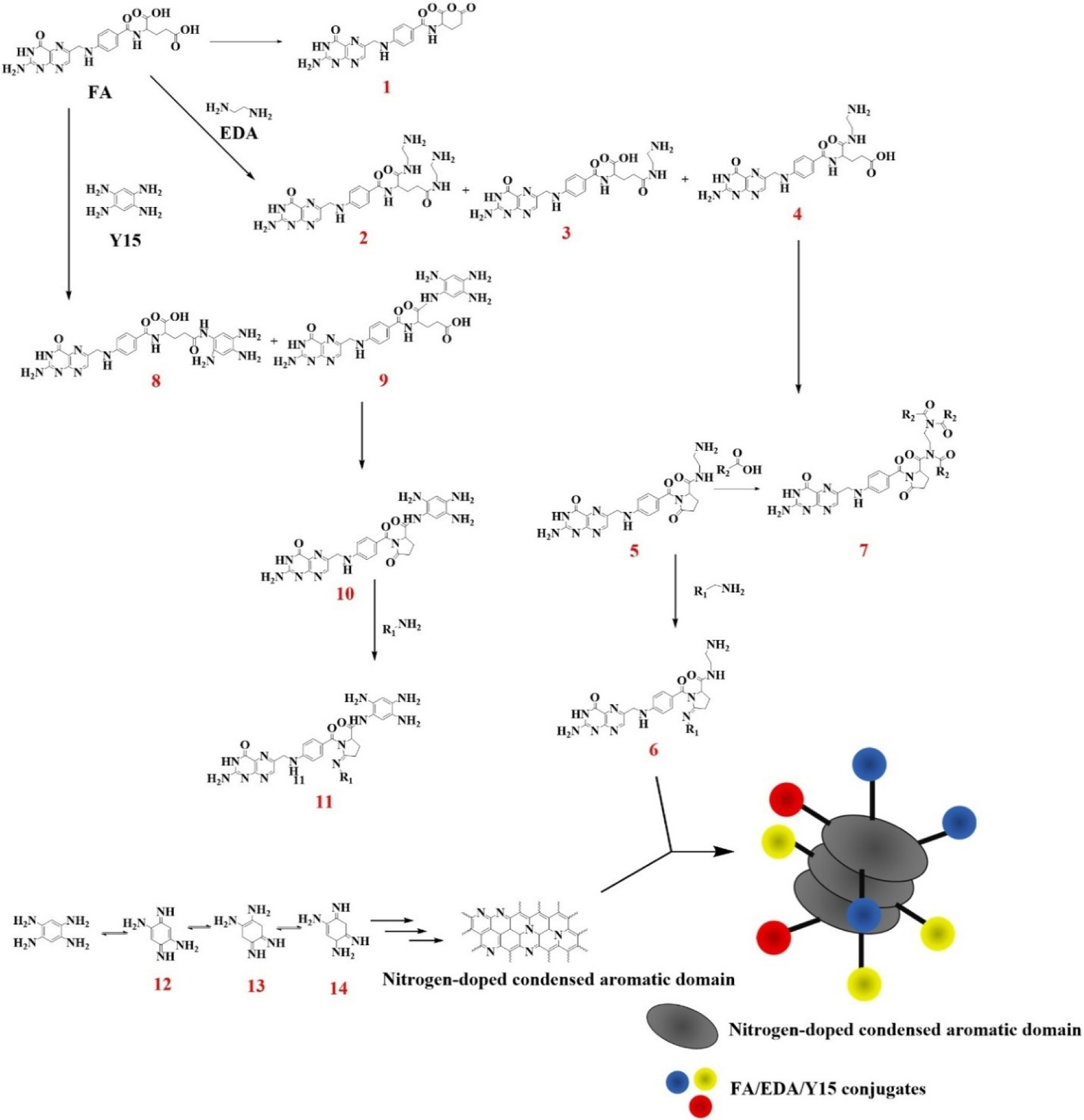
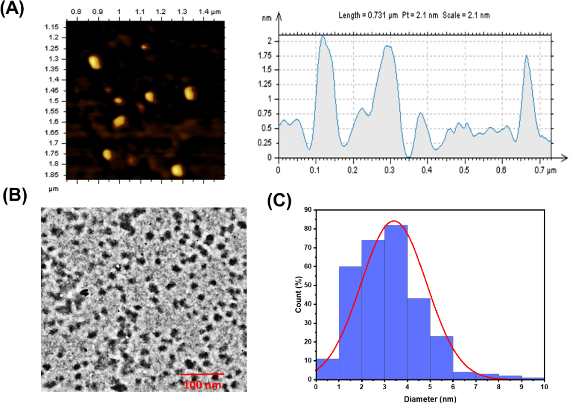
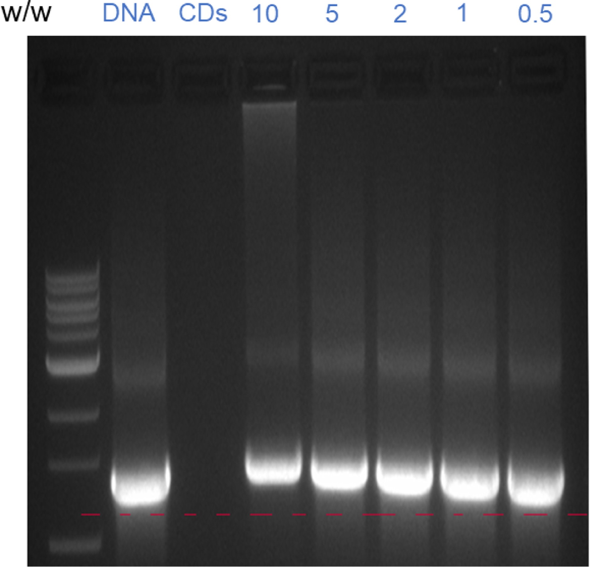
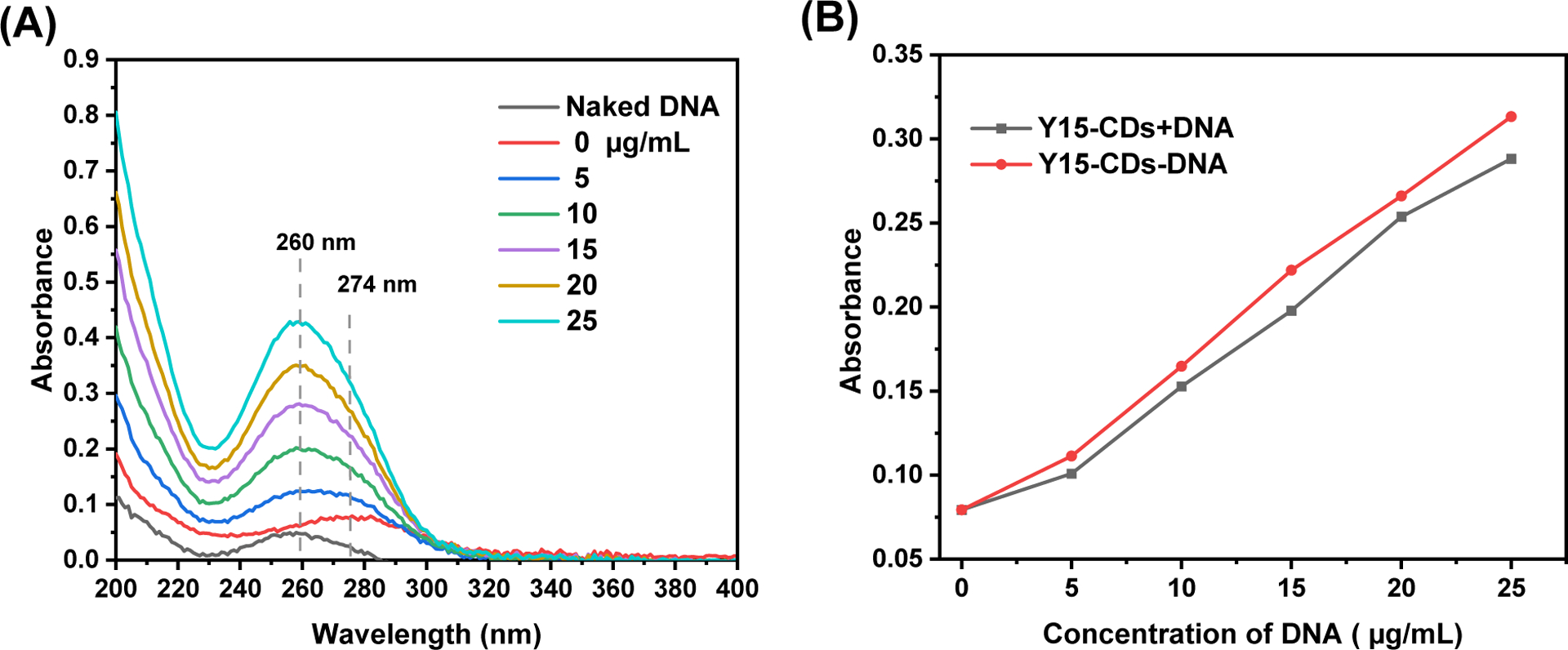
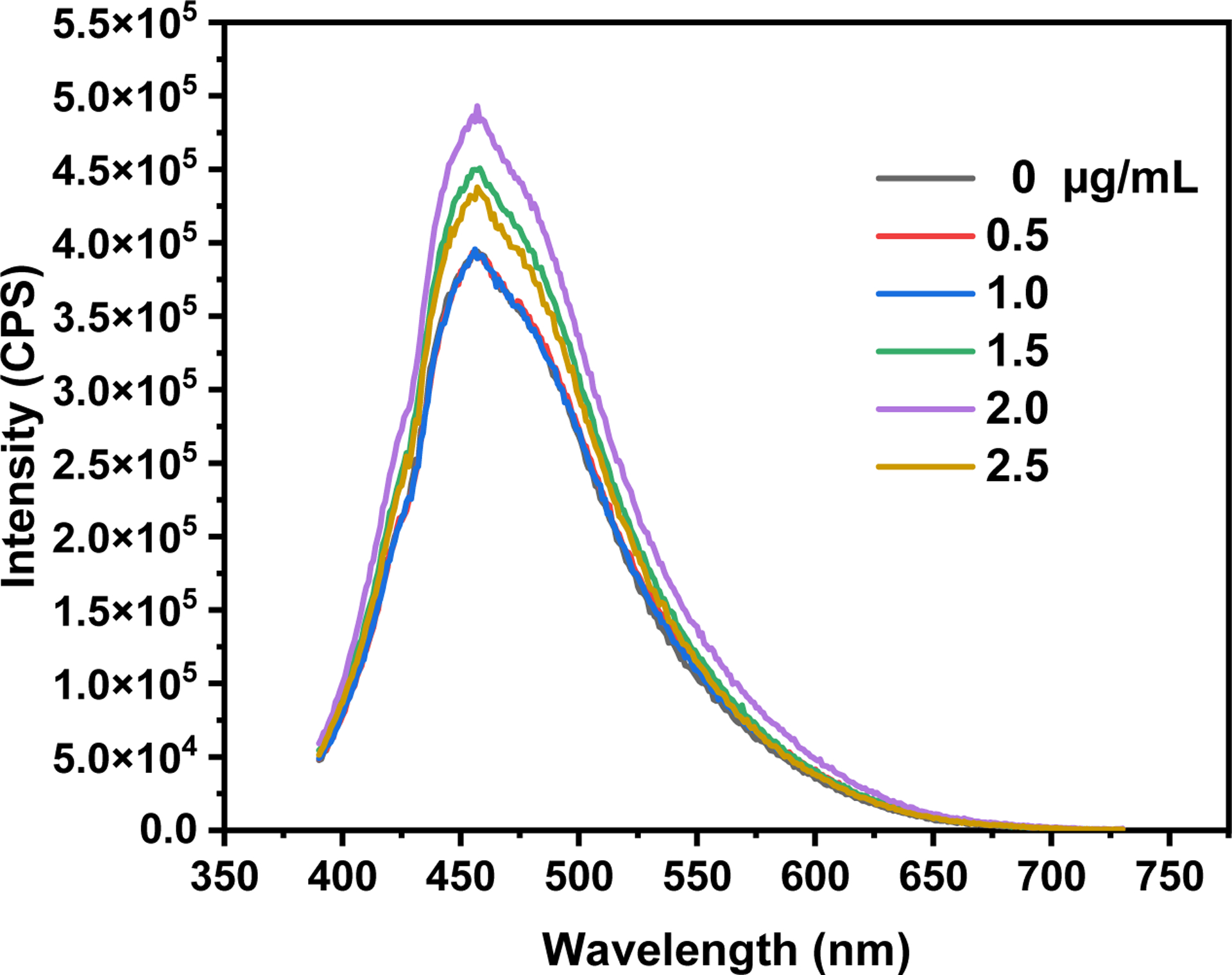
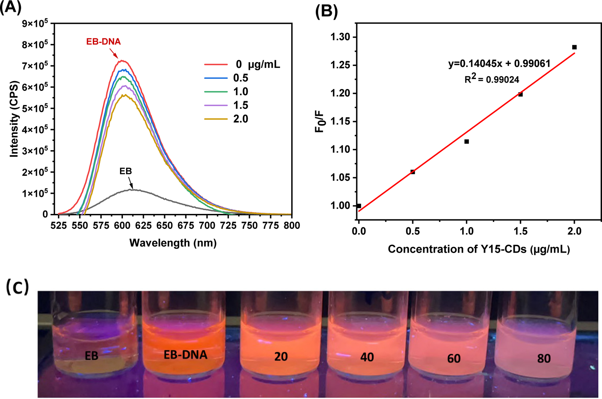
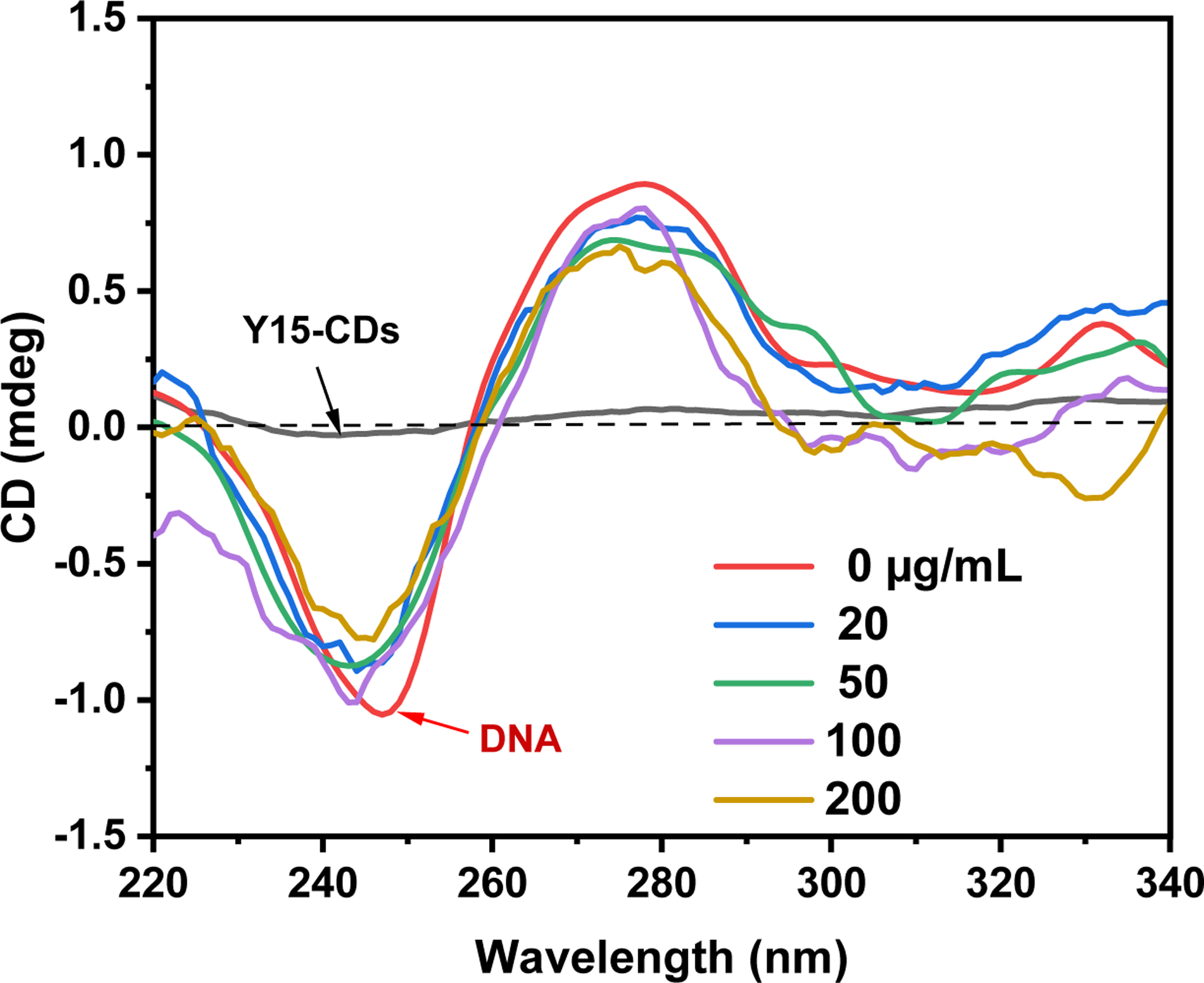
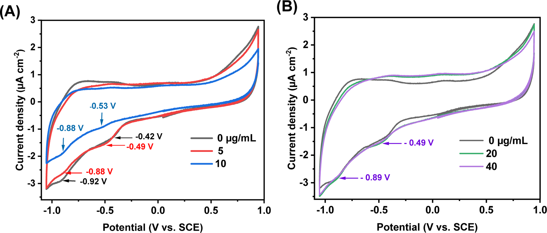
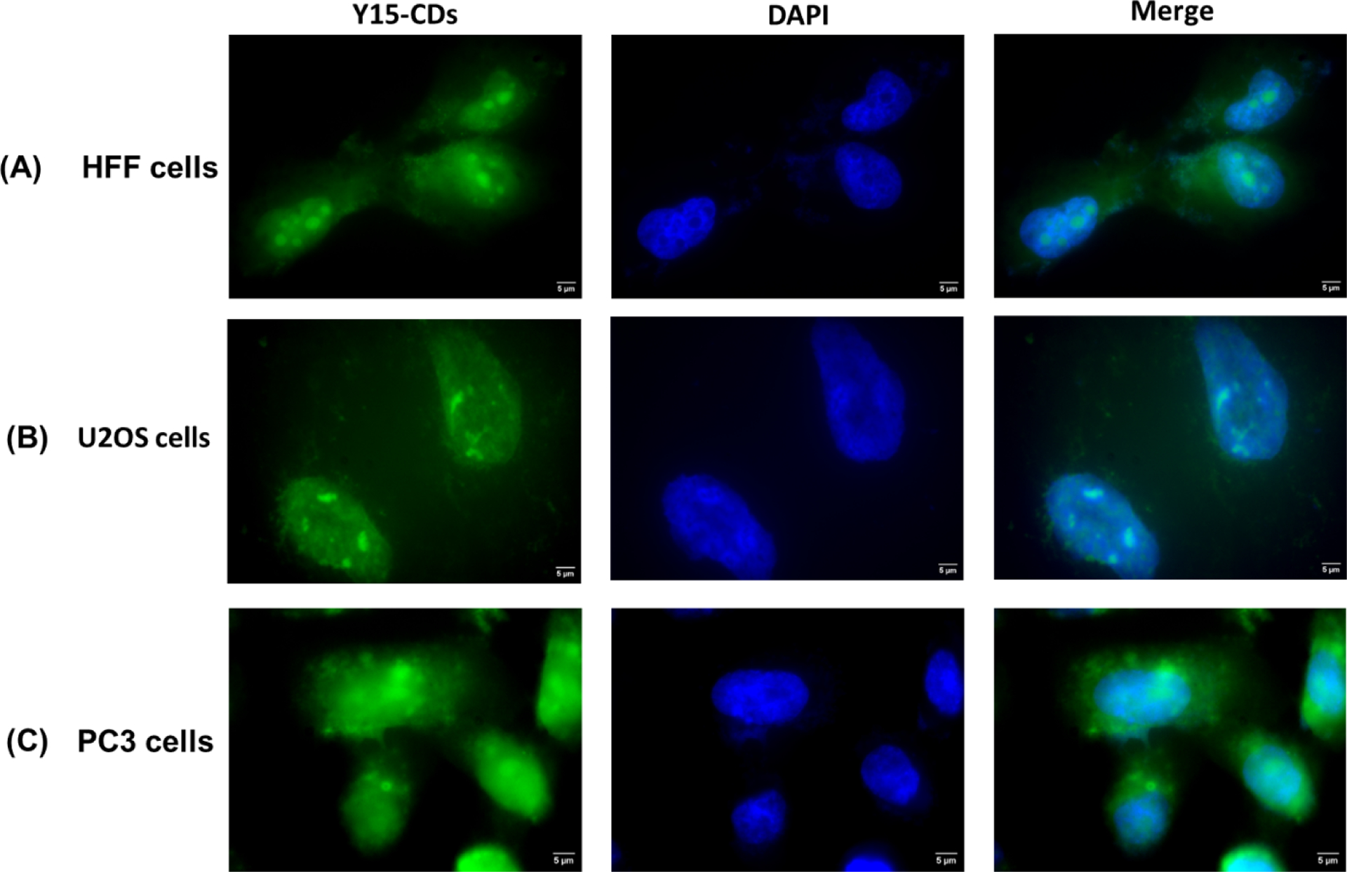
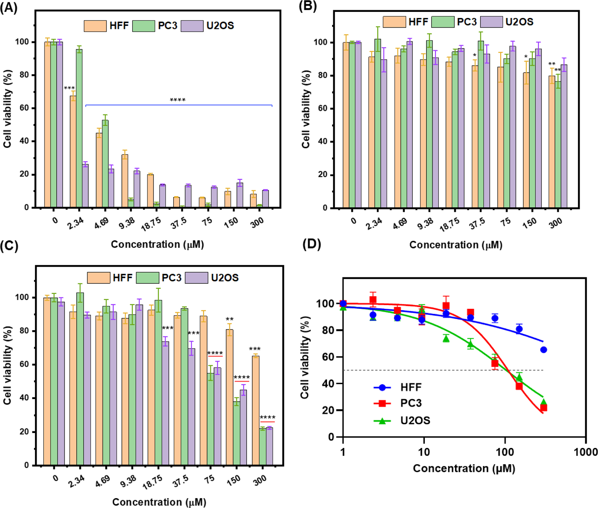
Nucleus targeting is tremendously important in cancer therapy. Cationic carbon dots (CCDs) are potential nanoparticles which might enter cells and penetrate nuclear membranes. Although some CCDs have been investigated in nucleus targeting and applied in nuclear imaging, the CCDs derived from drugs, that are able to target the nucleus, bind with DNA and inhibit the growth of cancer cells have not been reported. In this project, 1, 2, 4, 5-benzenetetramine (Y15, a focal adhesion kinase inhibitor) derived cationic carbon dots (Y15-CDs) were prepared via a hydrothermal approach utilizing Y15, folic acid and 1,2-ethylenediamine as precursors. Based on the structural, optical, and morphologic characterizations, Y15-CDs possess rich amine groups and nitrogen in structure, an excitation-dependent photoluminescence emission, and a small particle size of 2 to 4 nm. The DNA binding experiments conducted through agarose gel electrophoresis, UV-vis absorption, fluorescence emission, and circular dichroism spectroscopies, prove that Y15-CDs might bind with DNA via electrostatic interactions and partially intercalative binding modes. In addition, the cell imaging and cytotoxicity studies in human foreskin fibroblasts (HFF), prostate cancer (PC3) and osteosarcoma cells (U2OS) indicate the nucleus targeting and anticancer abilities of Y15-CDs. Most interestingly, Y15-CDs exhibit a higher cytotoxicity to cancer cells (PC3 and U2OS) than to normal cells (HFF), inferring that Y15-CDs might be potentially applied in cancer therapy.

Keywords: 1,2,4,5-benzenetetramine; Cancer therapy; Cationic carbon dots; DNA binding; Nucleus targeting.


Conflict of interest statement

Declaration of Competing Interest The authors declare that they have no known competing financial interests or personal relationships that could have appeared to influence the work reported in this paper.

Figures

Figure 1.
Figure 1.
(A) UV-vis spectrum of FA (10 μg/mL), Y15 (10 μg/mL) and Y15-CDs (50 μg/mL) and (B) fluorescence spectrum of Y15-CDs (2.5 μg/mL). Inset are the normalized fluorescence spectra.
Figure 2.
Figure 2.
FTIR spectra of FA, EDA, Y15 and Y15-CDs.
Figure 3.
Figure 3.
(A) TGA and (B) DTG of FA, EDA, Y15 and Y15-CDs.
Figure 4.
Figure 4.
High-resolution XPS spectra of (A) C1s, (B) O1s and (C) N1s of Y15-CDs.
Figure 5.
Figure 5.
A hypothetical mechanism of Y15-CDs formation.
Figure 6.
Figure 6.
(A) AFM, (B) TEM, and (C) TEM size distribution histogram of Y15-CDs.
Figure 7.
Figure 7.
The agarose gel electrophoresis analyses of Y15-CDs-DNA complexes at different weight ratio of Y15-CDs to DNA.
Figure 8.
Figure 8.
(A) UV-vis absorption spectra of 20 μg/mL Y15-CDs in the presence of different concentration of DNA (0, 5, 10, 15, 20 or 25 μg/mL) in DI water; (B) The absorbances of Y15-CDs-DNA complex solutions (the solution contains both of Y15-CDs at concentration of 0, 5, 10, 15, 20 or 25 μg/mL and DNA at 20 μg/mL), and the sum values of absorbance (Y15-CDs + DNA) of free DNA (the solution only contains DNA at 5, 10, 15, 20 or 25 μg/mL) and free Y15-CDs (the solution only contains Y15-CDs at 20 μg/mL).
Figure 9.
Figure 9.
Fluorescence emission spectra of 2 μg/mL Y15-CDs solution upon the excitation at 375 nm with addition of different concentration of DNA (0, 0.5, 1.0, 1.5, 2.0 and 2.5 μg/mL).
Figure 10.
Figure 10.
(A) Fluorescence emission spectra of 0.5 μg/mL EB bound with 1 μg/mL DNA in the presence of Y15-CDs (0, 0.5, 1.0, 1.5 and 2.0 μg/mL); (B) Stem-Volmer quenching plot of EB bound with DNA in the presence of different concentration of Y15-CDs. The excitation wavelength was 510 nm; (C) fluorescence of 20 μg/mL EB bound with 40 μg/mL DNA in the presence of Y15-CDs (20, 40, 60 and 80 μg/mL) under the UV lamp at 365 nm.
Figure 11.
Figure 11.
Circular dichroism spectra of 50 μg/mL DNA with addition of 20, 50, 100 and 200 μg/mL of Y15-CDs.
Figure 12.
Figure 12.
Cyclic voltammograms of 10 μg/mL of Y15-CDs (A) in the presence of 0, 5, and 10 μg/mL of DNA and (B) in the presence of 0, 20, and 40 μg/mL of DNA recorded in H2O using 0.1 M NaCl as supporting electrolyte, glassy carbon (GC) electrode as working electrode, Ag/AgCl as reference electrode, and a Pt wire as a counter electrode.
Figure 13.
Figure 13.
Fluorescence images of (A) human foreskin fibroblasts (HFF), (B) osteosarcoma (U2OS) and (C) prostate cancer (PC3) cells incubated with Y15-CDs (1 mg/mL) for 30 min.
Figure 14.
Figure 14.
In vitro cytotoxicity evaluation of (A) Y15, (B) FA-EDA and (C) Y15-CDs at different concentrations in HFF, PC3 and U2OS cells incubated for 72 h at 37 °C; (D) comparation of the cell viabilities of Y15-CDs to HFF, PC3, and U2OS cells. Data are showing as mean ± SEM. Statistical significance was determined by using a one-way ANOVA analysis. *p<0.05, **p<0.01, ***p<0.001, ****p<0.0001 compared to non-treated controls.

References

    1. Duncan R, Nanomedicine gets clinical, Materials Today 8(8) (2005) 16–17. 10.1016/S1369-7021(05)71032-4. - DOI
    1. Wang B, Song H, Qu X, Chang J, Yang B, Lu S, Carbon dots as a new class of nanomedicines: Opportunities and challenges, Coordination Chemistry Reviews 442 (2021) 1–18. 10.1016/j.ccr.2021.214010. - DOI
    1. Raj S, Khurana S, Choudhari R, Kesari KK, Kamal MA, Garg N, Ruokolainen J, Das BC, Kumar D, Specific targeting cancer cells with nanoparticles and drug delivery in cancer therapy, Semin Cancer Biol 69 (2021) 166–177. 10.1016/j.semcancer.2019.11.002. - DOI - PubMed
    1. Liu J, Li R, Yang B, Carbon Dots: A new type of carbon-based nanomaterial with wide applications, ACS Cent Sci 6(12) (2020) 2179–2195. 10.1021/acscentsci.0c01306. - DOI - PMC - PubMed
    1. Ji C, Zhou Y, Leblanc RM, Peng Z, Recent developments of carbon dots in biosensing: A review, ACS Sens 5(9) (2020) 2724–2741. 10.1021/acssensors.0c01556. - DOI - PubMed