. 2023 Feb 27;33(4):755-763.e3.
doi: 10.1016/j.cub.2023.01.004. Epub 2023 Jan 25.

Piebaldism and chromatophore development in reptiles are linked to the tfec gene

Affiliations

Piebaldism and chromatophore development in reptiles are linked to the tfec gene

Alan Garcia-Elfring et al. Curr Biol. .

Abstract

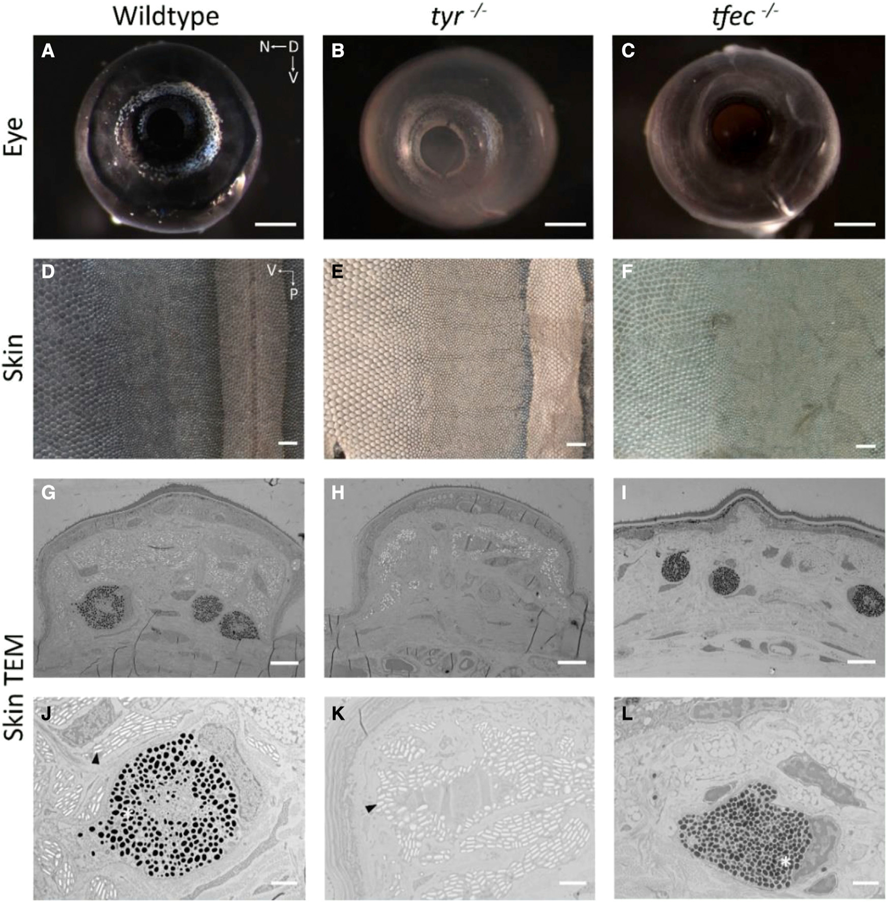
Reptiles display great diversity in color and pattern, yet much of what we know about vertebrate coloration comes from classic model species such as the mouse and zebrafish.1,2,3,4 Captive-bred ball pythons (Python regius) exhibit a remarkable degree of color and pattern variation. Despite the wide range of Mendelian color phenotypes available in the pet trade, ball pythons remain an overlooked species in pigmentation research. Here, we investigate the genetic basis of the recessive piebald phenotype, a pattern defect characterized by patches of unpigmented skin (leucoderma). We performed whole-genome sequencing and used a case-control approach to discover a nonsense mutation in the gene encoding the transcription factor tfec, implicating this gene in the leucodermic patches in ball pythons. We functionally validated tfec in a lizard model (Anolis sagrei) using the gene editing CRISPR/Cas9 system and TEM imaging of skin. Our findings show that reading frame mutations in tfec affect coloration and lead to a loss of iridophores in Anolis, indicating that tfec is required for chromatophore development. This study highlights the value of captive-bred ball pythons as a model species for accelerating discoveries on the genetic basis of vertebrate coloration.

Keywords: Mendelian phenotype; ball python; color morph; genomics; pigmentation.


Conflict of interest statement

Declaration of interests The authors declare no competing interests.

Figures

Figure 1.
Figure 1.. A small sample of the phenotypic variation found in captive-bred ball pythons (Python regius)
(A) Wild type, (B) piebald, (C) banana piebald, (D) pastel piebald, (E) pastel HRA enhancer, (F) ultramel clown, and (G) banana champagne. Photo credit: pethelpful.com (A) and Designing Morphs (B–G).
Figure 2.
Figure 2.. Inheritance patterns and genomic differentiation
(A) Clutch records (2008–2018) from a commercial breeder (KINOVA) indicate piebald has a recessive mode of inheritance. (B) FST plot between piebald and non-piebald samples using a chromosome-length genome assembly. The FST peak on chromosome 7 delineates the region of interest containing the putative causal gene for the piebald phenotype.
Figure 3.
Figure 3.. Phenotypic comparisons of Anolis sagrei
Wild type (A) and F0 tfec mutant (B). The mutant showed reduced body coloration, particularly in the snout, forelimbs, and hindlimbs (arrows).
Figure 4.
Figure 4.. tfec is required for iridophore development in Anolis sagrei
Presented are eye and skin samples from wild type (A, D, G, and J) and mutants with reading frame mutations in tyr (B, E, H, and K) and tfec (C, F, I, and L). (A–C) Anterior view of hatchling eyes. (D–F) Dissected skin from the trunk of hatchlings. For these panels, anterior surface is up, and the posterior surface is down. Ventral surface is on the left side of the image and the dorsal surface is on the right. The dorsal stripe can be seen in (D) and (E) while (F) exhibits a lack of this back pattern. (G–L) TEM images of individual dorsal scales (G–I) and higher-magnification images of melanophores and iridophores (J–L). Melanophores hold pigmented melanosomes while iridophore reflectiveness arises from guanine crystals. For tyr−/− note the absence of melanosomes and the presence of guanine crystals. For tfec−/− note the presence of melanosomes and the absence of guanine crystals. tyr samples are from F2 lizards; tfec samples are from F1 lizards. Asterisks show melanosomes while arrowheads point to guanine crystals. Scale bars, (A–F) 500 μm, (G–I) 6 μm, (J–L) 2 μm.

Similar articles

Cited by

References

    1. Caro T (2017). Wallace on coloration: contemporary perspective and unresolved insights. Trends Ecol. Evol 32, 23–30. 10.1016/j.tree.2016.10.003. - DOI - PubMed
    1. Endler JA, and Mappes J (2017). The current and future state of animal coloration research. Philos. Trans. R. Soc. Lond. B Biol. Sci 372, 20160352. 10.1098/rstb.2016.0352. - DOI - PMC - PubMed
    1. Davison A, Jackson HJ, Murphy EW, and Reader T (2019). Discrete or indiscrete? Redefining the colour polymorphism of the land snail Cepaea nemoralis. Heredity 123, 162–175. - PMC - PubMed
    1. Patterson LB, and Parichy DM (2013). Interactions with iridophores and the tissue environment required for patterning melanophores and xanthophores during zebrafish adult pigment stripe formation. PLoS Genet 9, e1003561. - PMC - PubMed
    1. Jahanbakhsh E, and Milinkovitch MC (2022). Modeling convergent scale-by-scale skin color patterning in multiple species of lizards. Curr. Biol 32, 5069–5082.e13. - PMC - PubMed

Publication types

Substances

LinkOut - more resources